-
 Best Disease
Mar 9 2013 by Hamid Ahmadieh, MD
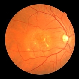
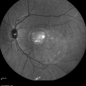
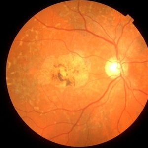
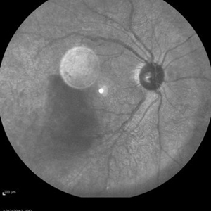
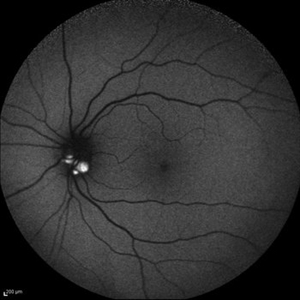
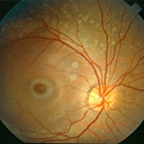
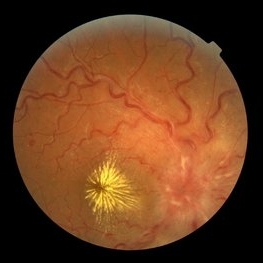
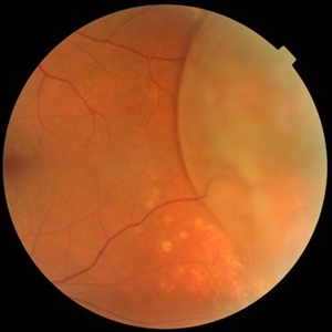
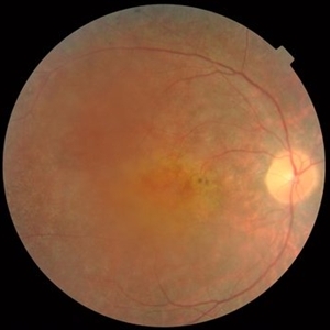
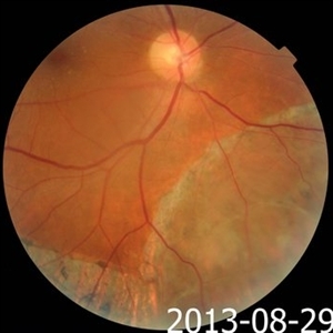
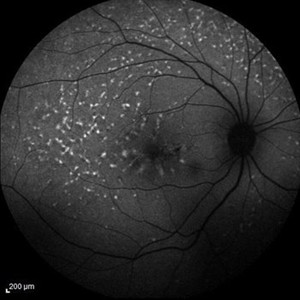
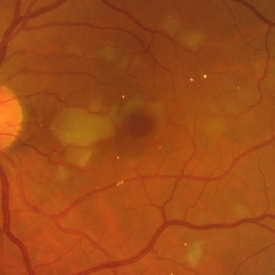
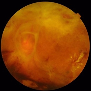
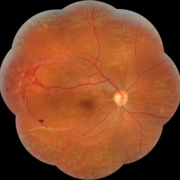
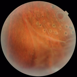
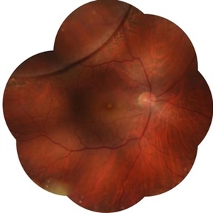
Color fundus photograph the right eye of a 49-year-old man with decreased VA due to advanced Best disease.
Photographer: Soodabeh Fooladin, Negah Eye Center, Tehran
Condition/keywords: Best disease
-
 Best Disease
Mar 9 2013 by Hamid Ahmadieh, MD
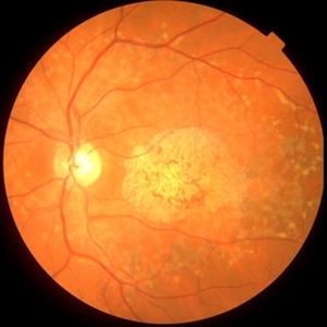
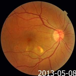
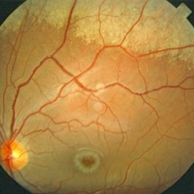
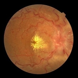
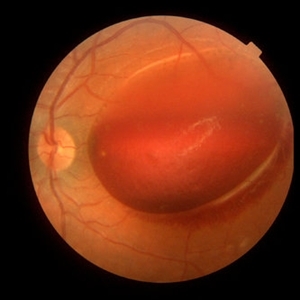
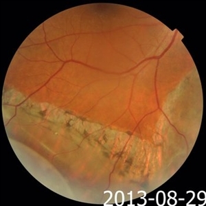
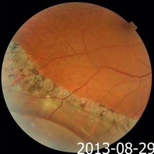
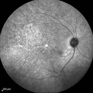
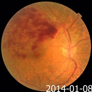
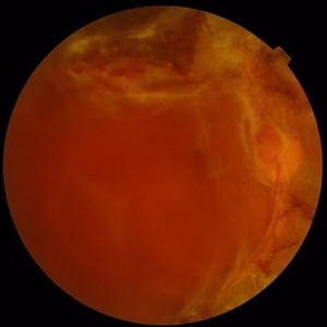
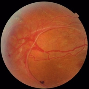
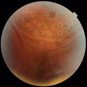
Color fundus photograph the left eye of a 49-year-old man with decreased VA due to advanced Best disease.
Photographer: Soodabeh Fooladin, Negah Eye Center, Tehran
Condition/keywords: Best disease
-
 Best Disease
Mar 9 2013 by Hamid Ahmadieh, MD
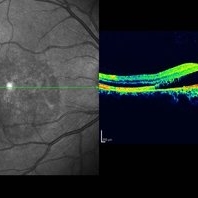
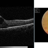
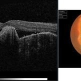
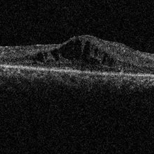
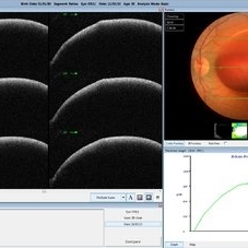
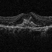
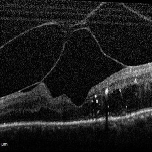
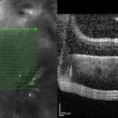
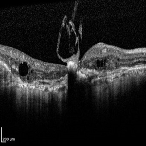
OCT of the right eye of a 49-year-old man with decreased VA due to advanced Best disease.
Photographer: Soodabeh Fooladin, Negah Eye Center, Tehran
Imaging device: Heidelberg Spectralis
Condition/keywords: Best disease, optical coherence tomography (OCT)
-
 Best Disease
Mar 9 2013 by Hamid Ahmadieh, MD
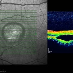
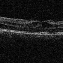
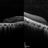
OCT of the left eye of a 49-year-old man with decreased VA due to advanced Best disease.
Photographer: Soodabeh Fooladin, Negah Eye Center, Tehran
Imaging device: Heidelberg Spectralis
Condition/keywords: Best disease, optical coherence tomography (OCT)
-
 Best Disease
Mar 9 2013 by Hamid Ahmadieh, MD
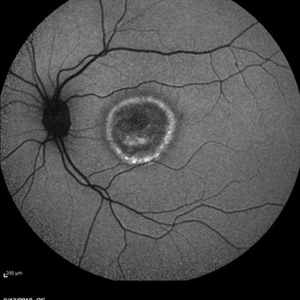
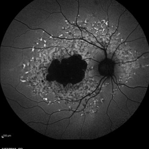
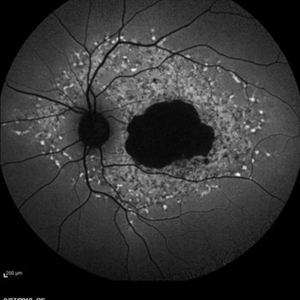
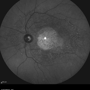
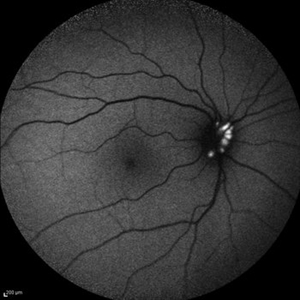
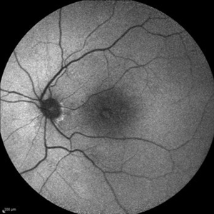
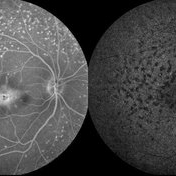
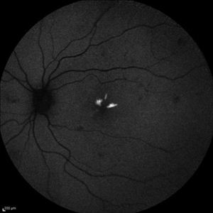
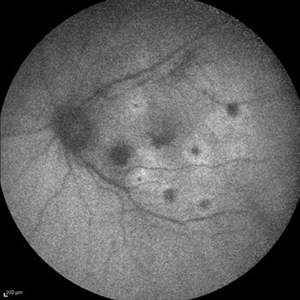
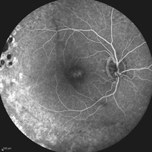
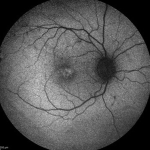
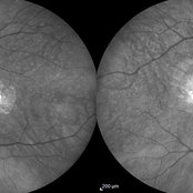
Autofluorescence Imaging of the left eye of a 49-year-old man with decreased VA due to advanced Best disease.
Photographer: Soodabeh Fooladin, Negah Eye Center, Tehran
Imaging device: Heidelberg Spectralis
Condition/keywords: autofluorescence imaging, Best disease
-
 Best Disease
Mar 9 2013 by Hamid Ahmadieh, MD
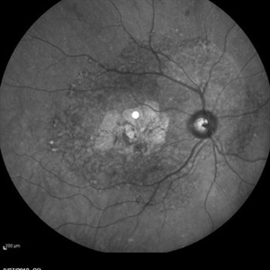
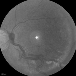
Infrared imaging of the left eye of a 49-year-old man with decreased VA due to advanced Best disease.
Photographer: Soodabeh Fooladin, Negah Eye Center, Tehran
Imaging device: Heidelberg Spectralis
Condition/keywords: Best disease, infrared image
-
 Best Disease
Mar 9 2013 by Hamid Ahmadieh, MD
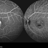
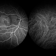
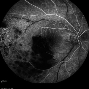
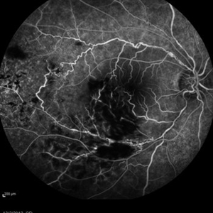
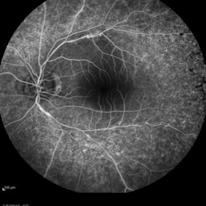
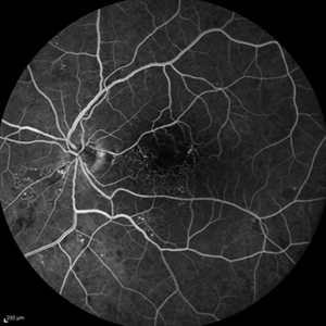
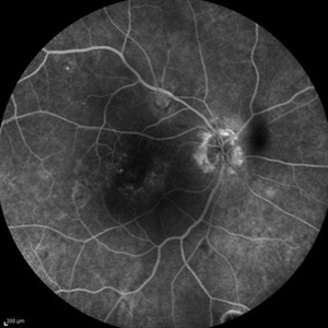
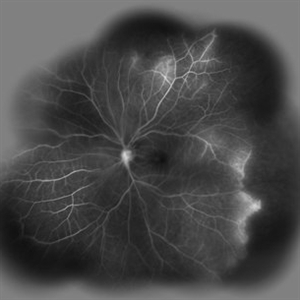
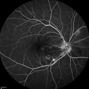
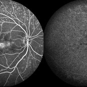
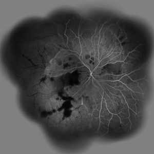
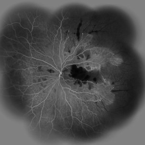
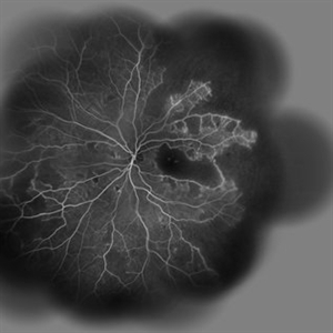
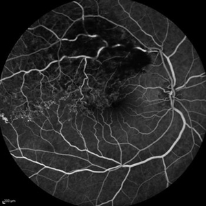
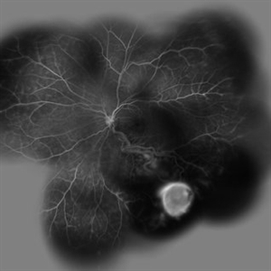
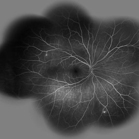
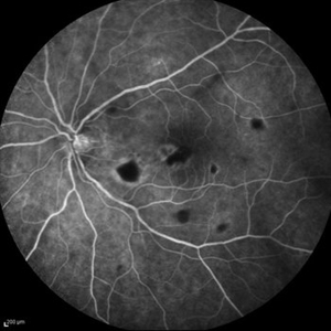
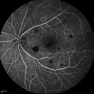
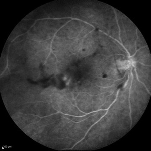
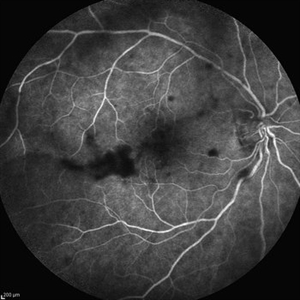
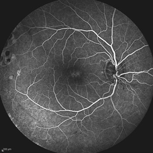
FA and ICG Angiography of the left eye of a 49-year-old man with advanced Best disease.
Photographer: Soodabeh Fooladin, Negah Eye Center, Tehran
Imaging device: Heidelberg Spectralis
Condition/keywords: Best disease, indocyanine green (ICG) angiography
-
 Best Disease
Mar 9 2013 by Hamid Ahmadieh, MD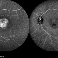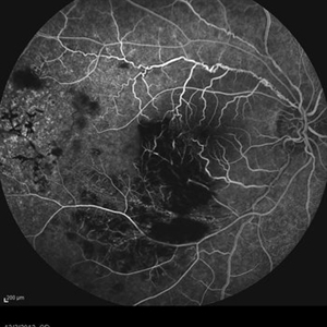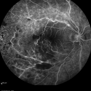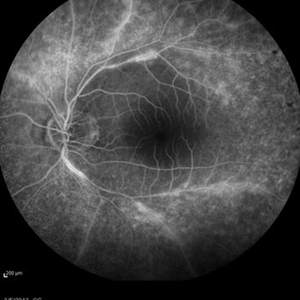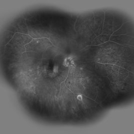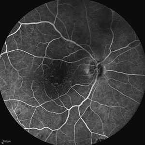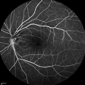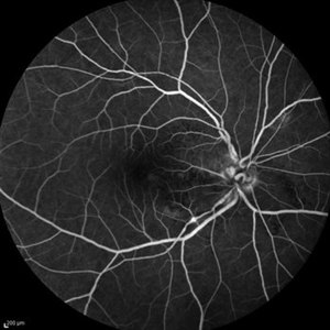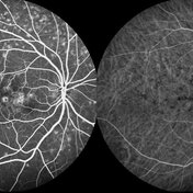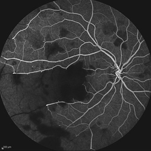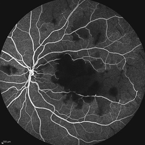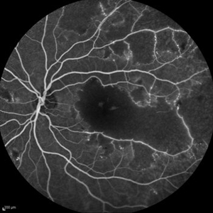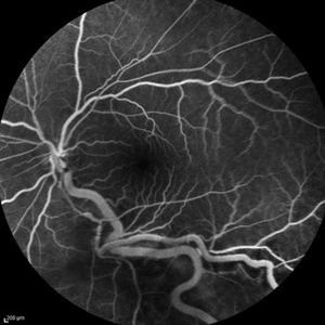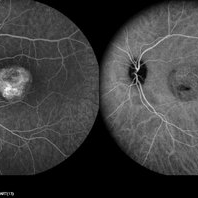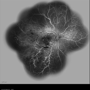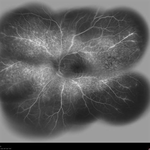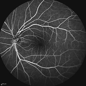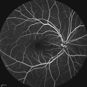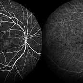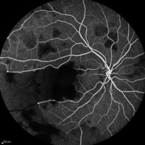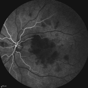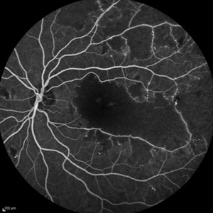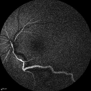
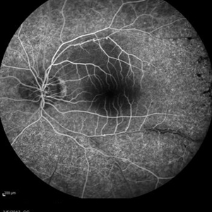
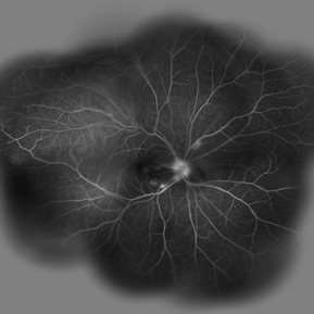
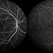
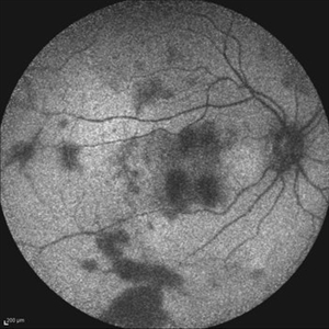
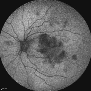
FA and ICG Angiography of the left eye of a 49-year-old man with advanced Best disease.
Photographer: Soodabeh Fooladin, Negah Eye Center, Tehran
Imaging device: Heidelberg Spectralis
Condition/keywords: Best disease, indocyanine green (ICG) angiography
-
 Late Stage Stargardt's Disease
Mar 13 2013 by Hamid Ahmadieh, MD
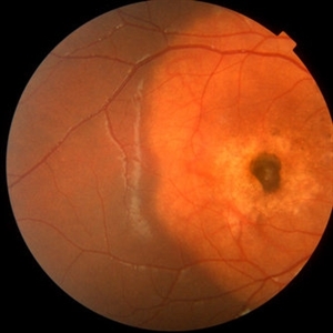
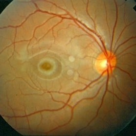
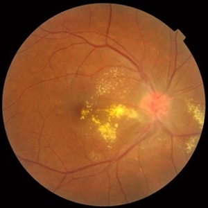
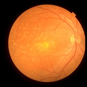
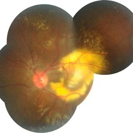
Color fundus photograph of the right eye of a 46-year-old man with decreased VA due to advanced Stargardt's disease.
Photographer: Nayereh Hadipoor, Negah Eye Center, Tehran
Imaging device: Heidelberg Spectralis
Condition/keywords: Stargardt disease
-
 Stargardt's Disease
Mar 13 2013 by Hamid Ahmadieh, MD
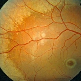
Color fundus photograph of the left eye of a 46-year-old man with decreased VA due to advanced Stargardt's disease.
Photographer: Nayereh Hadipoor, Negah Eye Center, Tehran
Imaging device: Heidelberg Spectralis
Condition/keywords: Stargardt disease
-
 Late Stage Stargardt's Disease
Mar 13 2013 by Hamid Ahmadieh, MD
Autofluorescence imaging of the right eye of a 46-year-old man with decreased VA due to advanced Stargardt's disease.
Photographer: Nayereh Hadipoor, Negah Eye Center, Tehran
Imaging device: Heidelberg Spectralis
Condition/keywords: autofluorescence imaging, Stargardt disease
-
 Late Stage Stargardt's Disease
Mar 13 2013 by Hamid Ahmadieh, MD
Autofluorescence imaging of the left eye of a 46-year-old man with decreased VA due to advanced Stargardt's disease.
Photographer: Nayereh Hadipoor, Negah Eye Center, Tehran
Imaging device: Heidelberg Spectralis
Condition/keywords: autofluorescence imaging, Stargardt disease
-
 Stargardt's Disease
Mar 13 2013 by Hamid Ahmadieh, MD
Infrared image of the right eye of a 46-year-old man with decreased VA due to advanced Stargardt's disease.
Photographer: Nayereh Hadipoor, Negah Eye Center, Tehran
Imaging device: Heidelberg Spectralis
Condition/keywords: infrared image, Stargardt disease
-
 Stargardts Disease
Mar 13 2013 by Hamid Ahmadieh, MD
Infrared image of the right eye of a 46-year-old man with decreased VA due to advanced Stargardts disease.
Photographer: Nayereh Hadipoor, Negah Eye Center, Tehran
Imaging device: Heidelberg Spectralis
Condition/keywords: infrared image, Stargardt disease
-
 Behcet's Disease
Mar 13 2013 by Hamid Ahmadieh, MD
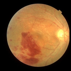
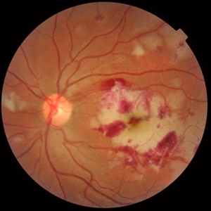
Color fundus photograph of the right eye of a 23-year-old man with retinal vasculitis and branch retinal vein occlusion (BRVO) due to Behcet's disease .
Photographer: Solmaz Shahmohammad, Negah Eye Center, Tehran
Imaging device: Heidelberg Spectralis
Condition/keywords: branch retinal vein occlusion (BRVO), retinal vasculitis
-
 Behcet's Disease
Mar 13 2013 by Hamid Ahmadieh, MD
OCT of the right eye of a 23-year-old man with retinal vasculitis and branch retinal vein occlusion (BRVO) due to Behcet's disease .
Photographer: Solmaz Shahmohammad, Negah Eye Center, Tehran
Imaging device: Topcon OCT
Condition/keywords: branch retinal vein occlusion (BRVO), optical coherence tomography (OCT), retinal vasculitis
-
 Behcet's Disease
Mar 13 2013 by Hamid Ahmadieh, MD
Infrared image of the right eye of a 23-year-old man with retinal vasculitis and branch retinal vein occlusion (BRVO) due to Behcet's disease .
Photographer: Solmaz Shahmohammad, Negah Eye Center, Tehran
Imaging device: Heidelberg Spectralis
Condition/keywords: branch retinal vein occlusion (BRVO), infrared image, retinal vasculitis
-
 Behcet's Disease
Mar 13 2013 by Hamid Ahmadieh, MD
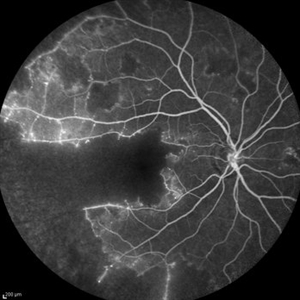
Early phase FA of the right eye of a 23-year-old man with retinal vasculitis and branch retinal vein occlusion (BRVO) due to Behcet's disease .
Photographer: Solmaz Shahmohammad, Negah Eye Center, Tehran
Imaging device: Heidelberg Spectralis
Condition/keywords: branch retinal vein occlusion (BRVO), retinal vasculitis
-
 Behcet's Disease
Mar 13 2013 by Hamid Ahmadieh, MD
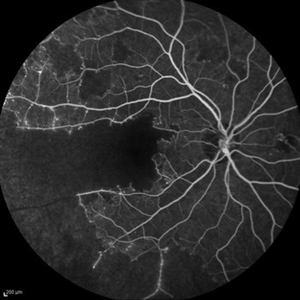
Mid phase FA of the right eye of a 23-year-old man with retinal vasculitis and branch retinal vein occlusion (BRVO) due to Behcet's disease .
Photographer: Solmaz Shahmohammad, Negah Eye Center, Tehran
Imaging device: Heidelberg Spectralis
Condition/keywords: branch retinal vein occlusion (BRVO), retinal vasculitis
-
 Behcet's Disease
Mar 13 2013 by Hamid Ahmadieh, MD
Late phase FA of the right eye of a 23-year-old man with retinal vasculitis and branch retinal vein occlusion (BRVO) due to Behcet's disease .
Photographer: Solmaz Shahmohammad, Negah Eye Center, Tehran
Imaging device: Heidelberg Spectralis
Condition/keywords: branch retinal vein occlusion (BRVO), retinal vasculitis
-
 Behcet's Disease
Mar 13 2013 by Hamid Ahmadieh, MD
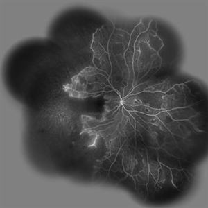
Wide field FA of the right eye of a 23-year-old man with retinal vasculitis and branch retinal vein occlusion (BRVO) due to Behcet's disease .
Photographer: Solmaz Shahmohammad, Negah Eye Center, Tehran
Imaging device: Heidelberg Spectralis
Condition/keywords: branch retinal vein occlusion (BRVO), retinal vasculitis
-
 Behcet's Disease
Mar 13 2013 by Hamid Ahmadieh, MD
Early phase FA of the left eye of a 23-year-old man with retinal vasculitis due to Behcet's disease .
Photographer: Solmaz Shahmohammad, Negah Eye Center, Tehran
Imaging device: Heidelberg Spectralis
Condition/keywords: retinal vasculitis
-
 Behcet's Disease
Mar 13 2013 by Hamid Ahmadieh, MD
Mid phase FA of the left eye of a 23-year-old man with retinal vasculitis due to Behcet's disease .
Photographer: Solmaz Shahmohammad , Negah Eye Center, Tehran
Imaging device: Heidelberg Spectralis
Condition/keywords: retinal vasculitis
-
 Behcet's Disease
Mar 13 2013 by Hamid Ahmadieh, MD
Late phase FA of the left eye of a 23-year-old man with retinal vasculitis due to Behcet's disease .
Photographer: Solmaz Shahmohammad, Negah Eye Center, Tehran
Imaging device: Heidelberg Spectralis
Condition/keywords: retinal vasculitis
-
 Behcet's Disease
Mar 13 2013 by Hamid Ahmadieh, MD
Wide field FA of the left eye of a 23-year-old man with retinal vasculitis due to Behcet's disease .
Photographer: Solmaz Shahmohammadi , Negah Eye Center, Tehran
Imaging device: Heidelberg Spectralis
Condition/keywords: retinal vasculitis
-
 Choroidal Osteoma + CNV
Mar 13 2013 by Hamid Ahmadieh, MD
Color fundus photograph the right eye of a 13-year-old girl with decreased VA due to CNV secondary to choroidal osteoma.
Photographer: Naghmeh Nozhat, Negah Eye Center, Tehran
Imaging device: Topcon
Condition/keywords: choroidal neovascularization (CNV), choroidal osteoma
-
 Choroidal Osteoma + CNV
Mar 13 2013 by Hamid Ahmadieh, MD
Optical coherence tomography (OCT) of the right eye of a 13-year-old girl with decreased VA due to CNV secondary to choroidal osteoma.
Photographer: Naghmeh Nozhat, Negah Eye Center, Tehran
Imaging device: Topcon
Condition/keywords: choroidal neovascularization (CNV), choroidal osteoma, optical coherence tomography (OCT)
-
 Optic Disc Drusen
Jul 10 2013 by Hamid Ahmadieh, MD
Fundus autofluorescence image of the right eye of a 24-year-old woman with optic disc drusen and VA 20/20.
Photographer: Solmaz Shahmohammadi, Negah Eye Center, Tehran
Imaging device: Heidelberg Spectralis
Condition/keywords: fundus autofluorescence (FAF), optic disc drusen
-
 Optic Disc Drusen
Jul 10 2013 by Hamid Ahmadieh, MD
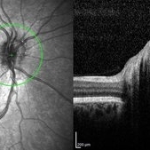
SD-OCT image of the right eye of a 24-year-old woman with optic disc drusen and VA 20/20.
Photographer: Solmaz Shahmohammadi, Negah Eye Center, Tehran
Imaging device: Heidelberg Spectralis
Condition/keywords: optic disc drusen, optical coherence tomography (OCT)
-
 Optic Disc Drusen
Jul 10 2013 by Hamid Ahmadieh, MD
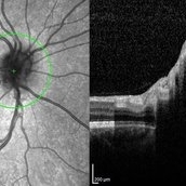
SD-OCT image of the right eye of a 24-year-old woman with optic disc drusen and VA 20/20.
Photographer: Solmaz Shahmohammadi, Negah Eye Center, Tehran
Imaging device: Heidelberg Spectralis
Condition/keywords: optic disc drusen, optical coherence tomography (OCT)
-
 Optic Disc Drusen
Jul 10 2013 by Hamid Ahmadieh, MD
Fundus autofluorescence image of the left eye of a 24-year-old woman with optic disc drusen and VA 20/20.
Photographer: Solmaz Shahmohammad, Negah Eye Center, Tehran
Imaging device: Heidelberg Spectralis
Condition/keywords: fundus autofluorescence (FAF), optic disc drusen
-
---thumb.jpg/image-square;max$300,300.ImageHandler) Optic Disc Drusen
Optic Disc Drusen
Jul 10 2013 by Hamid Ahmadieh, MD
SD-OCT image of the left eye of a 24-year-old woman with optic disc drusen and VA 20/20.
Photographer: Solmaz Shahmohammadi, Negah Eye Center, Tehran
Imaging device: Heidelberg Spectralis
Condition/keywords: optic disc drusen, optical coherence tomography (OCT)
-
---thumb.jpg/image-square;max$300,300.ImageHandler) Polypoidal Choroidal Vasculopathy
Polypoidal Choroidal Vasculopathy
Jul 13 2013 by Hamid Ahmadieh, MD
FAF image of the right eye of a 55-year-old woman with decreased vision and metamorphopsia due to PCV.
Photographer: Elham Salehi, Negah Eye Center, Tehran
Imaging device: Heidelberg Spectralis
Condition/keywords: fundus autofluorescence (FAF), polypoidal choroidal vasculopathy (PCV)
-
---thumb.jpg/image-square;max$300,300.ImageHandler) Polypoidal Choroidal Vasculopathy
Polypoidal Choroidal Vasculopathy
Jul 13 2013 by Hamid Ahmadieh, MD
Late phase FA and ICG images of the right eye of a 55-year-old woman with decreased vision and metamorphopsia due to PCV.
Photographer: Elham Salehi, Negah Eye Center, Tehran
Imaging device: Heidelberg Spectralis
Condition/keywords: indocyanine green (ICG) angiography, polypoidal choroidal vasculopathy (PCV)
-
 Polypoidal Choroidal Vasculopathy
Jul 13 2013 by Hamid Ahmadieh, MD
FA and ICG images of the right eye of a 55-year-old woman with decreased vision and metamorphopsia due to PCV.
Photographer: Elham Salehi, Negah Eye Center, Tehran
Imaging device: Heidelberg Spectralis
Condition/keywords: indocyanine green (ICG) angiography, polypoidal choroidal vasculopathy (PCV)
-
 Polypoidal Choroidal Vasculopathy
Jul 13 2013 by Hamid Ahmadieh, MD
Color fundus photograph of the right eye of a 55-year-old woman with decreased vision and metamorphopsia due to PCV.
Photographer: Elham Salehi, Negah Eye Center, Tehran
Imaging device: Topcon Fundus Camera
Condition/keywords: polypoidal choroidal vasculopathy (PCV)
-
---thumb.jpg/image-square;max$300,300.ImageHandler) Cystoid Macular Edema due to Retinitis Pigmentosa
Cystoid Macular Edema due to Retinitis Pigmentosa
Jul 13 2013 by Hamid Ahmadieh, MD
Color fundus photograph of the left eye of a 30-year-old woman with cystoid macular edema due to retinitis pigmentosa.
Photographer: Elham Salehi, Negah Eye Center, Tehran
Imaging device: Topcon Fundus Camera
Condition/keywords: cystoid macular edema (CME), retinitis pigmentosa
-
---thumb.jpg/image-square;max$300,300.ImageHandler) Cystoid Macular Edema due to Retinitis Pigmentosa
Cystoid Macular Edema due to Retinitis Pigmentosa
Jul 13 2013 by Hamid Ahmadieh, MD
Color fundus photograph of the right eye of a 30-year-old woman with cystoid macualr edema due to retinitis pigmentosa.
Photographer: Elham Salehi, Negah Eye Center, Tehran
Imaging device: Topcon Fundus Camera
Condition/keywords: cystoid macular edema (CME), retinitis pigmentosa
-
---thumb.jpg/image-square;max$300,300.ImageHandler) Cystoid Macular Edema due to Retinitis Pigmentosa
Cystoid Macular Edema due to Retinitis Pigmentosa
Jul 13 2013 by Hamid Ahmadieh, MD
OCT image of the left eye of a 30-year-old woman with cystoid macualr edema due to retinitis pigmentosa.
Photographer: Elham Salehi, Negah Eye Center, Tehran
Imaging device: Topcon OCT
Condition/keywords: cystoid macular edema (CME), optical coherence tomography (OCT), retinitis pigmentosa
-
 Cystoid Macular Edema due to Retinitis Pigmentosa
Jul 13 2013 by Hamid Ahmadieh, MD
OCT image of the right eye of a 30-year-old woman with cystoid macualr edema due to retinitis pigmentosa.
Photographer: Elham Salehi, Negah Eye Center, Tehran
Imaging device: Topcon OCT
Condition/keywords: cystoid macular edema (CME), optical coherence tomography (OCT), retinitis pigmentosa
-
---thumb.jpg/image-square;max$300,300.ImageHandler) Primary Hyperoxaluria and Oxalosis
Primary Hyperoxaluria and Oxalosis
Jul 24 2013 by Hamid Ahmadieh, MD
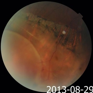
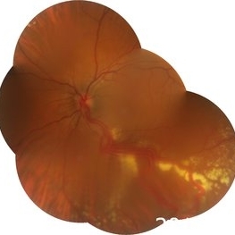
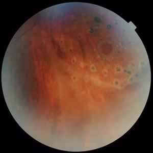
Color fundus photograph of the left eye of a 55-year-old man with primary hyperoxaluria and oxalosis. Vitreous hemorrhage originating from NVD due to vasoproliferative retinopathy is seen.
Photographer: Hanieh Payab, Ophthalmic Research Center, Tehran
Imaging device: Topcon Fundus Camera
Condition/keywords: neovascularization of the disc (NVD), oxalosis, primary hyperoxaluria, vasoproliferative retinopathy
-
---thumb.jpg/image-square;max$300,300.ImageHandler) Primary Hyperoxaluria and Oxalosis
Primary Hyperoxaluria and Oxalosis
Jul 24 2013 by Hamid Ahmadieh, MD
Late phase FA image of the left eye of a 55-year-old man with primary hyperoxaluria and oxalosis. Profound leakage from disc due to NVD is visible. Vasoproliferative retinopathy has occurred secondary to retinal ischemia due to intravascular deposition of calcium oxalate crystals.
Photographer: Hanieh Payab, Ophthalmic Research Center, Tehran
Imaging device: Topcon Fundus Camera
Condition/keywords: oxalosis, primary hyperoxaluria, vasoproliferative retinopathy
-
---thumb.jpg/image-square;max$300,300.ImageHandler) Primary Hyperoxaluria and Oxalosis
Primary Hyperoxaluria and Oxalosis
Jul 24 2013 by Hamid Ahmadieh, MD
Mid phase FA image of the left eye of a 55-year-old man with primary hyperoxaluria and oxalosis, Delayed filling of retinal vessels due to intravascular deposition of calcium oxalate crystals and non-perfusion of the temporal retina are visible.
Photographer: Hanieh Payab, Ophthalmic Research Center, Labbafinejad Medical Center, Tehran
Imaging device: Topcon Fundus Camera
Condition/keywords: ocular manifestation, oxalosis, primary hyperoxaluria
-
---thumb.jpg/image-square;max$300,300.ImageHandler) Primary Hyperoxaluria and Oxalosis
Primary Hyperoxaluria and Oxalosis
Jul 24 2013 by Hamid Ahmadieh, MD
Early phase FA image of the left eye of a 55-year-old man with primary hyperoxaluria and oxalosis, Delayed filling of retinal vessels due to intravascular deposition of calcium oxalate crystals and non-perfusion of the temporal retina are visible.
Photographer: Hanieh Payab, Ophthalmic Research Center, Labbafinejad Medical Center, Tehran
Imaging device: Topcon Fundus Camera
Condition/keywords: ocular manifestation, oxalosis, primary hyperoxaluria
-
---thumb.jpg/image-square;max$300,300.ImageHandler) Primary Hyperoxaluria and Oxalosis
Primary Hyperoxaluria and Oxalosis
Jul 24 2013 by Hamid Ahmadieh, MD
Red-free image of the left eye of a 55-year-old man with primary hyperoxaluria and oxalosis. Extensive deposition of calcium oxalate crystals are demonstrated in the RPE . Deposition of crystals in retinal arteries as well as venous caliber abnormality are also visible.
Photographer: Hanieh Payab, Ophthalmic Research Center, Labbafinejad Medical Center, Tehran
Imaging device: Topcon Fundus Camera
Condition/keywords: ocular manifestation, oxalosis, primary hyperoxaluria, red-free
-
---thumb.jpg/image-square;max$300,300.ImageHandler) Primary Hyperoxaluria and Oxalosis
Primary Hyperoxaluria and Oxalosis
Jul 24 2013 by Hamid Ahmadieh, MD
Red-free image of the left eye of a 55-year-old man with primary hyperoxaluria and oxalosis. Extensive deposition of calcium oxalate crystals are demonstrated in the RPE . Deposition of crystals in retinal arteries as well as venous caliber abnormality are also visible.
Photographer: Hanieh Payab, Ophthalmic Research Center, Labbafinejad Medical Center, Tehran
Imaging device: Topcon Fundus Camera
Condition/keywords: oxalosis, primary hyperoxaluria, red-free
-
---thumb.jpg/image-square;max$300,300.ImageHandler) Primary Hyperoxaluria and Oxalosis
Primary Hyperoxaluria and Oxalosis
Jul 24 2013 by Hamid Ahmadieh, MD
Color fundus photograph of the right eye of a 55-year-old man with primary hyperoxaluria and oxalosis. Characteristic crystalline retinopathy (flecked retina), black geographic maculopathy, and partial optic atrophy are visible. In addition, occluded branches of central retinal artery due to calcium oxalate deposition are visible.
Photographer: Hanieh Payab, Ophthalmic Research Center, Labbafinejad Medical Center, Tehran
Imaging device: Topcon Fundus Camera
Condition/keywords: oxalosis, primary hyperoxaluria
-
---thumb.jpg/image-square;max$300,300.ImageHandler) Carotid Cavernous Fistula
Carotid Cavernous Fistula
Jul 29 2013 by Hamid Ahmadieh, MD
Color fundus photograph of the right eye of a 40-year-old man with venous stasis retinopathy secondary to carotid cavernous fistula.
Condition/keywords: carotid-cavernous fistula, venous stasis retinopathy
-
---thumb.JPG/image-square;max$300,300.ImageHandler) Carotid Cavernous Fistula
Carotid Cavernous Fistula
Jul 29 2013 by Hamid Ahmadieh, MD
Photo slit lamp biomicroscope image of the right eye of a 40-year-old man with engorgement of a episcleral vessels due to carotid cavernous fistula.
Condition/keywords: carotid-cavernous fistula, episcleral vessel dilation
-
---thumb.JPG/image-square;max$300,300.ImageHandler) Carotid Cavernous Fistula
Carotid Cavernous Fistula
Jul 29 2013 by Hamid Ahmadieh, MD
Photo slit lamp biomicroscope image of the right eye of a 40-year-old man with engorgement of a episcleral vessels due to carotid cavernous fistula.
Condition/keywords: carotid-cavernous fistula, episcleral vessel dilation, slit lamp photo
-
 Niemann Pick Disease Type B
Aug 6 2013 by Hamid Ahmadieh, MD
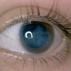
Photo slit lamp photograph the left eye of a patient with Niemann Pick Type B with corneal stromal depositions.
Photographer: Ali Mohammad-Rabie, Ophthalmic Research Center, Labbafinejad Medical Center, Tehran
Condition/keywords: slit lamp photo
-
 Niemann Pick Disease Type B
Aug 6 2013 by Hamid Ahmadieh, MD
Color fundus photograph of the right eye of a patient with Niemann Pick Type B with typical macular halo and confluent deposits in mid-periphery.
Photographer: Ali Mohammad-Rabie, Ophthalmic Research Center, Labbafinejad Medical Center, Tehran
Condition/keywords: fleck retinopathy, macular halo
-
 Niemann Pick Disease Type B
Aug 6 2013 by Hamid Ahmadieh, MD
Color fundus photograph of the left eye of a patient with Niemann Pick Type B with typical macular halo and confluent deposits in mid-periphery.
Photographer: Ali Mohammad-Rabie, Ophthalmic Research Center, Labbafinejad Medical Center, Tehran
Condition/keywords: fleck retinopathy, macular halo
-
 Niemann Pick Disease Type B
Aug 6 2013 by Hamid Ahmadieh, MD
Color fundus photograph of the right eye of a patient with Niemann Pick Type B with typical macular halo and confluent deposits in mid-periphery.
Photographer: Ali Mohammad-Rabie, Ophthalmic Research Center, Labbafinejad Medical Center, Tehran
Condition/keywords: macular halo
-
 Niemann Pick Disease Type B
Aug 6 2013 by Hamid Ahmadieh, MD
Color fundus photograph of the right eye of a patient with Niemann Pick Type B with typical macular halo and confluent deposits in mid-periphery.
Photographer: Ali Mohammad-Rabie, Ophthalmic Research Center, Labbafinejad Medical Center, Tehran
Condition/keywords: fleck retinopathy, macular halo
-
 Florid Type PDR and Venous Loop
Aug 6 2013 by Hamid Ahmadieh, MD
Color fundus photograph of the left eye of a patient with florid type PDR and venous loops.
Photographer: Hanieh Payab, Ophthalmic Research Center, Labbafinejad Medical Center, Tehran
Condition/keywords: florid type PDR, venous loop
-
 Neuroretinitis
Aug 6 2013 by Hamid Ahmadieh, MD
Color fundus photograph of the right eye of a patient with dropped vision due to acute neuroretinitis.
Photographer: Hanieh Payab, Ophthalmic Research Center, Labbafinejad Medical Center, Tehran
Condition/keywords: neuroretinitis
-
 Neuroretinitis
Aug 6 2013 by Hamid Ahmadieh, MD
Color fundus photograph of the right eye of a patient with dropped vision due to acute neuroretinitis.
Photographer: Hanieh Payab, Ophthalmic Research Center, Labbafinejad Medical Center, Tehran
Condition/keywords: neuroretinitis
-
---thumb.jpg/image-square;max$300,300.ImageHandler) Non-perfused BRVO with macular infarction
Non-perfused BRVO with macular infarction
Aug 20 2013 by Hamid Ahmadieh, MD
Late venous phase angiogram of the right eye of a 55-year-old woman with decreased vision due to BRVO. Notice capillary nonperfusion involving the macula.
Photographer: Naghmeh Nozhat, Negah Eye Center, Tehran
Condition/keywords: macular infarction, non-perfused branch retinal vein occlusion (BRVO)
-
---thumb.jpg/image-square;max$300,300.ImageHandler) Non-perfused BRVO with macular infarction
Non-perfused BRVO with macular infarction
Aug 20 2013 by Hamid Ahmadieh, MD
Mid arterio venous phase angiogram of the right eye of a 55-year-old woman with decreased vision due to BRVO. Notice capillary nonperfusion involving the macula.
Photographer: Naghmeh Nozhat, Negah Eye Center, Tehran
Condition/keywords: non-perfused branch retinal vein occlusion (BRVO)
-
---thumb.jpg/image-square;max$300,300.ImageHandler) Non-perfused BRVO with macular infarction
Non-perfused BRVO with macular infarction
Aug 20 2013 by Hamid Ahmadieh, MD
Color fundus photograph of the right eye of a 55-year-old woman with decreased vision due to BRVO. There are multiple cotton wool patches and retinal hemorrhages involving the macular center.
Photographer: Naghmeh Nozhat, Negah Eye Center, Tehran
Condition/keywords: non-perfused branch retinal vein occlusion (BRVO)
-
 Radiation Maculopathy
Aug 28 2013 by Hamid Ahmadieh, MD
Mid arteriovenous phase FA image of the left eye of a 65-year-old man with radiation maculopathy and retinopathy.
Photographer: Solmaz Shahmohammad, Negah Eye Center, Tehran
Imaging device: Heidelberg Spectralis
Condition/keywords: radiation maculopathy, radiation retinopathy
-
---thumb.jpg/image-square;max$300,300.ImageHandler) Radiation Maculopathy
Radiation Maculopathy
Aug 28 2013 by Hamid Ahmadieh, MD
OCT image of the right eye of a 65-year-old man with radiation maculopathy.
Photographer: Solmaz Shahmohammad, Negah Eye Center, Tehran
Imaging device: Topcon OCT
Condition/keywords: optical coherence tomography (OCT), radiation maculopathy
-
---thumb.jpg/image-square;max$300,300.ImageHandler) Radiation Maculopathy
Radiation Maculopathy
Aug 28 2013 by Hamid Ahmadieh, MD
OCT image of the left eye of a 65-year-old man with radiation maculopathy.
Photographer: Solmaz Shahmohammad, Negah Eye Center, Tehran
Imaging device: Topcon OCT
Condition/keywords: optical coherence tomography (OCT), radiation maculopathy
-
 Radiation Maculopathy
Aug 28 2013 by Hamid Ahmadieh, MD
Late phase wide- field FA image of the right eye of a 65-year-old man with radiation maculopathy and retinopathy.
Photographer: Solmaz Shahmohammad, Negah Eye Center, Tehran
Imaging device: Heidelberg Spectralis
Condition/keywords: radiation maculopathy, radiation retinopathy
-
 Radiation Maculopathy
Aug 28 2013 by Hamid Ahmadieh, MD
Late venous phase FA image of the right eye of a 65-year-old man with radiation maculopathy and retinopathy.
Photographer: Solmaz Shahmohammad, Negah Eye Center, Tehran
Imaging device: Heidelberg Spectralis
Condition/keywords: radiation maculopathy, radiation retinopathy
-
 Radiation Maculopathy
Aug 28 2013 by Hamid Ahmadieh, MD
Mid arteriovenous phase FA image of the right eye of a 65-year-old man with radiation maculopathy and retinopathy.
Photographer: Solmaz Shahmohammad, Negah Eye Center, Tehran
Imaging device: Heidelberg Spectralis
Condition/keywords: radiation maculopathy, radiation retinopathy
-
---thumb.bmp/image-square;max$300,300.ImageHandler) Radiation Maculopathy
Radiation Maculopathy
Aug 28 2013 by Hamid Ahmadieh, MD
Late venous phase FA image of the left eye of a 65-year-old man with radiation maculopathy and retinopathy.
Photographer: Solmaz Shahmohammad, Negah Eye Center, Tehran
Imaging device: Heidelberg Spectralis
Condition/keywords: radiation maculopathy, radiation retinopathy
-
 Radiation Maculopathy
Aug 28 2013 by Hamid Ahmadieh, MD
FAF image of the left eye of a 65-year-old man with radiation maculopathy.
Photographer: Solmaz Shahmohammad, Negah Eye Center, Tehran
Imaging device: Heidelberg Spectralis
Condition/keywords: fundus autofluorescence (FAF), radiation maculopathy
-
---thumb.jpg/image-square;max$300,300.ImageHandler) Macular Pucker
Macular Pucker
Sep 24 2013 by Hamid Ahmadieh, MD
Mid-arteriovenous phase angiogram of the left eye of a patient with vascular tortousity secondary to idiopathic epiretinal membrane.
Photographer: Naghmeh Nozhat, Negah Eye Center, Tehran
Imaging device: Heidelberg Spectralis
Condition/keywords: idiopathic epiretinal membrane, macular pucker, vascular tortousity
-
---thumb.jpg/image-square;max$300,300.ImageHandler) Macular Pucker
Macular Pucker
Sep 24 2013 by Hamid Ahmadieh, MD
Color fundus photograph of the left eye of a patient with macular pucker secondary to idiopathic epiretinal membrane.
Photographer: Naghmeh Nozhat, Negah Eye Center, Tehran
Condition/keywords: idiopathic epiretinal membrane, macular pucker
-
---thumb.jpg/image-square;max$300,300.ImageHandler) Vitreomacular Traction (VMT)
Vitreomacular Traction (VMT)
Sep 24 2013 by Hamid Ahmadieh, MD
OCT image of the right eye of a 70-year-old woman with visual disturbance due to VMT.
Photographer: Soodabeh Fooladin, Negah Eye Center, Tehran, Iran
Imaging device: Topcon OCT
Condition/keywords: optical coherence tomography (OCT), vitreomacular traction (VMT)
-
 Retinoschisis
Nov 13 2013 by Hamid Ahmadieh, MD
Color fundus photograph of the left eye of a 60-year-old woman with retinoschisis.
Photographer: Naghmeh Nozhat, Negah Eye Center, Tehran
Condition/keywords: retinoschisis
-
 Retinitis Pigmentosa
Nov 13 2013 by Hamid Ahmadieh, MD
Color fundus photograph of the right eye of a 30-year-old man with retinitis pigmentosa involving the center of the macula.
Photographer: Elham Salehi , Negah Eye Center, Tehran
Condition/keywords: retinitis pigmentosa
-
 Primary Subhyaloid Hemorrhage due to Valsalva Retinopathy
Nov 13 2013 by Hamid Ahmadieh, MD
OCT images of the left eye of a 25-year-old man with sudden drop of vision due to subhyaloid hemorrhage secondary to Valsalva retinopathy.
Photographer: Soodabeh Fooladin , Negah Eye Center, Tehran
Imaging device: TOPCON OCT
Condition/keywords: optical coherence tomography (OCT), subhyaloid hemorrhage, valsalva retinopathy
-
 Primary Subhyaloid Hemorrhage Due to Valsalva Retinopathy
Nov 13 2013 by Hamid Ahmadieh, MD
Color fundus photograph of the left eye of a 25-year-old man with sudden drop of vision due to subhyaloid hemorrhage secondary to Valsalva retinopathy.
Photographer: Soodabeh Fooladin , Negah Eye Center, Tehran
Imaging device: TOPCON OCT
Condition/keywords: subhyaloid hemorrhage, valsalva retinopathy
-
---thumb.jpg/image-square;max$300,300.ImageHandler) Primary Subhyaloid Hemorrhage Due to Valsalva Retinopathy
Primary Subhyaloid Hemorrhage Due to Valsalva Retinopathy
Nov 13 2013 by Hamid Ahmadieh, MD
Infrared image of the left eye of a 25-year-old man with primary subhyaloid hemorrhage due to Valsalva retinopathy.
Photographer: Nayereh Hadipour, Negah Eye Center, Tehran
Imaging device: Heidelberg Spectralis
Condition/keywords: infrared image, subhyaloid hemorrhage, valsalva retinopathy
-
---thumb.jpg/image-square;max$300,300.ImageHandler) Primary Subhyaloid Hemorrhage Due to Valsalva Retinopathy
Primary Subhyaloid Hemorrhage Due to Valsalva Retinopathy
Nov 13 2013 by Hamid Ahmadieh, MD
FAF image of the left eye of a 25-year-old man with primary subhyaloid hemorrhage due to Valsalva retinopathy.
Photographer: Nayereh Hadipour, Negah Eye Center, Tehran
Condition/keywords: fundus autofluorescence (FAF), subhyaloid hemorrhage, valsalva retinopathy
-
---thumb.jpg/image-square;max$300,300.ImageHandler) Primary Subhyaloid Hemorrhage Due to Valsalva Retinopathy
Primary Subhyaloid Hemorrhage Due to Valsalva Retinopathy
Nov 13 2013 by Hamid Ahmadieh, MD
Arterial phase angiogram of the left eye of a 25-year-old man with primary subhyaloid hemorrhage due to Valsalva retinopathy.
Photographer: Nayereh Hadipour, Negah Eye Center, Tehran
Condition/keywords: subhyaloid hemorrhage, valsalva retinopathy
-
---thumb.jpg/image-square;max$300,300.ImageHandler) Primary Subhyaloid Hemorrhage Due to Valsalva Retinopathy
Primary Subhyaloid Hemorrhage Due to Valsalva Retinopathy
Nov 13 2013 by Hamid Ahmadieh, MD
Early venous phase angiogram of the left eye of a 25-year-old man with primary subhyaloid hemorrhage due to Valsalva retinopathy.
Photographer: Nayereh Hadipour, Negah Eye Center, Tehran
Condition/keywords: subhyaloid hemorrhage, valsalva retinopathy
-
---thumb.jpg/image-square;max$300,300.ImageHandler) Primary Subhyaloid Hemorrhage Due to Valsalva Retinopathy
Primary Subhyaloid Hemorrhage Due to Valsalva Retinopathy
Nov 13 2013 by Hamid Ahmadieh, MD
Wide-field angiogram of the left eye of a 25-year-old man with primary subhyaloid hemorrhage due to Valsalva retinopathy.
Photographer: Nayereh Hadipour, Negah Eye Center, Tehran
Condition/keywords: subhyaloid hemorrhage, valsalva retinopathy
-
---thumb.jpg/image-square;max$300,300.ImageHandler) Primary Subhyaloid Hemorrhage Due to Valsalva Retinopathy
Primary Subhyaloid Hemorrhage Due to Valsalva Retinopathy
Nov 13 2013 by Hamid Ahmadieh, MD
Midvenous phase angiogram of the left eye of a 25-year-old man with primary subhyaloid hemorrhage due to Valsalva retinopathy.
Photographer: Nayereh Hadipour, Negah Eye Center, Tehran
Condition/keywords: subhyaloid hemorrhage, valsalva retinopathy
-
 IRVAN
IRVAN
Nov 13 2013 by Hamid Ahmadieh, MD
Funduscopic signs of acive IRVAN.
Condition/keywords: aneurysm, neuroretinitis, retinal vasculitis, retinitis
-
 Idiopathic Retinitis, Vasculitis, Aneurysms, and Neuroretinitis (IRVAN)
Nov 13 2013 by Hamid Ahmadieh, MD
Wide-field late phase FA image of the left eye of a 35-year-old woman with idiopathic retinitis, vasculitis, aneurysms, and neuroretinitis (IRVAN). Large areas of capillary non-perfusion are visible temporally.
Photographer: Solmaz Shahmohammad , Negah Eye Center , Tehran
Imaging device: Heidelberg Spectralis
Condition/keywords: aneurysm, neuroretinitis, retinal vasculitis, retinitis
-
 Idiopathic Retinitis, Vasculitis, Aneurysms, and Neuroretinitis (IRVAN)
Nov 13 2013 by Hamid Ahmadieh, MD
Late venous phase FA image of the left eye of a 35-year-old woman with idiopathic retinitis, vasculitis, aneurysms, and neuroretinitis (IRVAN).
Photographer: Solmaz Shahmohammad , Negah Eye Center , Tehran
Imaging device: Heidelberg Spectralis
Condition/keywords: aneurysm, neuroretinitis, retinal vasculitis, retinitis
-
 Idiopathic Retinitis, Vasculitis, Aneurysms, and Neuroretinitis (IRVAN)
Nov 13 2013 by Hamid Ahmadieh, MD
FA image of the left eye of a 35-year-old woman with idiopathic retinitis, vasculitis, aneurysms, and neuroretinitis (IRVAN).
Photographer: Solmaz Shahmohammad , Negah Eye Center , Tehran
Condition/keywords: aneurysm, neuroretinitis, retinal vasculitis, retinitis
-
---thumb.jpg/image-square;max$300,300.ImageHandler) Idiopathic Retinitis, Vasculitis, Aneurysms, and Neuroretinitis (IRVAN)
Idiopathic Retinitis, Vasculitis, Aneurysms, and Neuroretinitis (IRVAN)
Nov 13 2013 by Hamid Ahmadieh, MD
Color fundus photograph of the left eye of a 35-year-old woman with idiopathic retinitis, vasculitis, aneurysms, and neuroretinitis (IRVAN).There are bleedings originating from the neovascularization on disc.
Photographer: Solmaz Shahmohammad , Negah Eye Center , Tehran
Imaging device: Heidelberg Spectralis
Condition/keywords: aneurysm, neuroretinitis, retinal vasculitis, retinitis
-
 Idiopathic Retinitis, Vasculitis, Aneurysms, and Neuroretinitis (IRVAN)
Nov 13 2013 by Hamid Ahmadieh, MD
Wide -field late phase FA image of the right eye of a 35-year-old woman with idiopathic retinitis, vasculitis, aneurysms, and neuroretinitis (IRVAN).
Photographer: Solmaz Shahmohammad , Negah Eye Center , Tehran
Imaging device: Heidelberg Spectralis
Condition/keywords: aneurysm, neuroretinitis, retinal vasculitis, retinitis
-
 Idiopathic Retinitis, Vasculitis, Aneurysms, and Neuroretinitis (IRVAN)
Nov 13 2013 by Hamid Ahmadieh, MD
Late venous phase FA image of the right eye of a 35-year-old woman with idiopathic retinitis, vasculitis, aneurysms, and neuroretinitis (IRVAN).
Photographer: Solmaz Shahmohammad , Negah Eye Center , Tehran
Imaging device: Heidelberg Spectralis
Condition/keywords: aneurysm, neuroretinitis, retinal vasculitis, retinitis
-
 Idiopathic Retinitis, Vasculitis, Aneurysms, and Neuroretinitis (IRVAN)
Nov 13 2013 by Hamid Ahmadieh, MD
Mid-arteriovenous phase FA image of the right eye of a 35-year-old woman with idiopathic retinitis, vasculitis, aneurysms, and neuroretinitis (IRVAN).
Photographer: Solmaz Shahmohammad , Negah Eye Center , Tehran
Imaging device: Heidelberg Spectralis
Condition/keywords: aneurysm, neuroretinitis, retinal vasculitis, retinitis
-
---thumb.jpg/image-square;max$300,300.ImageHandler) Idiopathic Retinitis, Vasculitis, Aneurysms, and Neuroretinitis (IRVAN)
Idiopathic Retinitis, Vasculitis, Aneurysms, and Neuroretinitis (IRVAN)
Nov 13 2013 by Hamid Ahmadieh, MD
Color fundus photograph of the right eye of a 35-year-old woman with idiopathic retinitis, vasculitis, aneurysms, and neuroretinitis (IRVAN).
Photographer: Solmaz Shahmohammad , Negah Eye Center , Tehran
Condition/keywords: aneurysm, neuroretinitis, retinal vasculitis, retinitis
-
---thumb.jpg/image-square;max$300,300.ImageHandler) Silicone Oil Surface
Silicone Oil Surface
Nov 14 2013 by Hamid Ahmadieh, MD
OCT image of the silicone oil surface bridging an enhanced foveal depression. There is the history of a rhegmatogenous retinal detachment associated with full thickness macular hole repair.
Photographer: Naghmeh Nozhat, Negah Eye Center, Tehran
Condition/keywords: optical coherence tomography (OCT), silicone oil
-
---thumb.jpg/image-square;max$300,300.ImageHandler) Pigmented Demarcation Line and A Blood - containing Retinal Macrocyst
Pigmented Demarcation Line and A Blood - containing Retinal Macrocyst
Nov 14 2013 by Hamid Ahmadieh, MD
Color fundus photograph of the right eye of a 40-year-old man with longstanding retinal detachment showing a broad pigmented demarcation line and a retinal macrocyst . Notice blood inside the retinal macrocyst.
Photographer: Elham Salehi , Negah Eye Center, Tehran
Condition/keywords: demarcation line, fundus photograph, retinal macrocyst
-
 Pigmented Demarcation Line and Retinal Macrocyst
Nov 14 2013 by Hamid Ahmadieh, MD
Color fundus photograph of the right eye of a 40-year-old man with longstanding retinal detachment showing a broad pigmented demarcation line and a retinal macrocyst as well as patches of retinal hemorrhages.
Photographer: Elham Salehi , Negah Eye Center, Tehran
Condition/keywords: demarcation line, fundus photograph, retinal macrocyst
-
 Pigmented Demarcation Line and Retinal Macrocyst
Nov 14 2013 by Hamid Ahmadieh, MD
Color fundus photograph of the right eye of a 40-year-old man with longstanding retinal detachment showing a broad pigmented demarcation line and a retinal macrocyst.
Photographer: Elham Salehi , Negah Eye Center, Tehran
Condition/keywords: demarcation line, fundus photograph, retinal macrocyst
-
 Fundus Flavimaculatus and CNV
Nov 14 2013 by Hamid Ahmadieh, MD
Late phase FA and ICG angiography images of the right eye of a 35-year-old woman with subfoveal CNV secondary to fundus flavimaculatus .
Photographer: Nayereh Hadipour, Negah Eye Center, Tehran
Condition/keywords: choroidal neovascularization (CNV), fundus flavimaculatus, indocyanine green (ICG) angiography, retinal flecks
-
 Fundus Flavimaculatus and CNV
Nov 14 2013 by Hamid Ahmadieh, MD
FA and ICG angiography images of the right eye of a 35-year-old woman with subfoveal CNV secondary to fundus flavimaculatus .
Photographer: Nayereh Hadipour, Negah Eye Center, Tehran
Condition/keywords: choroidal neovascularization (CNV), fundus flavimaculatus, indocyanine green (ICG) angiography, retinal flecks
-
 Fundus Flavimaculatus and CNV
Nov 14 2013 by Hamid Ahmadieh, MD
Early FA and ICG angiography images of the right eye of a 35-year-old woman with subfoveal CNV secondary to fundus flavimaculatus .
Photographer: Nayereh Hadipour, Negah Eye Center, Tehran
Condition/keywords: choroidal neovascularization (CNV), fundus flavimaculatus, indocyanine green (ICG) angiography, retinal flecks
-
 Fundus Flavimaculatus and CNV
Nov 14 2013 by Hamid Ahmadieh, MD
FAF image of the right eye of a 35-year-old woman with subfoveal CNV secondary to fundus flavimaculatus .
Photographer: Nayereh Hadipour, Negah Eye Center, Tehran
Condition/keywords: choroidal neovascularization (CNV), fundus autofluorescence (FAF), fundus flavimaculatus, retinal flecks
-
 Fundus Flavimaculatus and CNV
Nov 14 2013 by Hamid Ahmadieh, MD
Infrared image of the right eye of a 35-year-old woman with subfoveal CNV secondary to fundus flavimaculatus .
Photographer: Nayereh Hadipour, Negah Eye Center, Tehran
Condition/keywords: choroidal neovascularization (CNV), fundus flavimaculatus, infrared image, retinal flecks
-
 Fundus Flavimaculatus and CNV
Nov 14 2013 by Hamid Ahmadieh, MD
OCT image of the right eye of a 35-year-old woman with subfoveal CNV secondary to fundus flavimaculatus .
Photographer: Elham Salehi , Negah Eye Center, Tehran
Condition/keywords: choroidal neovascularization (CNV), fundus flavimaculatus, optical coherence tomography (OCT)
-
 Fundus Flavimaculatus and CNV
Nov 14 2013 by Hamid Ahmadieh, MD
Color fundus photograph of the right eye of a 35-year-old woman with subfoveal CNV secondary to fundus flavimaculatus .
Photographer: Nayereh Hadipour, Negah Eye Center, Tehran
Condition/keywords: choroidal neovascularization (CNV), fundus flavimaculatus, fundus photograph, retinal flecks
-
---thumb.jpg/image-square;max$300,300.ImageHandler) Bergmeister's Papilla
Bergmeister's Papilla
Mar 22 2014 by Hamid Ahmadieh, MD
Color fundus photograph of the right eye of a 50-year-old man with Bergmeister's papilla.
Photographer: Naghmeh Nozhat, Negah Eye Center, Tehran
Imaging device: Topcon Fundus Camera
Condition/keywords: Bergmeister's Papillae, color photo
-
---thumb.jpg/image-square;max$300,300.ImageHandler) Myopic Traction Maculopathy
Myopic Traction Maculopathy
Mar 23 2014 by Hamid Ahmadieh, MD
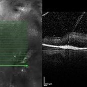
OCT image of the right eye of a 55-year-old woman with marked visual reduction due to myopic traction maculopathy manifesting as foveoschisis and neurosensory retinal detachment.
Photographer: Solmaz Shahmohammad , Negah Eye Center , Tehran
Imaging device: Specteralis
Condition/keywords: myopic foveoschisis, myopic traction maculopathy, neurosensory detachment of retina, optical coherence tomography (OCT)
-
---thumb.jpg/image-square;max$300,300.ImageHandler) Myopic Traction Maculopathy
Myopic Traction Maculopathy
Mar 23 2014 by Hamid Ahmadieh, MD
OCT image of the left eye of a 55-year-old woman with decreased vision secondary to myopic traction maculopathy.
Photographer: Solmaz Shahmohammad , Negah Eye Center , Tehran
Imaging device: Specteralis
Condition/keywords: myopic foveoschisis, myopic traction maculopathy, optical coherence tomography (OCT)
-
---thumb.jpg/image-square;max$300,300.ImageHandler) Vitrectomy for Asteroid Hyalosis
Vitrectomy for Asteroid Hyalosis
Mar 23 2014 by Hamid Ahmadieh, MD
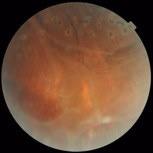
A 70-year-old woman undergoing 23-gauge vitrectomy for a severe form of asteroid hyalosis.
Condition/keywords: asteroid hyalosis, vitrectomy
-
---thumb.jpg/image-square;max$300,300.ImageHandler) Vitrectomy for Asteroid Hyalosis
Vitrectomy for Asteroid Hyalosis
Mar 23 2014 by Hamid Ahmadieh, MD
A 70-year-old woman undergoing vitrectomy for a severe form of asteroid hyalosis.
Condition/keywords: asteroid hyalosis, vitrectomy
-
 Acute Idiopathic Occlusive Retinal Vasculitis
May 31 2014 by Hamid Ahmadieh, MD
Wide- field fluorescein angiogram of the right eye of a 28-year-old woman with acute drop of vision due to occlusive retinal vasculitis leading to extensive capillary nonperfusion and macular infarction.
Photographer: Naghmeh Nozhat, Negah Eye Center, Tehran
Imaging device: Heidelberg Spectralis
Condition/keywords: capillary nonperfusion, retinal infarction, retinal vasculitis
-
 Acute Idiopathic Occlusive Retinal Vasculitis
May 31 2014 by Hamid Ahmadieh, MD
Mid phase fluorescein angiogram of the right eye of a 28-year-old woman with acute drop of vision due to occlusive retinal vasculitis leading to extensive capillary nonperfusion and macular infarction.
Photographer: Naghmeh Nozhat, Negah Eye Center, Tehran
Imaging device: Heidelberg Spectralis
Condition/keywords: capillary nonperfusion, retinal vasculitis
-
 Acute Idiopathic Occlusive Retinal Vasculitis
May 31 2014 by Hamid Ahmadieh, MD
Early phase fluorescein angiogram of the right eye of a 28-year-old woman with acute drop of vision due to occlusive retinal vasculitis leading to extensive capillary nonperfusion and macular infarction.
Photographer: Naghmeh Nozhat, Negah Eye Center, Tehran
Imaging device: Heidelberg Spectralis
Condition/keywords: retinal vasculitis
-
 Acute Idiopathic Occlusive Retinal Vasculitis
May 31 2014 by Hamid Ahmadieh, MD
Fundus autofluorescence image of the right eye of a 28-year-old woman with acute drop of vision due to occlusive retinal vasculitis leading to extensive nerve fiber layer infarction and retinal hemorrhages.
Photographer: Naghmeh Nozhat, Negah Eye Center, Tehran
Imaging device: Heidelberg Spectralis
Condition/keywords: fundus autofluorescence (FAF), retinal vasculitis
-
 Acute Idiopathic Occlusive Retinal Vasculitis
May 31 2014 by Hamid Ahmadieh, MD
Color fundus photograph of the right eye of a 28-year-old woman with sudden drop of vision due to acute occlusive retinal vasculitis leading to extensive nerve fiber layer infarction and retinal hemorrhages.
Photographer: Naghmeh Nozhat, Negah Eye Center, Tehran
Condition/keywords: color fundus photograph, cotton wool spots, retinal hemorrhage, retinal ischemia
-
 Acute Idiopathic Occlusive Retinal Vasculitis
May 31 2014 by Hamid Ahmadieh, MD
Wide- field fluorescein angiogram of the left eye of a 28-year-old woman with acute drop of vision due to occlusive retinal vasculitis leading to extensive capillary nonperfusion and macular infarction.
Photographer: Naghmeh Nozhat, Negah Eye Center, Tehran
Imaging device: Heidelberg Spectralis
Condition/keywords: capillary nonperfusion, retinal infarction, retinal vasculitis
-
 Acute Idiopathic Occlusive Retinal Vasculitis
May 31 2014 by Hamid Ahmadieh, MD
Mid- phase fluorescein angiogram of the left eye of a 28-year-old woman with acute drop of vision due to occlusive retinal vasculitis leading to extensive capillary nonperfusion and macular infarction.
Photographer: Naghmeh Nozhat, Negah Eye Center, Tehran
Imaging device: Heidelberg Spectralis
Condition/keywords: capillary nonperfusion, retinal vasculitis
-
 Acute Idiopathic Occlusive Retinal Vasculitis
May 31 2014 by Hamid Ahmadieh, MD
Early phase fluorescein angiogram of the left eye of a 28-year-old woman with acute drop of vision due to occlusive retinal vasculitis leading to extensive capillary nonperfusion and macular infarction.
Photographer: Naghmeh Nozhat, Negah Eye Center, Tehran
Imaging device: Heidelberg Spectralis
Condition/keywords: retinal vasculitis
-
 Acute Idiopathic Occlusive Retinal Vasculitis
May 31 2014 by Hamid Ahmadieh, MD
Fundus autofluorescence image of the left eye of a 28-year-old woman with acute drop of vision due to occlusive retinal vasculitis leading to extensive nerve fiber layer infarction and retinal hemorrhages.
Photographer: Naghmeh Nozhat, Negah Eye Center, Tehran
Imaging device: Heidelberg Spectralis
Condition/keywords: fundus autofluorescence (FAF), retinal vasculitis
-
 Acute Idiopathic Occlusive Retinal Vasculitis
May 31 2014 by Hamid Ahmadieh, MD
Color fundus photograph of the left eye of a 28-year-old woman with acute drop of vision due to occlusive retinal vasculitis leading to extensive nerve fiber layer infarction and retinal hemorrhages.
Photographer: Naghmeh Nozhat, Negah Eye Center, Tehran
Condition/keywords: color fundus photograph, cotton wool spots, retinal hemorrhage, retinal ischemia
-
 Idiopathic Occlusive Retinal Vasculitis (Late Stage)
May 31 2014 by Hamid Ahmadieh, MD
Wide- field image of the right eye of a 28-year-old woman with idiopathic occlusive retinal vasculitis 6 months after the onset.
Photographer: Solmaz Shahmohammad, Negah Eye Center, Tehran
Imaging device: Heidelberg Spectralis
Condition/keywords: capillary closure, macular infarction
-
 Idiopathic Occlusive Retinal Vasculitis (Late Stage)
May 31 2014 by Hamid Ahmadieh, MD
Late phase FA image of the right eye of a 28-year-old woman with idiopathic occlusive retinal vasculitis 6 months after the onset.
Photographer: Solmaz Shahmohammad, Negah Eye Center, Tehran
Imaging device: Heidelberg Spectralis
Condition/keywords: capillary closure, macular infarction
-
 Idiopathic Occlusive Retinal Vasculitis (Late Stage)
May 31 2014 by Hamid Ahmadieh, MD
FAF image of the left eye of a 28-year-old woman with idiopathic occlusive retinal vasculitis 6 months after the onset. Patches of hard exudate are present superior to the fovea.
Photographer: Elham Salehi, Negah Eye Center, Tehran
Imaging device: Heidelberg Spectralis
Condition/keywords: fundus autofluorescence (FAF)
-
 Idiopathic Occlusive Retinal Vasculitis (Late Stage)
May 31 2014 by Hamid Ahmadieh, MD
Wide-field FA image of the left eye of a 28-year-old woman with idiopathic occlusive retinal vasculitis 6 months after the onset.
Photographer: Elham Salehi, Negah Eye Center, Tehran
Imaging device: Heidelberg Spectralis
Condition/keywords: capillary closure, macular infarction
-
 Idiopathic Occlusive Retinal Vasculitis (Late Stage)
May 31 2014 by Hamid Ahmadieh, MD
Late phase FA image of the left eye of a 28-year-old woman with idiopathic occlusive retinal vasculitis 6 months after the onset.
Photographer: Elham Salehi, Negah Eye Center, Tehran
Imaging device: Heidelberg Spectralis
Condition/keywords: capillary closure
-
 Idiopathic Occlusive Retinal Vasculitis (Late Stage)
May 31 2014 by Hamid Ahmadieh, MD
Mid- venous phase FA image of the left eye of a 28-year-old woman with idiopathic occlusive retinal vasculitis leading to extensive capillary closure macular infarction and collaterals 6 month after the onset.
Photographer: Elham Salehi, Negah Eye Center, Tehran
Imaging device: Heidelberg Spectralis
Condition/keywords: capillary closure
-
 Multiple Branch Retinal Artery Occlusion ( BRAO)
May 31 2014 by Hamid Ahmadieh, MD
Color fundus photograph of the left eye of a 65-year-old man with sudden drop of vision due to multiple BRAO following carotid artery stenting leading to multiple emboli.
Photographer: Solmaz Shahmohammad, Negah Eye Center, Tehran
Condition/keywords: branch retinal artery occlusion (BRAO), color fundus photograph, retinal cloudy swelling
-
 Recurrent Branch Retinal Vein Occlusion
May 31 2014 by Hamid Ahmadieh, MD
FA image of the right eye of a 60-year-old man with recurrent BRVO. Multiple collaterals are visible secondary to previous BRVO.
Photographer: Soodabeh Fooladin, Negah Eye Center, Tehran, Iran
Imaging device: Heidelberg Spectralis
Condition/keywords: collaterals
-
 Recurrent Branch Retinal Vein Occlusion
May 31 2014 by Hamid Ahmadieh, MD
Color fundus photograph of the right eye of a 60-year-old man with recurrent BRVO.
Photographer: Soodabeh Fooladin, Negah Eye Center, Tehran, Iran
Condition/keywords: branch retinal vein occlusion (BRVO), color fundus photograph, flame shaped retinal hemorrhage
-
 Vitreoschisis ( Spider-like)
May 31 2014 by Hamid Ahmadieh, MD
OCT image of the left eye of a 70-year-old woman with proliferative diabetic retinopathy associated with vitreoschisis and diabetic macular edema. The vitreal changes simulate the appearance of a spider on the retina !
Photographer: Nayereh Hadipour, Negah Eye Center, Tehran
Condition/keywords: diabetic macular edema, optical coherence tomography (OCT), vitreoschisis
-
 Venous Loop & Venous Beading
May 31 2014 by Hamid Ahmadieh, MD
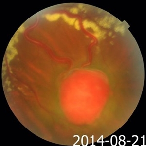
Color fundus photograph of the left eye of a diabetic patient with NVD, NVE, venous loop and venous beading.
Photographer: Elham Salehi, Negah Eye Center, Tehran
Condition/keywords: color fundus photograph, neovascularization elsewhere (NVE), neovascularization of the disc (NVD), proliferative diabetic retinopathy (PDR), venous beading, venous loop
-
 Venous Loop & Venous Beading
May 31 2014 by Hamid Ahmadieh, MD
Color fundus photograph of the left eye of a diabetic patient with NVD, NVE, venous loop and venous beading.
Photographer: Elham Salehi, Negah Eye Center, Tehran
Condition/keywords: color fundus photograph, neovascularization elsewhere (NVE), neovascularization of the disc (NVD), proliferative diabetic retinopathy (PDR), venous beading, venous loop
-
 Von Hippel-Lindau
Sep 1 2014 by Hamid Ahmadieh, MD
Wide filed late phase FA image of the left eye of a 30-year-old woman with Von Hippel Lindau.
Photographer: Solmaz Shahmohammad, Negah Eye Center, Tehran, Iran
Condition/keywords: Von Hippel-Lindau
-
 Von Hippel-Lindau
Sep 1 2014 by Hamid Ahmadieh, MD
Mid venous phase FA image of the left eye of a 30-year-old woman with Von Hippel-Lindau. The afferent and efferent vessels have been filled.
Photographer: Solmaz Shahmohammad, Negah Eye Center, Tehran, Iran
Condition/keywords: Von Hippel-Lindau
-
 Von Hippel-Lindau
Sep 1 2014 by Hamid Ahmadieh, MD
Arterial phase FA image of the left eye of a 30-year-old woman with Von Hippel-Lindau. The afferent vessel has been filled.
Photographer: Solmaz Shahmohammad, Negah Eye Center, Tehran, Iran
Condition/keywords: Von Hippel-Lindau
-
 Von Hippel-Lindau
Sep 1 2014 by Hamid Ahmadieh, MD
Infrared image of the left eye of a 30-year-old woman with Von Hippel-Lindau.
Photographer: Solmaz Shahmohammad, Negah Eye Center, Tehran, Iran
Condition/keywords: infrared image, Von Hippel-Lindau
-
 Von Hippel-Lindau
Sep 1 2014 by Hamid Ahmadieh, MD
Two small retinal capillary hemangiomas detected by wide field FA in the symptom-free right eye of 30-year-old woman with Von Hippel-Lindau.
Photographer: Solmaz Shahmohammad, Negah Eye Center, Tehran, Iran
Condition/keywords: Von Hippel-Lindau
-
 Von Hippel-Lindau
Sep 1 2014 by Hamid Ahmadieh, MD
Advanced retinal capillary hemangioma in the left eye of a 30-year-old woman with Von Hippel-Lindau.
Photographer: Solmaz Shahmohammad, Negah Eye Center, Tehran, Iran
Condition/keywords: color fundus photograph, Von Hippel-Lindau
-
 Von Hippel-Lindau
Sep 1 2014 by Hamid Ahmadieh, MD
Advanced retinal capillary hemangioma in the left eye of a 30-year-old woman with Von Hippel-Lindau.
Photographer: Solmaz Shahmohammad, Negah Eye Center, Tehran, Iran
Condition/keywords: color fundus photograph, Von Hippel-Lindau
-
 Advanced PDR
Sep 1 2014 by Hamid Ahmadieh, MD
OCT image of the right eye of a 50-year-old woman with advanced PDR.
Photographer: Soodabeh Fooladian, Negah Eye Center, Tehran, Iran
Condition/keywords: optical coherence tomography (OCT), proliferative diabetic retinopathy (PDR)
-
 Advanced PDR
Sep 1 2014 by Hamid Ahmadieh, MD
OCT image of the left eye of a 50-year-old woman with advanced PDR.
Photographer: Soodabeh Fooladian, Negah Eye Center, Tehran, Iran
Condition/keywords: optical coherence tomography (OCT), proliferative diabetic retinopathy (PDR)
-
 Advanced PDR
Sep 1 2014 by Hamid Ahmadieh, MD
Color fundus photograph of the left eye of a 50-year-old woman with advanced PDR.
Photographer: Soodabeh Fooladian, Negah Eye Center, Tehran, Iran
Condition/keywords: color fundus photograph, proliferative diabetic retinopathy (PDR)
-
 Advanced PDR
Sep 1 2014 by Hamid Ahmadieh, MD
Color fundus photograph of the right eye of a 50-year-old woman with advanced PDR.
Photographer: Soodabeh Fooladian, Negah Eye Center, Tehran, Iran
Condition/keywords: color fundus photograph, proliferative diabetic retinopathy (PDR), subhyaloid hemorrhage
-
 Coats' Disease
Sep 1 2014 by Hamid Ahmadieh, MD
Color fundus photograph of the left eye of a 10-year-old boy with accumulation of hard exudates in the macula due to Coats' Disease.
Photographer: Nayereh Hadipour, Negah Eye Center, Tehran, Iran
Condition/keywords: color fundus photograph, exudative maculopathy
-
 Endogenous Endophthalmitis
Sep 3 2014 by Hamid Ahmadieh, MD
Infrared image of the left eye of a 45-year-old diabetic man with the history of urinary tract infection. The most probable diagnosis was candida endogenous endophthalmitis.
Photographer: Nayereh Hadipour, Negah Eye Center, Tehran, Iran
Condition/keywords: candida endophthalmitis, endogenous endophthalmitis, infrared image
-
 Endogenous Endophthalmitis
Sep 3 2014 by Hamid Ahmadieh, MD
Late phase FA image of the left eye of a 45-year-old diabetic man with the history of urinary tract infection. The most probable diagnosis was candida endogenous endophthalmitis.
Photographer: Nayereh Hadipour, Negah Eye Center, Tehran, Iran
Condition/keywords: candida endophthalmitis, endogenous endophthalmitis
-
 Endogenous Endophthalmitis
Sep 3 2014 by Hamid Ahmadieh, MD
FA image of the left eye of a 45-year-old diabetic man with the history of urinary tract infection. The most probable diagnosis was candida endogenous endophthalmitis.
Photographer: Nayereh Hadipour, Negah Eye Center, Tehran, Iran
Condition/keywords: candida endophthalmitis, endogenous endophthalmitis
-
 Endogenous Endophthalmitis
Sep 3 2014 by Hamid Ahmadieh, MD
Late phase FA image of the right eye of a 45-year-old diabetic man with the history of urinary tract infection. The most probable diagnosis was candida endogenous endophthalmitis.
Photographer: Nayereh Hadipour, Negah Eye Center, Tehran, Iran
Condition/keywords: candida endophthalmitis, endogenous endophthalmitis
-
 Endogenous Endophthalmitis
Sep 3 2014 by Hamid Ahmadieh, MD
FA image of the right eye of a 45-year-old diabetic man with the history of urinary tract infection. The most probable diagnosis was candida endogenous endophthalmitis.
Photographer: Nayereh Hadipour, Negah Eye Center, Tehran, Iran
Condition/keywords: candida endophthalmitis, endogenous endophthalmitis
-
 Endogenous Endophthalmitis
Sep 3 2014 by Hamid Ahmadieh, MD
Color fundus photograph of the left eye of a 45-year-old diabetic man with the history of urinary tract infection. The most probable diagnosis was candida endogenous endophthalmitis.
Photographer: Nayereh Hadipour, Negah Eye Center, Tehran, Iran
Condition/keywords: candida endophthalmitis, color fundus photograph, endogenous endophthalmitis
-
 Endogenous Endophthalmitis
Sep 3 2014 by Hamid Ahmadieh, MD
Color fundus photograph of the right eye of a 45-year-old diabetic man with the history of urinary tract infection. The most probable diagnosis was candida endogenous endophthalmitis.
Photographer: Nayereh Hadipour, Negah Eye Center, Tehran, Iran
Condition/keywords: candida endophthalmitis, color fundus photograph, endogenous endophthalmitis
-
 Severe Neovascularization Secondary to Idiopathic Occlusive Retinal Vasculitis
Jan 17 2015 by Hamid Ahmadieh, MD
Color fundus photograph of the right eye of a 28-year-old woman with severe retinal neovascularization secondary to idiopatic occlusive retinal vasculitis.
Photographer: Solmaz Shahmohammad, Negah Eye Center, Tehran
Condition/keywords: color fundus photograph, neovascularization (NV), retinal vasculitis
-
 Severe Neovascularization Secondary to Idiopathic Occlusive Retinal Vasculitis
Jan 17 2015 by Hamid Ahmadieh, MD
Wide- field color fundus photograph of the right eye of a 28-year-old woman with severe retinal neovascularization secondary to idiopatic occlusive retinal vasculitis.
Photographer: Solmaz Shahmohammad, Negah Eye Center, Tehran
Condition/keywords: color fundus photograph, neovascularization (NV), retinal vasculitis
-
 X-Linked Retinoschisis
Jan 31 2015 by Hamid Ahmadieh, MD
Late phase FA of the right eye of a 35-year-old man with x-linked retinoschisis. Please notice the foveal schisis.
Photographer: Solmaz Shahmohammad, Negah Eye Center, Tehran, Iran
Imaging device: Heidelberg
Condition/keywords: foveal schisis, x-linked retinoschisis (XLRS)
-
 X-Linked Retinoschisis
Jan 31 2015 by Hamid Ahmadieh, MD
Mid venous phase FA of the right eye of a 35-year-old man with x-linked retinoschisis. Please notice the foveal schisis.
Photographer: Solmaz Shahmohammad, Negah Eye Center, Tehran, Iran
Imaging device: Heidelberg
Condition/keywords: foveal schisis, x-linked retinoschisis (XLRS)
-
 X-Linked Retinoschisis
Jan 31 2015 by Hamid Ahmadieh, MD
FAF of the right eye of a 35-year-old man with x-linked retinoschisis. Please notice the foveal schisis.
Photographer: Solmaz Shahmohammad, Negah Eye Center, Tehran, Iran
Imaging device: Heidelberg
Condition/keywords: foveal schisis, fundus autofluorescence (FAF), x-linked retinoschisis (XLRS)
-
 X-Linked Retinoschisis
Jan 31 2015 by Hamid Ahmadieh, MD
OCT of the left eye of a 35-year-old man with x-linked retinoschisis. Please notice the foveal schisis.
Photographer: Solmaz Shahmohammad, Negah Eye Center, Tehran, Iran
Imaging device: Topcon OCT system
Condition/keywords: foveal schisis, optical coherence tomography (OCT), x-linked retinoschisis (XLRS)
-
 X-Linked Retinoschisis
Jan 31 2015 by Hamid Ahmadieh, MD
Color fundus photograph of the left eye of a 35-year-old man with x-linked retinoschisis. Please notice peripheral retinoschisis with large inner layer holes and laser scars around an outer layer hole.
Photographer: Shabnam Poureh, Negah Eye Center, Tehran, Iran
Condition/keywords: color fundus photograph, retinoschisis, x-linked retinoschisis (XLRS)
-
 X-Linked Retinoschisis
Jan 31 2015 by Hamid Ahmadieh, MD
Color fundus photograph of the left eye of a 35-year-old man with x-linked retinoschisis. Please notice the foveal schisis.
Photographer: Shabnam Poureh, Negah Eye Center, Tehran, Iran
Condition/keywords: color fundus photograph, foveal schisis, x-linked retinoschisis (XLRS)
-
 Peripheral Senile Retinoschisis
Jun 20 2015 by Hamid Ahmadieh, MD
Wide filed color fundus photograph of the right eye of a 70-year-old man. A peripheral retinoschisis was incidentally noticed in his fundus examination.
Photographer: Shabnam Poureh, Negah Eye Center, Tehran, Iran
Condition/keywords: color fundus photograph, retinoschisis
-
 Vitreomacular Adhesion in AMD
Jul 7 2015 by Hamid Ahmadieh, MD
OCT of the right eye of a 75-year-old man with advanced wet type AMD.
Photographer: Nayereh Hadipour, Negah Eye Center, Tehran, Iran
Imaging device: Specteralis
Condition/keywords: age-related macular degeneration (AMD), optical coherence tomography (OCT), vitreomacular adhesion
-
 Central Areolar Choroidal Dystrophy
Jul 7 2015 by Hamid Ahmadieh, MD
OCT images of both eyes of a 58-year-old man with progressive loss of vision. VA OD is 20/60 and VA OS is 20/400.
Photographer: Soulmaz Shahmohammad, Negah Eye Center, Tehran, Iran
Imaging device: Specteralis
Condition/keywords: central areolar choroidal dystrophy (CACD), optical coherence tomography (OCT)
-
 Central Areolar Choroidal Dystrophy
Jul 7 2015 by Hamid Ahmadieh, MD
Infrared image of both eyes of a 58-year-old man with progressive loss of vision. VA OD is 20/60 and VA OS is 20/400.
Photographer: Soulmaz Shahmohammad, Negah Eye Center, Tehran, Iran
Imaging device: Specteralis
Condition/keywords: central areolar choroidal dystrophy (CACD), infrared image
-
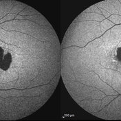 Central Areolar Choroidal Dystrophy
Jul 7 2015 by Hamid Ahmadieh, MD
Fundus autofluorescence of both eyes of a 58-year-old man with progressive loss of vision. VA OD is 20/60 and VA OS is 20/400.
Photographer: Soulmaz Shahmohammad, Negah Eye Center, Tehran, Iran
Imaging device: Specteralis
Condition/keywords: central areolar choroidal dystrophy (CACD), fundus autofluorescence (FAF)
-
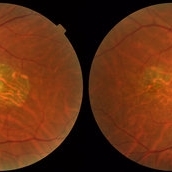 Central Areolar Choroidal Dystrophy
Jul 7 2015 by Hamid Ahmadieh, MD
Color fundus photograph of both eyes of a 58-year-old man with progressive loss of vision. VA OD is 20/60 and VA OS is 20/400.
Photographer: Soulmaz Shahmohammad, Negah Eye Center, Tehran, Iran
Imaging device: Topcon
Condition/keywords: central areolar choroidal dystrophy (CACD), color fundus photograph
-
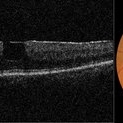 Macular Pseudohole
Jul 7 2015 by Hamid Ahmadieh, MD
Color fundus photograph and optical coherence tomography of the left eye of a 72-year-old woman with blurred vision due to epiretinal membrane. VA OS is 20/40 . Macular pseudohole is visible.
Photographer: Shabnam Poureh, Negah Eye Center, Tehran, Iran
Imaging device: Topcn
Condition/keywords: color fundus photograph, macular pseudohole, optical coherence tomography (OCT)
-
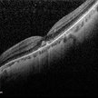 Spontaneous Macular Hemorrhage in High Myopia
Jul 7 2015 by Hamid Ahmadieh, MD
A highly myopic 30-year-old-man noticed sudden drop of vision and metamorphopsia in his left eye. OCT showed a macular haemorrhage (Fig. a) which partially resolved spontaneously after 6 weeks ( Fig. b). Four months later, VA improved to 20/40 and the image distortion markedly resolved ( Fig. c).
Photographer: Soulmaz Shahmohammad, Negah Eye Center, Tehran, Iran
Imaging device: Specteralis
Condition/keywords: high myopia, macular hemorrhage, optical coherence tomography (OCT)
-
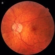 Spontaneous Macular Hemorrhage in High Myopia
Jul 7 2015 by Hamid Ahmadieh, MD
A highly myopic 30-year-old-man noticed sudden drop of vision and metamorphopsia in his left eye. Color fundus photograph showed a macular haemorrhage (Fig. a) which partially resolved spontaneously after 6 weeks ( Fig. b). Four months later, VA improved to 20/40 and the image distortion markedly resolved ( Fig. c). Lacquer cracks were visible.
Photographer: Soulmaz Shahmohammad, Negah Eye Center, Tehran, Iran
Condition/keywords: color fundus photograph, high myopia, macular hemorrhage
-
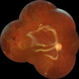 Relaxing Retinectomy
Jul 7 2015 by Hamid Ahmadieh, MD
Color fundus photograph of the right eye of a 25-year-old woman who underwent pars plana vitrectomy , relaxing retinectomy , endolaser photocoagulation and silicone oil injection for management of a very advanced vasoproliferative vitreoretinopathy with combined traction / rhegmatogenous RD secondary to a very severe occlusive retinal vasculitis. Retinal reattachment was achieved after surgery.
Photographer: Soulmaz Shahmohammad, Negah Eye Center, Tehran, Iran
Condition/keywords: color fundus photograph, combined retinal detachment, occlusive retinal vasculitis, relaxing retinectomy, vasoproliferative vitreoretinopathy
-
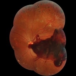 Subhyaloid Hemorrhage
Jul 7 2015 by Hamid Ahmadieh, MD
Color fundus photograph of the left eye of a 25-year-old woman with severe subhyaloid hemorrhage due to an advanced vasoproliferative vitreoretinopathy secondary to a severe idiopathic occlusive retinal vasculitis.
Photographer: Soulmaz Shahmohammad, Negah Eye Center, Tehran, Iran
Condition/keywords: color fundus photograph, occlusive vasculitis, subhyaloid hemorrhage
-
 Encircling Buckle Effect
Jul 7 2015 by Hamid Ahmadieh, MD
Late FA image of the right eye of a 30-year-old man who underwent pars plana vitrectomy , endolaser photocoagulation and an encircling band placement a couple of years before following a penetrating trauma at the vitreous base area at the 7 o'clock meridian.
Photographer: Nayereh Hadipour, Negah Eye Center,Tehran, Iran
Imaging device: Specteralis
Condition/keywords: pars plana vitrectomy (PPV)
-
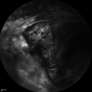 Retinal Detachment Secondary to FEVR
Jul 7 2015 by Hamid Ahmadieh, MD
FA image of the vasoproliferative tissue in a case of advanced FEVR.
Photographer: Soulmaz Shahmohammad, Negah Eye Center, Tehran, Iran
Imaging device: Specteralis
Condition/keywords: familial exudative vitreoretinopathy (FEVR), vasoproliferative vitreoretinopathy
-
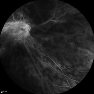 Retinal Detachment Secondary to FEVR
Jul 7 2015 by Hamid Ahmadieh, MD
FA image of the right eye of a 28-year-old man with total RD secondary to advanced FEVR.
Photographer: Soulmaz Shahmohammad, Negah Eye Center, Tehran, Iran
Imaging device: Specteralis
Condition/keywords: familial exudative vitreoretinopathy (FEVR)
-
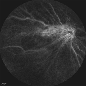 Retinal Detachment Secondary to FEVR
Jul 7 2015 by Hamid Ahmadieh, MD
Late FA image of the right eye of a 28-year-old man with total RD secondary to advanced FEVR.
Photographer: Soulmaz Shahmohammad, Negah Eye Center, Tehran, Iran
Imaging device: Specteralis
Condition/keywords: familial exudative vitreoretinopathy (FEVR)
-
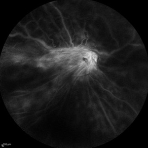 Retinal Detachment Secondary to FEVR
Jul 7 2015 by Hamid Ahmadieh, MD
Late FA image of the right eye of a 28-year-old man with total RD secondary to advanced FEVR.
Photographer: Soulmaz Shahmohammad, Negah Eye Center, Tehran, Iran
Imaging device: Specteralis
Condition/keywords: familial exudative vitreoretinopathy (FEVR)
-
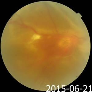 Retinal Detachment Secondary to FEVR
Jul 7 2015 by Hamid Ahmadieh, MD
Color fundus photograph of the right eye of a 28-year-old man with total retinal detachment secondary to advanced FEVR.
Photographer: Soulmaz Shahmohammad, Negah Eye Center, Tehran, Iran
Condition/keywords: color fundus photograph, familial exudative vitreoretinopathy (FEVR)
-
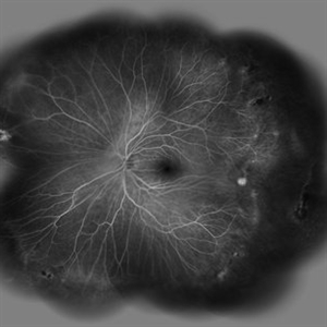 Vascular Abnormalities in an Asymptomatic Eye with FEVR
Jul 7 2015 by Hamid Ahmadieh, MD
Wide -field FA image of the asymptomatic left eye of a 28-year-old man with total RD secondary to FEVR in his right eye. Retinal vascular abnormalities including avascular zone in the extreme periphery associated with vasodilation and arteriovenous anastomosis are visible in the left eye.
Photographer: Soulmaz Shahmohammad, Negah Eye Center, Tehran, Iran
Imaging device: Specteralis
Condition/keywords: familial exudative vitreoretinopathy (FEVR), peripheral retinal nonperfusion
-
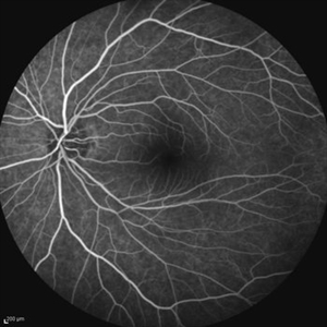 Asymptomatic Eye in FEVR
Jul 7 2015 by Hamid Ahmadieh, MD
FA image of the asymptomatic left eye of a 28-year-old man with total RD secondary to advanced FEVR in his right eye. Notice straightening of the retinal vessels.
Photographer: Soulmaz Shahmohammad, Negah Eye Center, Tehran, Iran
Imaging device: Specteralis
Condition/keywords: asymptomatic, familial exudative vitreoretinopathy (FEVR)
-
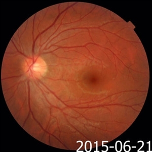 Asymptomatic Eye in FEVR
Jul 7 2015 by Hamid Ahmadieh, MD
Color fundus photograph of the asymptomatic eye of a patient with FEVR. Notice straightening of the retinal vessels.
Photographer: Soulmaz Shahmohammad, Negah Eye Center, Tehran, Iran
Condition/keywords: color fundus photograph, familial exudative vitreoretinopathy (FEVR)
-
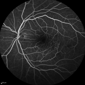 Old Macular BRVO
Oct 10 2015 by Hamid Ahmadieh, MD
Mid -venous phase FA of the left eye of a 58-year-old woman with the history of macular BRVO. Note microaneurysms, collaterals , tortuosity of venules and an enlarged irregular FAZ compatible with macular ischemia.
Photographer: Nayereh Hadipour, Negah Eye Center, Tehran, Iran
Condition/keywords: collaterals, macular branch retinal vein occlusion (BRVO), macular ischemia, microaneurysms
-
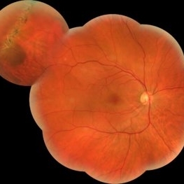 Lattice Degeneration and Choroidal Nevus
Oct 10 2015 by Hamid Ahmadieh, MD
Wide field color fundus photograph of the right eye of a 46-year-old woman with a typical lattice degeneration and an adjacent choroidal nevus.
Photographer: Solmaz Shahmohammad, Negah Eye Center, Tehran, Iran
Condition/keywords: choroidal nevus, color fundus photograph, lattice degeneration
-
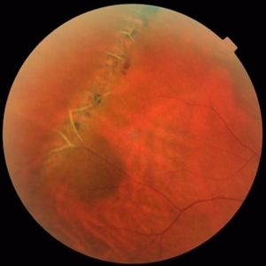 Lattice Degeneration and Choroidal Nevus
Oct 10 2015 by Hamid Ahmadieh, MD
Color fundus photograph of the right eye of a 46-year-old woman with a typical lattice degeneration and an adjacent choroidal nevus.
Photographer: Solmaz Shahmohammad, Negah Eye Center, Tehran, Iran
Condition/keywords: choroidal nevus, color fundus photograph, lattice degeneration
-
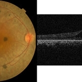 Retinitis Pigmentosa & CME
Oct 10 2015 by Hamid Ahmadieh, MD
Color fundus photograph of the right eye of a 40-year-old woman with cystoid macular edema ( CME) secondary to retinitis pigmentosa (RP).
Photographer: Shabnam Pooreh, Negah Eye Center, Tehran, Iran
Condition/keywords: color fundus photograph, cystoid macular edema (CME), retinitis pigmentosa
-
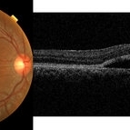 Best Disease
Oct 10 2015 by Hamid Ahmadieh, MD
Color fundus photograph and OCT image of the right eye of a 35-year-old man with decreased vision due to Best disease.
Photographer: Shabnam Pooreh, Negah Eye Center, Tehran, Iran
Condition/keywords: Best disease, color fundus photograph, optical coherence tomography (OCT), vitelliform lesion
-
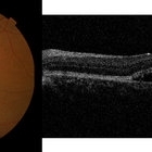 Best Disease
Oct 10 2015 by Hamid Ahmadieh, MD
Color fundus photograph and OCT image of the left eye of a 35-year-old man with decreased vision due to Best disease.
Photographer: Shabnam Pooreh, Negah Eye Center, Tehran, Iran
Condition/keywords: Best disease, color fundus photograph, optical coherence tomography (OCT), vitelliform lesion
-
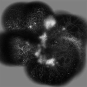 PDR
PDR
Oct 10 2015 by Hamid Ahmadieh, MD
Merged late phase FA image of the the right eye of a diabetic patient with PDR ; profound leakage from the NVD and NVE foci are present. Scars of previous scatter laser photocoagulation are visible.
Photographer: Shabnam Pooreh, Negah Eye Center, Tehran, Iran
Condition/keywords: proliferative diabetic retinopathy (PDR)
-
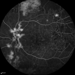 PDR
PDR
Oct 10 2015 by Hamid Ahmadieh, MD
Late venous phase FA image of the left eye of a diabetic patient with PDR. Leakage from the NVD and NVE foci are visible.
Photographer: Shabnam Pooreh, Negah Eye Center, Tehran, Iran
Condition/keywords: proliferative diabetic retinopathy (PDR)
-
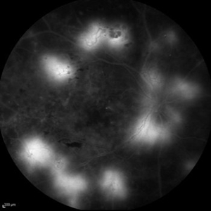 PDR
PDR
Oct 10 2015 by Hamid Ahmadieh, MD
Late phase FA image of the right eye of a diabetic patient with PDR. Profound leakage from the NVD and NVE foci are visible.
Photographer: Shabnam Pooreh, Negah Eye Center, Tehran, Iran
Condition/keywords: proliferative diabetic retinopathy (PDR)
-
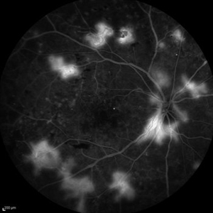 PDR
PDR
Oct 10 2015 by Hamid Ahmadieh, MD
FA image of the right eye of a diabetic patient with PDR . Leakage from the NVD and NVE foci are visible.
Photographer: Shabnam Pooreh, Negah Eye Center, Tehran, Iran
Condition/keywords: proliferative diabetic retinopathy (PDR)
-
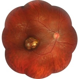 Congenital Toxoplasmosis
Oct 10 2015 by Hamid Ahmadieh, MD
Wide-field color fundus photograph of the right eye of a 15 -year-old boy with decreased vision due to a large chorioretinal scar involving the macula . The lesion is typical for a congenital ocular toxoplasmosis .
Photographer: Solmaz Shahmohammad, Negah Eye Center, Tehran, Iran
Condition/keywords: color fundus photograph, congenital toxoplasmosis
-
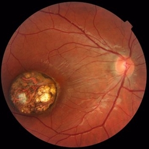 Congenital Toxoplasmosis
Oct 10 2015 by Hamid Ahmadieh, MD
Color fundus photograph of the right eye of a 15 -year-old boy with decreased vision due to a large chorioretinal scar involving the macula . The lesion is typical for a congenital ocular toxoplasmosis .
Photographer: Solmaz Shahmohammad, Negah Eye Center, Tehran, Iran
Condition/keywords: color fundus photograph, congenital toxoplasmosis
-
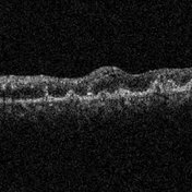 Primary Hyperoxaluria and Oxalosis
Oct 10 2015 by Hamid Ahmadieh, MD
OCT image of the left eye of a 55- year- old woman with oxalosis. Intraretinal and sub -retinal deposition of calcium oxalate crystals are seen.
Photographer: Shabnam Pooreh, Negah Eye Center, Tehran, Iran
Condition/keywords: optical coherence tomography (OCT), oxalosis
-
 Primary Hyperoxaluria and Oxalosis
Oct 10 2015 by Hamid Ahmadieh, MD
FA image of the left eye of a 55-year-old woman with primary hyperoxaluria and oxalosis showing occlusion of the retinal arteries. Staining of calcium oxalate crystals is visible.
Photographer: shabnam Pooreh, Negah Eye Center, Tehran, Iran
Condition/keywords: oxalosis, primary hyperoxaluria
-
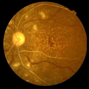 Primary Hyperoxaluria and Oxalosis
Oct 10 2015 by Hamid Ahmadieh, MD
Color fundus photograph of the left eye of a 55-year-old woman with primary hyperoxaluria and oxalosis leading to intraretinal and subretinal deposition of calcium oxalate crystals . In addition, deposition of these crystals in the retinal vessels has led to the occlusion of retinal arterioles and venules leading to multiple cotton wools and dot and blot retinal hemorrhages.
Photographer: Shabnam Pooreh, Negah Eye Center, Tehran, Iran
Condition/keywords: color fundus photograph, oxalosis, primary hyperoxaluria
-
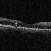 Primary Hyperoxaluria and Oxalosis
Oct 10 2015 by Hamid Ahmadieh, MD
OCT image of the right eye of a 55- year- old woman with primary hyperoxaluria and oxalosis. Intraretinal and sub- retinal deposition of calcium oxalate crystals are visible .
Photographer: Shabnam Pooreh, Negah Eye Center, Tehran, Iran
Condition/keywords: optical coherence tomography (OCT), oxalosis, primary hyperoxaluria
-
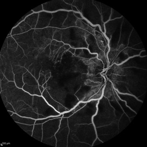 Primary Hyperoxaluria and Oxalosis
Oct 10 2015 by Hamid Ahmadieh, MD
Late venous phase FA image of the right of a 55-year-old woman with primary hyperoxaluria and oxalosis . Notice macular infarction and areas of capillary non -perfusion in retinal mid periphery due to the occlusion of retinal arterioles.
Photographer: Shabnam Pooreh, Negah Eye Center, Tehran, Iran
Condition/keywords: oxalosis, primary hyperoxaluria
-
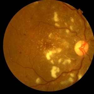 Primary Hyperoxaluria and Oxalosis
Oct 10 2015 by Hamid Ahmadieh, MD
Color fundus photograph of the right eye of a 55-year-old woman with primary hyperoxaluria and oxalosis leading to intraretinal and subretinal deposition of calcium oxalate crystals . In addition, deposition of these crystals in the retinal vessels has led to the occlusion of retinal arterioles and venules leading to multiple cotton wools and dot and blot retinal hemorrhages.
Photographer: shabnam Pooreh, Negah Eye Center, Tehran, Iran
Condition/keywords: color fundus photograph, oxalosis, primary hyperoxaluria
-
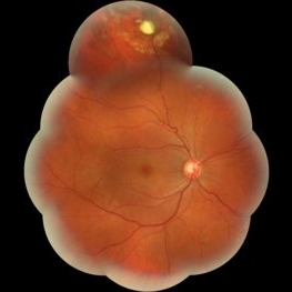 Intraretinal Foreign Body
Oct 10 2015 by Hamid Ahmadieh, MD
Merged color fundus photograph of the right eye of a patient with intraretinal metallic foreign body . Barrier laser photocoagulation was performed before vitrectomy and foreign body removal.
Photographer: Soodabeh Fouladin, Negah Eye Center, Tehran, Iran
Condition/keywords: color fundus photograph, intraocular foreign body
-
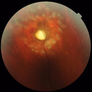 Intraretinal Foreign Body
Oct 10 2015 by Hamid Ahmadieh, MD
Color fundus photograph of the right eye of a patient with an intraretinal foreign body delimited by laser spots. An adjacent patch of retinal haemorrhage is visible.
Photographer: Soodabeh Fouladin, Negah Eye Center, Tehran, Iran
Condition/keywords: color fundus photograph, intraocular foreign body
-
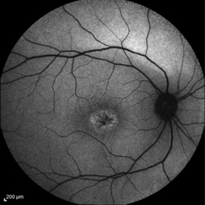 Juvenile Retinoschisis
Oct 10 2015 by Hamid Ahmadieh, MD
FAF image of the right eye of a 30-year-old man with juvenile retinoschisis. VA OD is 20/80.
Photographer: Shabnam Pooreh, Negah Eye Center, Tehran, Iran
Condition/keywords: fundus autofluorescence (FAF), juvenile retinoschisis
-
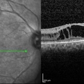 Juvenile Retinoschisis
Oct 10 2015 by Hamid Ahmadieh, MD
OCT image of the right eye of a 30-year-old man with juvenile retinoschisis. VA OD is 20/80.
Photographer: Shabnam Pooreh, Negah Eye Center, Tehran, Iran
Imaging device: Specteralis
Condition/keywords: juvenile retinoschisis, optical coherence tomography (OCT)
-
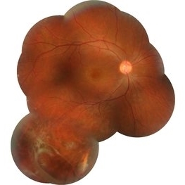 Juvenile Retinoschisis
Oct 10 2015 by Hamid Ahmadieh, MD
Merged color fundus photograph of the right eye of a 30-year-old man with juvenile retioschisis. Involvement of the retinal periphery with typical large inner layer retinal holes is visible.
Photographer: Shabnam Pooreh, Negah Eye Center, Tehran, Iran
Condition/keywords: color fundus photograph, inner layer holes, juvenile retinoschisis
-
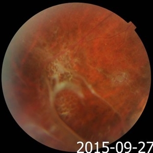 Juvenile Retinoschisis
Oct 10 2015 by Hamid Ahmadieh, MD
Typical changes including large inner layer retinal holes in the retinal periphery of a 30-year-old man with juvenile retinoschisis.
Photographer: Shabnam Pooreh, Negah Eye Center, Tehran, Iran
Condition/keywords: color fundus photograph, inner layer holes, juvenile retinoschisis
-
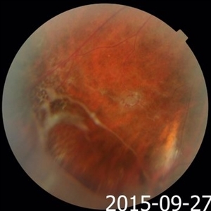 Juvenile Retinoschisis
Oct 10 2015 by Hamid Ahmadieh, MD
Color fundus photograph showing typical changes including large inner layer holes in the inferior temporal retinal periphery of the right eye a 30-year-old man with juvenile retinoschisis.
Photographer: Shabnam Pooreh, Negah Eye Center, Tehran, Iran
Condition/keywords: color fundus photograph, inner layer holes, juvenile retinoschisis
-
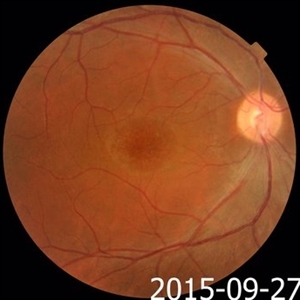 Juvenile Retinoschisis
Oct 10 2015 by Hamid Ahmadieh, MD
Color fundus photograph of the right eye of a 30-year-old man with juvenile retinoschisis and typical macular changes.
Photographer: Shabnam Pooreh, Negah Eye Center, Tehran , Iran
Condition/keywords: color fundus photograph, juvenile retinoschisis, macular schisis
-
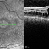 Juvenile Retinoschisis
Oct 10 2015 by Hamid Ahmadieh, MD
OCT image of the left eye of a 30-year-old man with juvenile retinoschisis. VA OS is 20/100.
Photographer: Shabnam Pooreh, Negah Eye Center, Tehran , Iran
Imaging device: Specteralis
Condition/keywords: juvenile retinoschisis, macular schisis, optical coherence tomography (OCT)
-
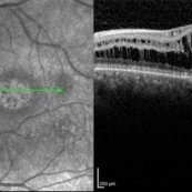 Juvenile Retinoschisis
Oct 10 2015 by Hamid Ahmadieh, MD
OCT image of the left eye of a 30-year-old man with juvenile retinoschisis. VA OS is 20/100.
Photographer: shabnam Pooreh, Negah Eye Center, Tehran , Iran
Imaging device: Specteralis
Condition/keywords: juvenile retinoschisis, macular schisis, optical coherence tomography (OCT)
-
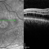 Juvenile Retinoschisis
Oct 10 2015 by Hamid Ahmadieh, MD
OCT image of the left eye of a 30-year-old man with juvenile retinoschisis. VA OS is 20/100.
Photographer: Shabnam Pooreh, Negah Eye Center, Tehran , Iran
Imaging device: Specteralis
Condition/keywords: juvenile retinoschisis, macular schisis, optical coherence tomography (OCT)
-
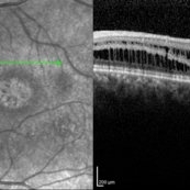 Juvenile Retinoschisis
Oct 10 2015 by Hamid Ahmadieh, MD
OCT image of the left eye of a 30-year-old man with juvenile retinoschisis. VA OS is 20/100.
Photographer: Shabnam Pooreh, Negah Eye Center, Tehran , Iran
Imaging device: Specteralis
Condition/keywords: juvenile retinoschisis, macular schisis, optical coherence tomography (OCT)
-
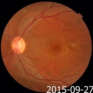 Juvenile Retinoschisis
Oct 10 2015 by Hamid Ahmadieh, MD
Color fundus photograph of the left eye of a 30-year-old man with juvenile retinoschisis and typical macular changes.
Photographer: Shabnam Pooreh, Negah Eye Center, Tehran , Iran
Condition/keywords: color fundus photograph, juvenile retinoschisis, macular schisis
-
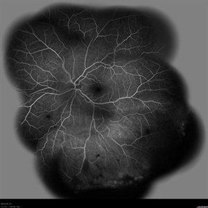 Vasoproliferative Tumor
-
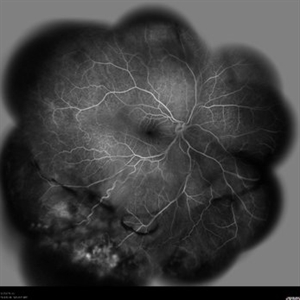 Vasoproliferative Tumor
-
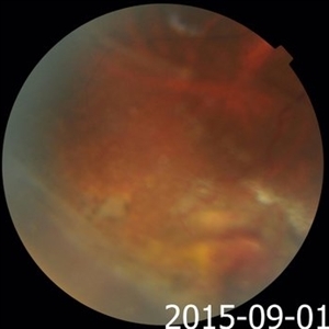 Vasoproliferative Tumor
-
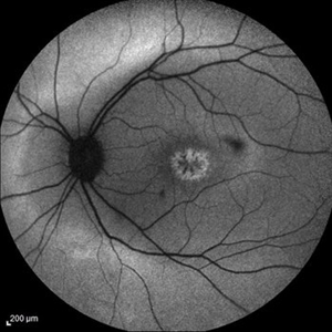 Juvenile Retinoschisis
May 14 2016 by Hamid Ahmadieh, MD
FAF image of the left eye of a 30-year-old man with juvenile retinoschisis. VA OD is 20/80.
Photographer: Shabnam Pooreh, Negah Eye Center, Tehran , Iran
Imaging device: Specteralis
Condition/keywords: juvenile retinoschisis
-
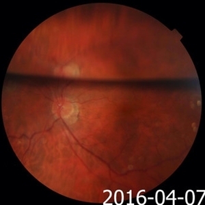 Gas-Filled Eye
Dec 27 2016 by Hamid Ahmadieh, MD
Color fundus photograph of the left eye showing partially absorbed intraocular gas following vitrectomy and fluid/gas exchange. Reflection of the disc image is visible.
Photographer: Solmaz Shahmohammad, Negah Eye Center, Tehran,Iran
Condition/keywords: color fundus photograph, intraocular gas
-
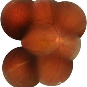 Longstanding Retinal Detachment Secondary to a Larg Retinal Tear
Dec 27 2016 by Hamid Ahmadieh, MD
Montaged color fundus photograph of the right eye of a patient with longstanding retinal detachment. Demarcation lines are visible.
Photographer: Shabnam Poureh, Negah Eye Center, Tehran, Iran
Condition/keywords: color fundus photograph, demarcation line
-
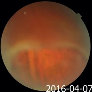 Longstanding Retinal Detachment Due to a Larg Retinal Tear
Dec 27 2016 by Hamid Ahmadieh, MD
Color fundus photograph of the right eye of a patient with longstanding retinal detachment. A large retinal break is visible.
Photographer: Shabnam Poureh, Negah Eye Center, Tehran, Iran
Condition/keywords: color fundus photograph, retinal break
-
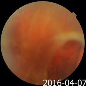 Longstanding Retinal Detachment Secondary to a Larg Retinal Tear
Dec 27 2016 by Hamid Ahmadieh, MD
Color fundus photograph of the right eye of a patient with post-traumatic longstanding retinal detachment. A large retinal break is visible.
Photographer: Shabnam Poureh, Negah Eye Center, Tehran, Iran
Condition/keywords: color fundus photograph, retinal break
-
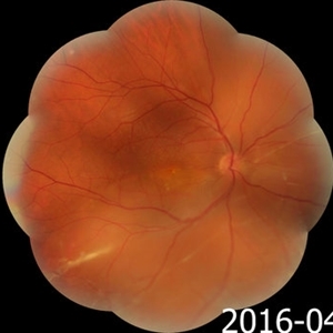 Longstanding Retinal Detachment Due to a Larg Retinal Tear
Dec 27 2016 by Hamid Ahmadieh, MD
Wide-field color fundus photograph of the right eye of a patient with longstanding retinal detachment. Demarcation lines are visible.
Photographer: Shabnam Poureh, Negah Eye Center, Tehran, Iran
Condition/keywords: color fundus photograph
-
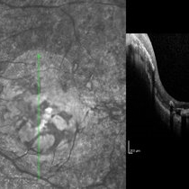 Macular Coloboma and Pigmentary Retinopathy
Feb 25 2017 by Hamid Ahmadieh, MD
Infrared and OCT images of the left eye of a 25-year-old woman with bilateral macular colobomata and pigmentary retinopathy similar to Leber's congenital amaurosis.
Photographer: Shabnam Poureh, Negah Eye Center, Tehran, Iran
Imaging device: Spectralis OCTc
Condition/keywords: infrared image
-
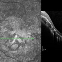 Macular Coloboma and Pigmentary Retinopathy
Feb 25 2017 by Hamid Ahmadieh, MD
Infrared and OCT images of the left eye of a 25-year-old woman with bilateral macular colobomata and pigmentary retinopathy similar to Leber's congenital amaurosis.
Photographer: Shabnam Poureh, Negah Eye Center, Tehran, Iran
Condition/keywords: bilateral pigmentary retinopathy, infrared image, macular coloboma, optical coherence tomography (OCT)
-
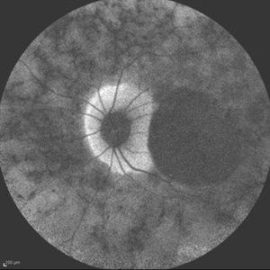 Macular Coloboma and Pigmentary Retinopathy
Feb 25 2017 by Hamid Ahmadieh, MD
Fundus autofluorescence (FAF) image of the left eye of a 25-year-old woman with bilateral macular colobomata and pigmentary retinopathy similar to Leber's congenital amaurosis.
Photographer: Shabnam Poureh, Negah Eye Center, Tehran, Iran
Condition/keywords: bilateral pigmentary retinopathy, fundus autofluorescence (FAF), macular coloboma
-
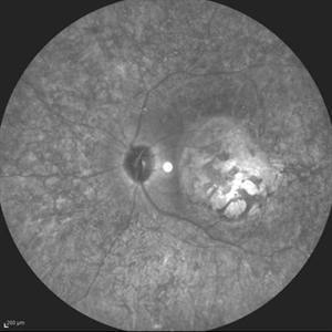 Macular Coloboma and Pigmentary Retinopathy
Feb 25 2017 by Hamid Ahmadieh, MD
Infrared image of the left eye of a 25-year-old woman with bilateral macular colobomata and pigmentary retinopathy similar to Leber's congenital amaurosis.
Photographer: Shabnam Poureh, Negah Eye Center, Tehran, Iran
Condition/keywords: bilateral pigmentary retinopathy, infrared image, macular coloboma
-
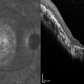 Macular Coloboma and Pigmentary Retinopathy
Feb 25 2017 by Hamid Ahmadieh, MD
Infrared and OCT images of the right eye of a 25-year-old woman with bilateral macular colobomata and pigmentary retinopathy similar to Leber's congenital amaurosis.
Photographer: Shabnam Poureh, Negah Eye Center, Tehran, Iran
Imaging device: Spectralis OCT
Condition/keywords: infrared image, macular coloboma, optical coherence tomography (OCT)
-
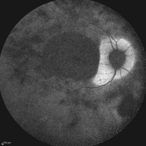 Macular Coloboma and Pigmentary Retinopathy
Feb 25 2017 by Hamid Ahmadieh, MD
Fundus autofluorescence (FAF) image of the right eye of a 25-year-old woman with bilateral macular colobomata and pigmentary retinopathy similar to Leber's congenital amaurosis.
Photographer: Shabnam Poureh, Negah Eye Center, Tehran, Iran
Condition/keywords: bilateral pigmentary retinopathy, fundus autofluorescence (FAF), macular coloboma
-
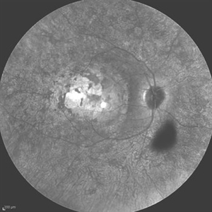 Leber's Congenital Amaurosis
Feb 25 2017 by Hamid Ahmadieh, MD
Infrared image of the right eye of a 25-year-old woman with bilateral macular colobomata and pigmentary retinopathy similar to Leber's congenital amaurosis.
Photographer: Shabnam Poureh, Negah Eye Center, Tehran, Iran
Condition/keywords: bilateral pigmentary retinopathy, infrared image, macular coloboma
-
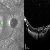 Macular Coloboma and Pigmentary Retinopathy
Feb 25 2017 by Hamid Ahmadieh, MD
Infrared and OCT images of the right eye of a 25-year-old woman with bilateral macular colobomata and pigmentary retinopathy similar to Leber's congenital amaurosis.
Photographer: Shabnam Poureh, Negah Eye Center, Tehran, Iran
Condition/keywords: infrared image, macular coloboma, optical coherence tomography (OCT)
-
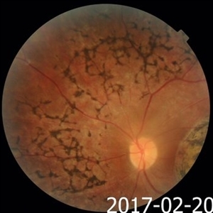 Macular Coloboma and Pigmentary Retinopathy
Feb 25 2017 by Hamid Ahmadieh, MD
Color fundus photograph of the left eye of a 25-year-old woman with the history of low vision since childhood. Bilateral macular colobomata and pigmentary retinopathy similar to Leber's congenital amaurosis are present.
Photographer: Shabnam Poureh, Negah Eye Center, Tehran, Iran
Condition/keywords: bilateral pigmentary retinopathy, color fundus photograph, macular coloboma
-
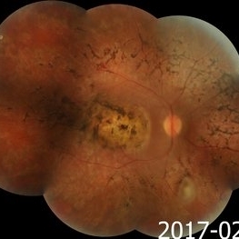 Macular Coloboma and Pigmentary Retinopathy
Feb 25 2017 by Hamid Ahmadieh, MD
Merged color fundus photograph of the right eye of a 25-year-old woman with the history of low vision since childhood. Bilateral macular colobomata and pigmentary retinopathy similar to Leber's congenital amaurosis are present.
Photographer: Shabnam Poureh, Negah Eye Center, Tehran, Iran
Condition/keywords: bilateral pigmentary retinopathy, color fundus photograph, macular coloboma, pigmentary retinal dystrophy
-
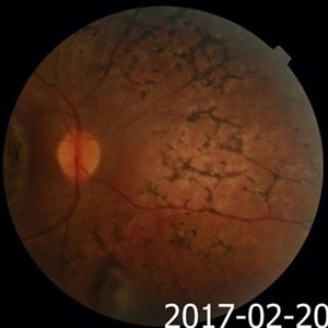 Macular Coloboma and Pigmentary Retinopathy
Feb 25 2017 by Hamid Ahmadieh, MD
Color fundus photograph of the right eye of a 25-year-old woman with the history of low vision since childhood. Bilateral macular colobomata and pigmentary retinopathy similar to Leber's congenital amaurosis are present.
Photographer: Shabnam Poureh, Negah Eye Center, Tehran, Iran
Condition/keywords: color fundus photograph, pigmentary retinal dystrophy
-
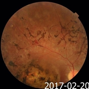 Macular Coloboma and Pigmentary Retinopathy
Feb 25 2017 by Hamid Ahmadieh, MD
Color fundus photograph of the right eye of a 25-year-old woman with the history of low vision since childhood. Bilateral macular colobomata and pigmentary retinopathy similar to Leber's congenital amaurosis are present.
Photographer: Shabnam Poureh, Negah Eye Center, Tehran, Iran
Condition/keywords: bilateral pigmentary retinopathy, color fundus photograph, macular coloboma
-
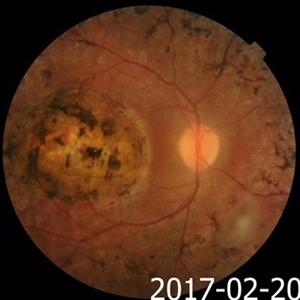 Macular Coloboma and Pigmentary Retinopathy
Feb 25 2017 by Hamid Ahmadieh, MD
Color fundus photograph of the right eye of a 25-year-old woman with the history of low vision since childhood. Bilateral macular colobomata and pigmentary retinopathy similar to Leber's congenital amaurosis are present.
Photographer: Shabnam Poureh, Negah Eye Center, Tehran, Iran
Condition/keywords: bilateral pigmentary retinopathy, color fundus photograph, macular coloboma, pigmentary retinal dystrophy
-
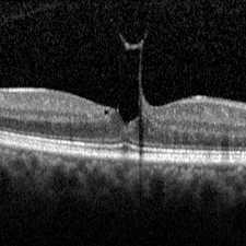 VMT
VMT
Mar 14 2017 by Hamid Ahmadieh, MD
OCT findings of a typical vitreomacular traction ( VMT) with lamellar macular hole appearance and a counterpart traction on the cortical vitreous.
Photographer: Solmaz Shahmohammad, Negah Eye Center, Tehran,Iran
Imaging device: Spectralis OCT
Condition/keywords: lamellar macular hole, vitreomacular traction (VMT)
-
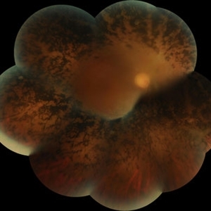 Advanced Retinitis Pigmentosa
Mar 14 2017 by Hamid Ahmadieh, MD
Merged color fundus photograph of the right eye of a patient with advanced retinitis pigmentosa sparing the posterior pole.
Photographer: Soodabeh Fouladin, Negah Eye Center, Tehran, Iran
Condition/keywords: color fundus photograph, retinitis pigmentosa (RP) dystrophy
-
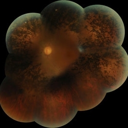 RP
RP
Mar 14 2017 by Hamid Ahmadieh, MD
Merged color fundus photograph of the left eye of a patient with advanced retinitis pigmentosa sparing the posterior pole.
Photographer: Soodabeh Fouladin, Negah Eye Center, Tehran, Iran
Condition/keywords: color fundus photograph, retinitis pigmentosa (RP) dystrophy
-
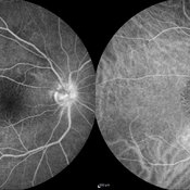 Leber's Idiopathic Stellate Neuroretinitis
Mar 14 2017 by Hamid Ahmadieh, MD
FA and ICG angiography findings in the right eye of a 25-year-old man with Leber's idiopathic stellate neuroretinitis. Leakage from disc surface is seen in the late phase of FA. Stellate maculopathy has resulted in a specific pattern in ICG angiography.
Photographer: Shabnam Poureh, Negah Eye Center, Tehran, Iran
Condition/keywords: indocyanine green (ICG) angiography, Leber's stellate maculopathy
-
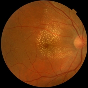 Leber's Idiopathic Stellate Neuroretinitis
Mar 14 2017 by Hamid Ahmadieh, MD
Color fundus photograph of the right eye of a 25-year-old man with decreased vision due to Leber's idiopathic stellate neuroretinitis.
Photographer: Ms Shabnam Poureh, Negah Eye Center, Tehran, Iran
Condition/keywords: color fundus photograph, Leber's stellate maculopathy
-
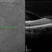 Cone-Rod Dystrophy
Mar 15 2017 by Hamid Ahmadieh, MD
Infrared and OCT images of the left eye of a 16-year-old boy with decreased visual acuity and color vision deficiency due to cone-rod dystrophy.
Photographer: Abazarnezhad , Negah Eye Center, Tehran, Iran
Imaging device: Spectralis OCT
Condition/keywords: cone dystrophy, infrared image, optical coherence tomography (OCT)
-
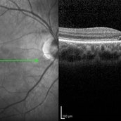 Cone-Rod Dystrophy
Mar 15 2017 by Hamid Ahmadieh, MD
Infrared and OCT images of the right eye of a 16-year-old boy with decreased visual acuity and color vision deficiency due to cone-rod dystrophy.
Photographer: Abazarnezhad , Negah Eye Center, Tehran, Iran
Imaging device: Spectralis OCT
Condition/keywords: cone dystrophy, infrared image, optical coherence tomography (OCT)
-
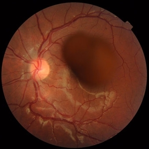 Valsalva Retinopathy
Mar 15 2017 by Hamid Ahmadieh, MD
Color fundus photograph of the left eye of a 30-year-old man with sudden drop of vision due to Valsalva retinopathy.
Photographer: Soodabeh Fouladin, Negah Eye Center, Tehran, Iran
Condition/keywords: color fundus photograph, valsalva retinopathy
-
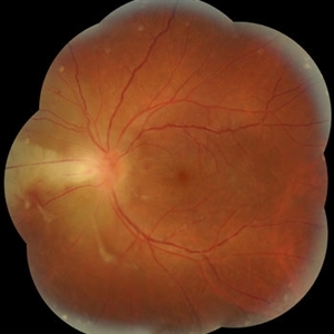 Acute Toxoplasmosis Neuroretinitis
Mar 15 2017 by Hamid Ahmadieh, MD
Color fundus photograph of the left eye of a 26-year-old man with clinical picture of acute neuroretinitis and serologic evidence of Toxoplasma gondii infection. Disc swelling, necrotizing retinitis with overlying vitreous inflammation, retinal vasculitis and localized exudative retinal detachment are visible.
Photographer: Solmaz Shahmohammad, Negah Eye Center, Tehran,Iran
Condition/keywords: color fundus photograph, exudative retinal detachment, neuroretinitis, retinal vasculitis, toxoplasmosis
-
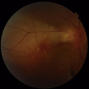 Acute Toxoplasmosis Neuroretinitis
Mar 15 2017 by Hamid Ahmadieh, MD
Color fundus photograph of the left eye of a 26-year-old man with clinical picture of acute neuroretinitis and serologic evidence of Toxoplasma gondii infection. Disc swelling, necrotizing retinitis with overlying vitreous inflammation, retinal vasculitis and localized exudative retinal detachment are visible.
Photographer: Solmaz Shahmohammad, Negah Eye Center, Tehran,Iran
Condition/keywords: color fundus photograph, exudative retinal detachment, neuroretinitis, retinal vasculitis
-
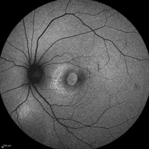 Traumatic Macular Hole and Choroidal Ruptures
Mar 15 2017 by Hamid Ahmadieh, MD
Fundus autofluorescence ( FAF) image of the left eye of a 30 -year-old woman with a recent history of closed eye injury leading to a large traumatic macular hole and two concentric choroidal ruptures.
Photographer: Solmaz Shahmohammad, Negah Eye Center, Tehran,Iran
Condition/keywords: choroidal rupture, fundus autofluorescence (FAF), traumatic macular hole
-
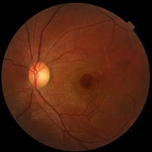 Traumatic Macular Hole and Choroidal Ruptures
Mar 15 2017 by Hamid Ahmadieh, MD
Color fundus photograph of the left eye of a 30 -year-old woman with a history of closed eye injury leading to a large traumatic macular hole and two concentric choroidal ruptures.
Photographer: Solmaz Shahmohammad, Negah Eye Center, Tehran,Iran
Condition/keywords: choroidal rupture, color fundus photograph, traumatic macular hole
-
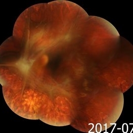 Advanced Proliferative Diabetic Retinopathy
Nov 4 2017 by Hamid Ahmadieh, MD
Merged color fundus photograph of the left eye of a 30-year-old woman with type1 diabetes since childhood. Note laser scars, severe fibrous proliferation, traction RD and macular dragging.
Photographer: Shabnam Poureh, Negah Eye Center, Tehran, Iran
Condition/keywords: color fundus photograph, diabetes, fibrous proliferation, proliferative diabetic retinopathy (PDR), severe traction
-
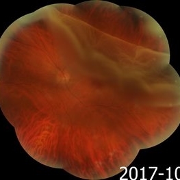 Giant Retinal Tear
Nov 9 2017 by Hamid Ahmadieh, MD
Merged color fundus photograph of the left eye of a 40-year-old man with rhegmatogenous retinal detachment due to a giant retinal tear with rolled edges.
Photographer: Soodabeh Fouladin, Negah Eye Center, Tehran, Iran
Condition/keywords: color fundus photograph, giant retinal tear
-
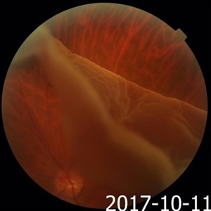 Giant Retinal Tear
Nov 9 2017 by Hamid Ahmadieh, MD
Color fundus photograph of the left eye of a 40-year-old man with rhegmatogenous retinal detachment due to a giant retinal tear with rolled edges.
Photographer: Soodabeh Fouladin, Negah Eye Center, Tehran, Iran
Condition/keywords: color fundus photograph, giant retinal tear
-
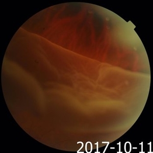 Giant Retinal Tear
Nov 9 2017 by Hamid Ahmadieh, MD
Color fundus photograph of the left eye of a 40-year-old man with rhegmatogenous retinal detachment due to a giant retinal tear with rolled edges.
Photographer: Soodabeh Fouladin, Negah Eye Center, Tehran, Iran
Condition/keywords: color fundus photograph, giant retinal tear
-
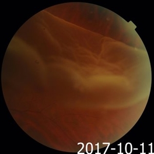 Giant Retinal Tear
Nov 9 2017 by Hamid Ahmadieh, MD
Color fundus photograph of the left eye of a 40-year-old man with rhegmatogenous retinal detachment due to a giant retinal tear with rolled edges.
Photographer: Soodabeh Fouladin, Negah Eye Center, Tehran, Iran
Condition/keywords: color fundus photograph, giant retinal tear
-
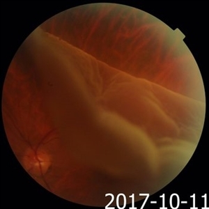 Giant Retinal Tear
Nov 9 2017 by Hamid Ahmadieh, MD
Color fundus photograph of the left eye of a 40-year-old man with rhegmatogenous retinal detachment due to a giant retinal tear with rolled edges.
Photographer: Soodabeh Fouladin, Negah Eye Center, Tehran, Iran
Condition/keywords: color fundus photograph, giant retinal tear, rolled edges of retina
-
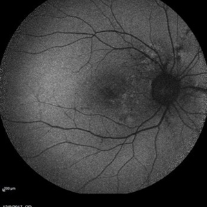 Angioid Streaks
Mar 17 2018 by Hamid Ahmadieh, MD
Fundus autofluorescence of the right eye of a 35-year-old woman with decreased vision due to CNV secondary to angioid streaks.
Photographer: Shabnam Pooreh, Negah Eyes Center, Tehran, Iran
Condition/keywords: angioid streaks, fundus autofluorescence (FAF)
-
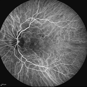 Angioid Streaks & CNV (Fig 3)
Aug 25 2012 by Hamid Ahmadieh, MD
Early phase ICG angiography imaging of a 53-year-old woman with a juxtafoveal CNV secondary to angioid streaks.
Photographer: Hamid Ahmadieh, Ophthalmic Research Center, Labbafinejad Medical Center
Imaging device: Heidelberg Spectralis
Condition/keywords: angioid streaks, choroidal neovascularization (CNV), indocyanine green (ICG) angiography
-
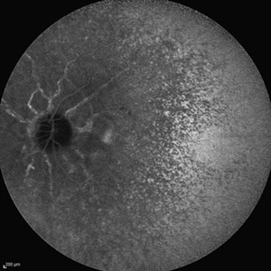 Angioid Streaks & CNV (Fig 4)
Aug 25 2012 by Hamid Ahmadieh, MD
Late phase ICG angiography imaging of a 53-year-old woman with a juxtafoveal CNV secondary to angioid streaks.
Photographer: Hamid Ahmadieh, Ophthalmic Research Center, Labbafinejad Medical Center
Imaging device: Heidelberg Spectralis
-
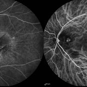 Polypoidal Choroidal Vasculopathy
Aug 25 2012 by Hamid Ahmadieh, MD
FA & ICG angiography imagings of a 73-year-old man with a peripapillary PCV.
Photographer: Hamid Ahmadieh, Ophthalmic Research Center, Labbafinejad Medical Center
Imaging device: Heidelberg Spectralis
Condition/keywords: indocyanine green (ICG) angiography, polypoidal choroidal vasculopathy (PCV)
-
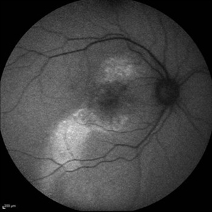 Multifocal CSCR 1
Sep 2 2012 by Hamid Ahmadieh, MD
Fundus autofluorescence of a 36-year-old man with an active multifocal CSCR.
Photographer: Hamid Ahmadieh, Ophthalmic Research Center, Labbafinejad Medical Center
Imaging device: Heidelberg Spectralis
Condition/keywords: central serous chorioretinopathy (CSCR)
-
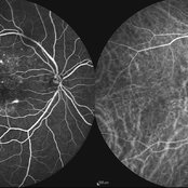 Multifocal CSCR 2
Sep 2 2012 by Hamid Ahmadieh, MD
Early-phase FA and ICG angiograms of a 36-year-old man with an active multifocal CSCR.
Photographer: Hamid Ahmadieh, Ophthalmic Research Center, Labbafinejad Medical Center
Imaging device: Heidelberg Spectralis
Condition/keywords: central serous chorioretinopathy (CSCR), indocyanine green (ICG) angiography
-
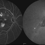 Multifocal CSCR
Sep 2 2012 by Hamid Ahmadieh, MD
Late-phase FA and ICG angiograms of a 36-year-old man with an active multifocal CSCR.
Photographer: Hamid Ahmadieh, Ophthalmic Research Center, Labbafinejad Medical Center
Imaging device: Heidelberg Spectralis
Condition/keywords: central serous chorioretinopathy (CSCR), indocyanine green (ICG) angiography
-
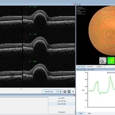 PED due to CSCR
Sep 2 2012 by Hamid Ahmadieh, MD
OCT image of a 37-year-old man with a serous PED secondary to CSCR.
Photographer: Hamid Ahmadieh, Ophthalmic Research Center, Labbafinejad Medical Center
Imaging device: Heidelberg Spectralis
Condition/keywords: central serous chorioretinopathy (CSCR), optical coherence tomography (OCT), pigment epithelial detachment (PED)
-
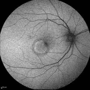 PED due to CSCR 2
Sep 2 2012 by Hamid Ahmadieh, MD
Autofluorescence imaging of a 37-year-old man with a serous PED secondary to CSCR.
Photographer: Hamid Ahmadieh, Ophthalmic Research Center, Labbafinejad Medical Center
Imaging device: Heidelberg Spectralis
Condition/keywords: autofluorescence imaging, central serous chorioretinopathy (CSCR), pigment epithelial detachment (PED)
-
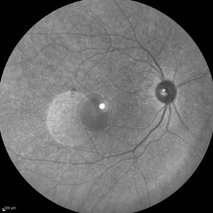 PED due to CSCR 3
Sep 2 2012 by Hamid Ahmadieh, MD
Infrared imaging of a 37-year-old man with a serous PED secondary to CSCR.
Photographer: Hamid Ahmadieh, Ophthalmic Research Center, Labbafinejad Medical Center
Imaging device: Heidelberg Spectralis
Condition/keywords: central serous chorioretinopathy (CSCR), pigment epithelial detachment (PED)
-
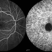 PED due to CSCR 4
Sep 2 2012 by Hamid Ahmadieh, MD
Early phase FA & ICG images of a 37-year-old man with a serous PED secondary to CSCR
Photographer: Hamid Ahmadieh, Ophthalmic Research Center, Labbafinejad Medical Center
Imaging device: Heidelberg Spectralis
Condition/keywords: central serous chorioretinopathy (CSCR), indocyanine green (ICG) angiography, pigment epithelial detachment (PED)
-
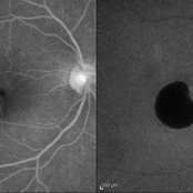 PED due to CSCR 5
Sep 2 2012 by Hamid Ahmadieh, MD
Late-phase FA and ICG images of a 37-year-old man with a serous PED secondary to CSCR.
Photographer: Hamid Ahmadieh, Ophthalmic Research Center, Labbafinejad Medical Center
Imaging device: Heidelberg Spectralis
Condition/keywords: central serous chorioretinopathy (CSCR), indocyanine green (ICG) angiography, pigment epithelial detachment (PED)
-
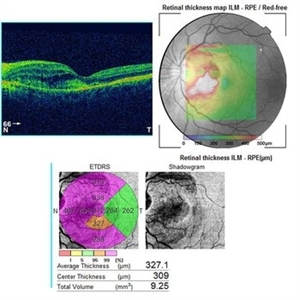 Angioid Streaks & CNV (Fig 5)
Sep 2 2012 by Hamid Ahmadieh, MD
OCT imaging of a 53-year-old woman with a juxtafoveal CNV secondary to angioid streaks.
Photographer: Hamid Ahmadieh, Ophthalmic Research Center, Labbafinejad Medical Center
Imaging device: Topcon
Condition/keywords: angioid streaks, choroidal neovascularization (CNV), optical coherence tomography (OCT)
-
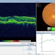 Choroidal Osteoma Plus CNV
Sep 2 2012 by Hamid Ahmadieh, MD
Color fundus photograph and OCT imaging of a 47-year-old man with a juxtafoveal CNV superimposed on a choroidal osteoma.
Photographer: Hamid Ahmadieh, Ophthalmic Research Center, Labbafinejad Medical Center
Imaging device: Topcon
Condition/keywords: choroidal neovascularization (CNV), choroidal osteoma, optical coherence tomography (OCT)
-
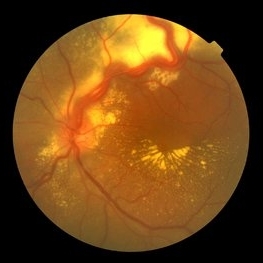 Von Hippel-Lindau
Sep 3 2012 by Hamid Ahmadieh, MD
Color fundus photograph of a 35-year-old woman with retinal angiomatosis.
Photographer: Hamid Ahmadieh, MD, Ophthalmic Research Center, Labbafinejad Medical Center, Shahid Beheshti University of Medical Sciences
Imaging device: Topcon Fundus Camera
Condition/keywords: retinal angiomatous proliferation (RAP), Von Hippel-Lindau
-
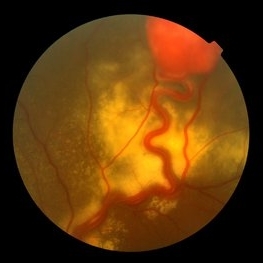 Von Hippel-Lindau
Sep 3 2012 by Hamid Ahmadieh, MD
Color fundus photograph of a 35-year-old woman with retinal angiomatosis.
Photographer: Hamid Ahmadieh, MD, Ophthalmic Research Center, Labbafinejad Medical Center, Shahid Beheshti University of Medical Sciences
Imaging device: Topcon Fundus Camera
Condition/keywords: retinal angiomatous proliferation (RAP)
-
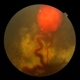 Von Hippel-Lindau
Sep 3 2012 by Hamid Ahmadieh, MD
Color fundus photograph of a 35-year-old woman with retinal angiomatosis.
Photographer: Hamid Ahmadieh, MD, Ophthalmic Research Center, Labbafinejad Medical Center, Shahid Beheshti University of Medical Sciences , Tehran
Imaging device: Topcon Fundus Camera
Condition/keywords: retinal angiomatous proliferation (RAP), Von Hippel-Lindau
-
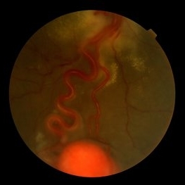 Von Hippel-Lindau
Sep 5 2012 by Hamid Ahmadieh, MD
Color fundus photograph of a 32-year-old man with retinal angiomatosis.
Photographer: Hamid Ahmadieh, MD, Ophthalmic Research Center, Labbafinejad Medical Center, Shahid Beheshti University of Medical Sciences
Imaging device: Topcon Fundus Camera
Condition/keywords: retinal angiomatous proliferation (RAP), Von Hippel-Lindau
-
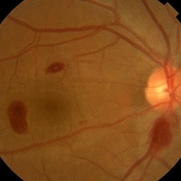 Ocular Manifestation of Acute Leukemia
Sep 5 2012 by Hamid Ahmadieh, MD
Color fundus photograph of a 26-year-old man with acute leukemia.
Photographer: Hamid Ahmadieh, MD, Ophthalmic Research Center, Labbafinejad Medical Center, Shahid Beheshti University of Medical Sciences
Imaging device: Topcon Fundus Camera
Condition/keywords: acute leukemia, white centered retinal hemorrhage (Roth Spot)
-
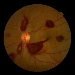 Ocular Manifestation of Acute Leukemia
Sep 8 2012 by Hamid Ahmadieh, MD
Color fundus photograph of a 26-year-old man with acute leukemia.
Photographer: Hamid Ahmadieh, MD, Ophthalmic Research Center, Labbafinejad Medical Center, Shahid Beheshti University of Medical Sciences , Tehran
Imaging device: Topcon Fundus Camera
Condition/keywords: acute leukemia, white centered retinal hemorrhage (Roth Spot)
-
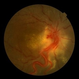 Von Hippel-Lindau
Sep 8 2012 by Hamid Ahmadieh, MD
Color fundus photograph of a 32-year-old man with retinal angiomatosis.
Photographer: Hamid Ahmadieh, MD, Ophthalmic Research Center, Labbafinejad Medical Center, Shahid Beheshti University of Medical Sciences
Imaging device: Topcon Fundus Camera
Condition/keywords: retinal angiomatous proliferation (RAP), Von Hippel-Lindau
-
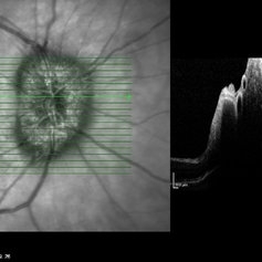 Papilledema
Sep 8 2012 by Hamid Ahmadieh, MD
OCT of the optic nerve head of the right eye of a 55-year-old woman with a malignant intracranial tumor.
Photographer: Hamid Ahmadieh, MD, Ophthalmic Research Center, Labbafinejad Medical Center, Shahid Beheshti University of Medical Sciences
Imaging device: Heidelberg Spectralis
Condition/keywords: malignant intracranial tumor, optical coherence tomography (OCT), papilledema
-
 Papilledema
Sep 8 2012 by Hamid Ahmadieh, MD
Color fundus photograph of the right eye of a 55-year-old woman with an intracranial malignant tumor.
Photographer: Hamid Ahmadieh, MD, Ophthalmic Research Center, Labbafinejad Medical Center, Shahid Beheshti University of Medical Sciences, Tehran, Iran
Imaging device: Topcon Fundus Camera
Condition/keywords: papilledema
-
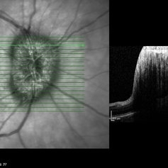 Papilledema
Sep 8 2012 by Hamid Ahmadieh, MD
OCT of the optic nerve head of the right eye of a 55-year-old woman with a malignant intracranial tumor.
Photographer: Hamid Ahmadieh, MD, Ophthalmic Research Center, Labbafinejad Medical Center, Shahid Beheshti University of Medical Sciences
Imaging device: Heidelberg Spectralis
Condition/keywords: optical coherence tomography (OCT), papilledema
-
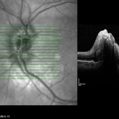 Papilledema
Sep 8 2012 by Hamid Ahmadieh, MD
OCT of the optic nerve head of the left eye of a 55-year-old woman with a malignant intracranial tumor.
Photographer: Hamid Ahmadieh, MD, Ophthalmic Research Center, Labbafinejad Medical Center, Shahid Beheshti University of Medical Sciences
Imaging device: Heidelberg Spectralis
Condition/keywords: malignant intracranial tumor, optical coherence tomography (OCT), papilledema
-
 Papilledema
Sep 8 2012 by Hamid Ahmadieh, MD
Color fundus photograph of the left eye of a 55-year-old woman with a malignant intracranial tumor.
Photographer: Hamid Ahmadieh, MD, Ophthalmic Research Center, Labbafinejad Medical Center, Shahid Beheshti University of Medical Sciences
Imaging device: Topcon Fundus Camera
Condition/keywords: papilledema
-
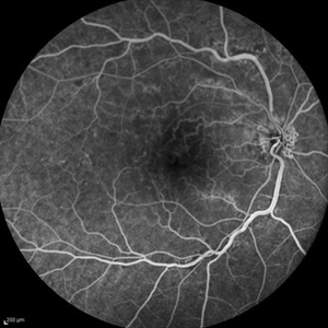 Optociliary Shunt Vessels in Old CRVO
Sep 8 2012 by Hamid Ahmadieh, MD
FA image of a 60-year-old woman with the history of central retinal vein occlusion.
Photographer: Hamid Ahmadieh, MD, Ophthalmic Research Center, Labbafinejad Medical Center, Shahid Beheshti University of Medical Sciences
Imaging device: Heidelberg Spectralis
Condition/keywords: central retinal vein occlusion (CRVO), shunts vessels
-
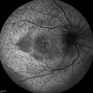 Cystoid Macular Edema (CME)
Sep 11 2012 by Hamid Ahmadieh, MD
Fundus autofluorescence (FAF) of the right eye a 17-year-old boy with chronic intermediate uveitis showing CME.
Photographer: Hamid Ahmadieh, MD, Ophthalmic Research Center, Labbafinejad Medical Center, Shahid Beheshti University of Medical Sciences
Imaging device: Heidelberg Spectralis
Condition/keywords: cystoid macular edema (CME), fundus autofluorescence (FAF), intermediate uveitis
-
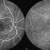 Cystoid Macular Edema (CME)
Sep 11 2012 by Hamid Ahmadieh, MD
FA & ICG angiography imagings of a 17-year-old boy with CME secondary to chronic intermediate uveitis.
Photographer: Hamid Ahmadieh, MD, Ophthalmic Research Center, Labbafinejad Medical Center, Shahid Beheshti University of Medical Sciences
Imaging device: Heidelberg Spectralis
Condition/keywords: cystoid macular edema (CME), indocyanine green (ICG) angiography, intermediate uveitis
-
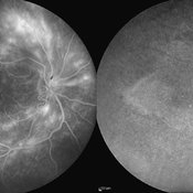 Cystoid Macular Edema (CME)
Sep 11 2012 by Hamid Ahmadieh, MD
Late phase FA & ICG angiography imagings of the right eye a 17-year-old boy with CME & retinal periphlebitis secondary to chronic intermediate uveitis.
Photographer: Hamid Ahmadieh, MD, Ophthalmic Research Center, Labbafinejad Medical Center, Shahid Beheshti University of Medical Sciences
Imaging device: Heidelberg Spectralis
Condition/keywords: cystoid macular edema (CME), indocyanine green (ICG) angiography, intermediate uveitis
-
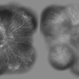 Cystoid Macular Edema (CME)
Sep 11 2012 by Hamid Ahmadieh, MD
Wide-field FA & ICG angiography imagings of a 17-year-old boy with CME & retinal periphlebitis secondary to chronic intermediate uveitis.
Photographer: Hamid Ahmadieh, MD, Ophthalmic Research Center, Labbafinejad Medical Center, Shahid Beheshti University of Medical Sciences
Imaging device: Heidelberg Spectralis
Condition/keywords: cystoid macular edema (CME), indocyanine green (ICG) angiography, intermediate uveitis
-
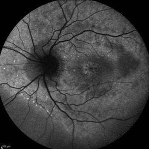 Cystoid Macular Edema (CME)
Sep 11 2012 by Hamid Ahmadieh, MD
Autofluorescence imaging of the left eye of a 17-year-old boy with chronic intermediate uveitis showing CME.
Photographer: Hamid Ahmadieh, MD, Ophthalmic Research Center, Labbafinejad Medical Center, Shahid Beheshti University of Medical Sciences
Imaging device: Heidelberg Spectralis
Condition/keywords: autofluorescence imaging, cystoid macular edema (CME), intermediate uveitis
-
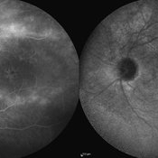 Cystoid Macular Edema (CME)
Sep 11 2012 by Hamid Ahmadieh, MD
Late phase FA & ICG angiography imagings of the left eye a 17-year-old boy with CME & retinal periphlebitis secondary to chronic intermediate uveitis.
Photographer: Hamid Ahmadieh, MD, Ophthalmic Research Center, Labbafinejad Medical Center, Shahid Beheshti University of Medical Sciences
Imaging device: Heidelberg Spectralis
Condition/keywords: cystoid macular edema (CME), indocyanine green (ICG) angiography, intermediate uveitis
-
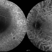 Retinitis Pigmentosa
Sep 11 2012 by Hamid Ahmadieh, MD
FA & ICG angiography images of a 40-year-old man with RP.
Photographer: Hamid Ahmadieh, MD, Ophthalmic Research Center, Labbafinejad Medical Center, Shahid Beheshti University of Medical Sciences
Imaging device: Heidelberg Spectralis
Condition/keywords: indocyanine green (ICG) angiography, retinitis pigmentosa
-
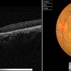 Pseudoxanthoma Elasticum (PXE)
Sep 11 2012 by Hamid Ahmadieh, MD
Color fundus photograph and OCT image of a 35-year-old woman with juxtafoveal CNV due to angioid streaks of choroid secondary to PXE.
Photographer: Hamid Ahmadieh, MD, Ophthalmic Research Center, Labbafinejad Medical Center, Shahid Beheshti University of Medical Sciences
Imaging device: Topcon
Condition/keywords: angioid streaks, choroidal neovascularization (CNV), optical coherence tomography (OCT), pseudoxanthoma elasticum (PXE)
-
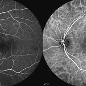 Chronic Active Central Serous Chorioretinopathy (CSCR)
Sep 11 2012 by Hamid Ahmadieh, MD
Early phase FA & ICG angiography images of a 30-year-old man with chronic active CSCR.
Photographer: Hamid Ahmadieh, MD, Ophthalmic Research Center, Labbafinejad Medical Center, Shahid Beheshti University of Medical Sciences
Imaging device: Heidelberg Spectralis
Condition/keywords: central serous chorioretinopathy (CSCR), indocyanine green (ICG) angiography
-
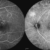 Chronic Active Central Serous Chorioretinopathy (CSCR)
Sep 11 2012 by Hamid Ahmadieh, MD
Late phase FA & ICG angiography images of a 30-year-old man with chronic active CSCR.
Photographer: Hamid Ahmadieh, MD, Ophthalmic Research Center, Labbafinejad Medical Center, Shahid Beheshti University of Medical Sciences
Imaging device: Heidelberg Spectralis
Condition/keywords: central serous chorioretinopathy (CSCR), indocyanine green (ICG) angiography
-
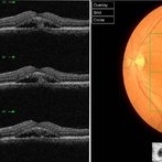 Chronic Active Central Serous Chorioretinopathy (CSCR)
Sep 11 2012 by Hamid Ahmadieh, MD
Color fundus photograph and OCT image of a 30-year-old man with chronic active CSCR.
Photographer: Hamid Ahmadieh, MD, Ophthalmic Research Center, Labbafinejad Medical Center, Shahid Beheshti University of Medical Sciences
Imaging device: Topcon
Condition/keywords: central serous chorioretinopathy (CSCR), optical coherence tomography (OCT)
-
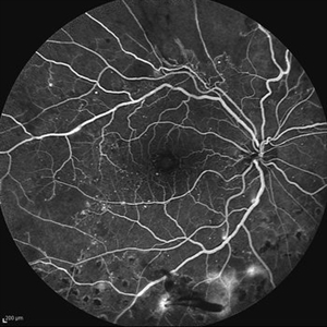 Proliferative Diabetic Retinopathy (PDR)
Sep 11 2012 by Hamid Ahmadieh, MD
FA image of a 55-year-old woman with active PDR.
Photographer: Hamid Ahmadieh, MD, Ophthalmic Research Center, Labbafinejad Medical Center, Shahid Beheshti University of Medical Sciences
Imaging device: Heidelberg HRA
Condition/keywords: retinal neovascularization
-
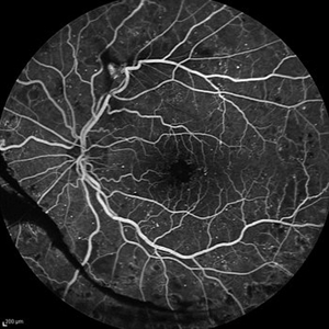 Proliferative Diabetic Retinopathy (PDR)
Sep 11 2012 by Hamid Ahmadieh, MD
FA image of a 55-year-old woman with active PDR.
Photographer: Hamid Ahmadieh, MD, Ophthalmic Research Center, Labbafinejad Medical Center, Shahid Beheshti University of Medical Sciences
Imaging device: Heidelberg HRA
Condition/keywords: preretinal hemorrhage, retinal neovascularization
-
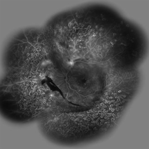 Proliferative Diabetic Retinopathy (PDR)
Sep 11 2012 by Hamid Ahmadieh, MD
Wide- field FA image of a 55-year-old woman with active PDR and the history of scatter laser photocoagulation.
Photographer: Hamid Ahmadieh, MD, Ophthalmic Research Center, Labbafinejad Medical Center, Shahid Beheshti University of Medical Sciences
Imaging device: Heidelberg HRA
Condition/keywords: preretinal hemorrhage, retinal neovascularization, scatter laser photocoagulation
-
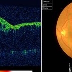 Branch Retinal Vein Occlusion (BRVO)
Sep 11 2012 by Hamid Ahmadieh, MD
Color fundus photograph and OCT image of a 60-year-old woman with a recent onset BRVO.
Photographer: Hamid Ahmadieh, MD, Ophthalmic Research Center, Labbafinejad Medical Center, Shahid Beheshti University of Medical Sciences
Imaging device: Topcon
Condition/keywords: branch retinal vein occlusion (BRVO), optical coherence tomography (OCT)
-
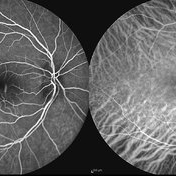 Acute Central Serous Chorioretinopathy
Sep 15 2012 by Hamid Ahmadieh, MD
Early phase FA & ICG angiography images of a 30-year-old man with acute CSCR.
Photographer: Hamid Ahmadieh, MD, Ophthalmic Research Center, Labbafinejad Medical Center, Shahid Beheshti University of Medical Sciences
Imaging device: Heidelberg HRA
Condition/keywords: central serous chorioretinopathy (CSCR), indocyanine green (ICG) angiography
-
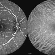 Acute Central Serous Chorioretinopathy
Sep 15 2012 by Hamid Ahmadieh, MD
Mid-phase FA & ICG angiography images of a 30-year-old man with acute CSCR.
Photographer: Hamid Ahmadieh, MD, Ophthalmic Research Center, Labbafinejad Medical Center, Shahid Beheshti University of Medical Sciences
Imaging device: Heidelberg HRA
Condition/keywords: central serous chorioretinopathy (CSCR), indocyanine green (ICG) angiography
-
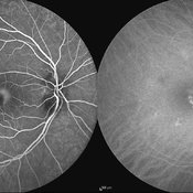 Acute Central Serous Chorioretinopathy
Sep 15 2012 by Hamid Ahmadieh, MD
Late phase FA & ICG angiography images of a 30-year-old man with acute CSCR.
Photographer: Hamid Ahmadieh, MD, Ophthalmic Research Center, Labbafinejad Medical Center, Shahid Beheshti University of Medical Sciences
Imaging device: Heidelberg HRA
Condition/keywords: central serous chorioretinopathy (CSCR), indocyanine green (ICG) angiography
-
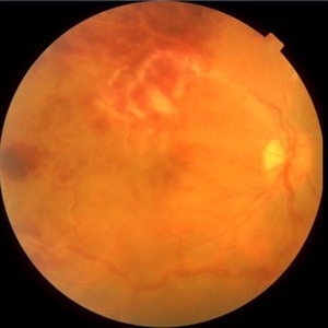 Frosted Branch Angiitis-1
Sep 15 2012 by Hamid Ahmadieh, MD
Color fundus photograph of a 25-year-old man with sudden onset visual loss due to frosted branch angiitis.
Photographer: Hamid Ahmadieh, MD, Ophthalmic Research Center, Labbafinejad Medical Center, Shahid Beheshti University of Medical Sciences
Imaging device: Topcon Fundus Camera
Condition/keywords: frosted branch angiitis
-
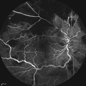 Frosted Branch Angiitis-2
Sep 15 2012 by Hamid Ahmadieh, MD
Early phase FA image of a 25-year-old man with sudden onset visual loss due to frosted branch angiitis.
Photographer: Hamid Ahmadieh, MD, Ophthalmic Research Center, Labbafinejad Medical Center, Shahid Beheshti University of Medical Sciences
Imaging device: Heidelberg HRA
-
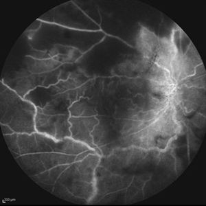 Frosted Branch Angiitis-3
Sep 15 2012 by Hamid Ahmadieh, MD
Late phase FA image of a 25-year-old man with sudden onset visual loss due to frosted branch angiitis.
Photographer: Hamid Ahmadieh, MD, Ophthalmic Research Center, Labbafinejad Medical Center, Shahid Beheshti University of Medical Sciences
Imaging device: Heidelberg HRA
Condition/keywords: frosted branch angiitis
-
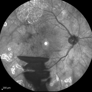 Proliferative Diabetic Retinopathy
Sep 15 2012 by Hamid Ahmadieh, MD
Infrared image of a 30-year-old woman with the history of scatter laser photocoagulation and a preretinal hemorrhage due to active PDR .
Photographer: Hamid Ahmadieh, MD, Ophthalmic Research Center, Labbafinejad Medical Center, Shahid Beheshti University of Medical Sciences
Imaging device: Heidelberg HRA
Condition/keywords: infrared image, preretinal hemorrhage
-
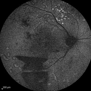 Proliferative Diabetic Retinopathy
Sep 15 2012 by Hamid Ahmadieh, MD
Fundus autofluorescence image of a 30-year-old woman with the history of scatter laser photocoagulation and a preretinal hemorrhage due to active PDR .
Photographer: Hamid Ahmadieh, MD, Ophthalmic Research Center, Labbafinejad Medical Center, Shahid Beheshti University of Medical Sciences
Imaging device: Heidelberg HRA
Condition/keywords: fundus autofluorescence (FAF), preretinal hemorrhage
-
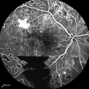 Proliferative Diabetic Retinopathy
Sep 15 2012 by Hamid Ahmadieh, MD
FA image of a 30-year-old woman with the history of scatter laser photocoagulation, NVE and a preretinal hemorrhage due to active PDR .
Photographer: Hamid Ahmadieh, MD, Ophthalmic Research Center, Labbafinejad Medical Center, Shahid Beheshti University of Medical Sciences
Imaging device: Heidelberg HRA
Condition/keywords: preretinal hemorrhage
-
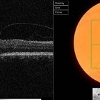 Vitreomacular Traction and AMD
Sep 15 2012 by Hamid Ahmadieh, MD
Fundus photograph and OCT image of a 70-year-old man with a subfoveal CNV scar secondary to neovascular AMD and a vitreomacular adhesion.
Photographer: Hamid Ahmadieh, MD, Ophthalmic Research Center, Labbafinejad Medical Center, Shahid Beheshti University of Medical Sciences
Imaging device: Topcon OCT
Condition/keywords: choroidal neovascularization (CNV), neovascular age-related macular degeneration (AMD), vitreomacular adhesion, vitreomacular traction (VMT)
-
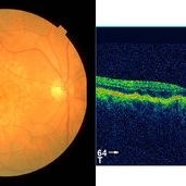 Dry Type AMD
Sep 22 2012 by Hamid Ahmadieh, MD
Color fundus photograph and OCT image of a 70-year-old woman with dry type AMD.
Photographer: Hamid Ahmadieh, MD, Ophthalmic Research Center, Labbafinejad Medical Center, Shahid Beheshti University of Medical Sciences
Imaging device: Topcon Fundus Camera & Topcon OCT
Condition/keywords: dry age-related macular degeneration (dry AMD), optical coherence tomography (OCT)
-
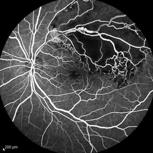 Non-perfused BRVO
Sep 22 2012 by Hamid Ahmadieh, MD
FA image of a 60-year-old woman with non-perfused occlusion of superior temporal branch of central retinal vein.
Photographer: Naghmeh Nozhat, Negah Eye Center, Tehran
Imaging device: HRA
Condition/keywords: branch retinal vein occlusion (BRVO)
-
 Macular Telangiectasia Type 2 & CNV
Sep 22 2012 by Hamid Ahmadieh, MD
Color fundus photograph & OCT imagings of the left eye of a 70-year-old man with idiopathic macular telangiectasia type 2 and CNV.
Photographer: Hamid Ahmadieh, MD, Ophthalmic Research Center, Labbafinejad Medical Center, Shahid Beheshti University of Medical Sciences
Imaging device: Topcon Fundus Camera & Topcon OCT
Condition/keywords: choroidal neovascularization (CNV), idiopathic macular telangiectasia, optical coherence tomography (OCT)
-
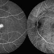 Macular Telangiectasia Type 2 & CNV
Sep 22 2012 by Hamid Ahmadieh, MD
Late phase FA and ICG angiography imagings of the left eye of a 70-year-old man with idiopathic macular telangiectasia type 2 and CNV.
Photographer: Hamid Ahmadieh, MD, Ophthalmic Research Center, Labbafinejad Medical Center, Shahid Beheshti University of Medical Sciences
Imaging device: HRA
Condition/keywords: choroidal neovascularization (CNV), idiopathic macular telangiectasia, indocyanine green (ICG) angiography
-
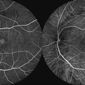 Macular Telangiectasia Type 2 & CNV
Sep 22 2012 by Hamid Ahmadieh, MD
FA and ICG angiography imagings of the left eye of a 70-year-old man with idiopathic macular telangiectasia type 2 and CNV.
Photographer: Hamid Ahmadieh, MD, Ophthalmic Research Center, Labbafinejad Medical Center, Shahid Beheshti University of Medical Sciences
Imaging device: HRA
Condition/keywords: choroidal neovascularization (CNV), idiopathic macular telangiectasia, indocyanine green (ICG) angiography
-
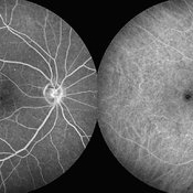 Macular Telangiectasia Type 2
Sep 22 2012 by Hamid Ahmadieh, MD
Late phase FA and ICG angiography imagings of the right eye of a 70-year-old man with idiopathic macular telangiectasia type 2.
Photographer: Hamid Ahmadieh, MD, Ophthalmic Research Center, Labbafinejad Medical Center, Shahid Beheshti University of Medical Sciences
Imaging device: HRA
Condition/keywords: idiopathic macular telangiectasia, indocyanine green (ICG) angiography
-
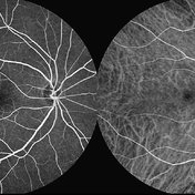 Macular Telangiectasia Type 2
Sep 22 2012 by Hamid Ahmadieh, MD
FA and ICG angiography imagings of the right eye of a 70-year-old man with idiopathic macular telangiectasia type 2.
Photographer: Hamid Ahmadieh, MD, Ophthalmic Research Center, Labbafinejad Medical Center, Shahid Beheshti University of Medical Sciences
Imaging device: HRA
Condition/keywords: idiopathic macular telangiectasia, indocyanine green (ICG) angiography
-
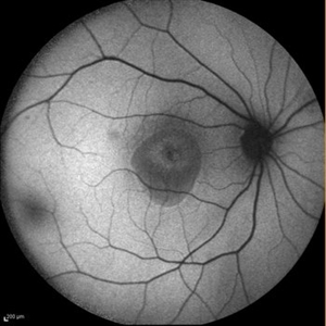 Chronic Central Serous Chorioretinopathy
Sep 26 2012 by Hamid Ahmadieh, MD
Autofluorescence imaging of the right eye of a 50-year-old man with active chronic CSCR and BCVA of 20/100.
Photographer: Hamid Ahmadieh, MD, Ophthalmic Research Center, Labbafinejad Medical Center, Shahid Beheshti University of Medical Sciences
Imaging device: Heidelberg Spectralis
Condition/keywords: autofluorescence imaging, chronic central serous chorioretinopathy (CSCR)
-
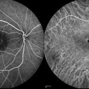 Chronic Central Serous Chorioretinopathy
Sep 26 2012 by Hamid Ahmadieh, MD
FA& ICG angiography of the right eye of a 50-year-old man with active chronic CSCR and BCVA of 20/100.
Photographer: Hamid Ahmadieh, MD, Ophthalmic Research Center, Labbafinejad Medical Center, Shahid Beheshti University of Medical Sciences
Imaging device: Heidelberg Spectralis
Condition/keywords: chronic central serous chorioretinopathy (CSCR), indocyanine green (ICG) angiography
-
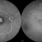 Chronic Central Serous Chorioretinopathy
Sep 26 2012 by Hamid Ahmadieh, MD
Late FA& ICG angiography imaging of the right eye of a 50-year-old man with active chronic CSCR and BCVA of 20/100.
Photographer: Hamid Ahmadieh, MD, Ophthalmic Research Center, Labbafinejad Medical Center, Shahid Beheshti University of Medical Sciences
Imaging device: Heidelberg Spectralis
Condition/keywords: chronic central serous chorioretinopathy (CSCR), indocyanine green (ICG) angiography
-
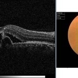 Chronic Central Serous Chorioretinopathy
Sep 26 2012 by Hamid Ahmadieh, MD
Color fundus photograph and OCT imaging of the right eye of a 50-year-old man with active chronic CSCR and BCVA of 20/100
Photographer: Hamid Ahmadieh, MD, Ophthalmic Research Center, Labbafinejad Medical Center, Shahid Beheshti University of Medical Sciences
Imaging device: Topcon OCT
Condition/keywords: chronic central serous chorioretinopathy (CSCR), optical coherence tomography (OCT)
-
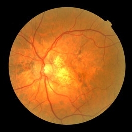 Angioid Streaks
Sep 29 2012 by Hamid Ahmadieh, MD
Color fundus photograph of a 59-year-old man with angioid streaks.
Photographer: Hamid Ahmadieh, MD; Ophthalmic Research Center, Labbafinejad Medical Center, Shahid Beheshti University of Medical Sciences
Imaging device: Topcon Fundus Camera
-
 Advanced PDR
Sep 29 2012 by Hamid Ahmadieh, MD
Color fundus photograph of a 32-year-old man with insulin-dependent diabetes mellitus and regressed advanced PDR with severe fibrous proliferation and traction retinal detachment sparing the macula.
Photographer: Hamid Ahmadieh, MD; Ophthalmic Research Center, Labbafinejad Medical Center, Shahid Beheshti University of Medical Sciences
Condition/keywords: fibrous proliferation, tractional retinal detachment
-
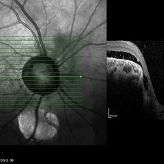 Coloboma of Disc & Choroid
Oct 6 2012 by Hamid Ahmadieh, MD
OCT image of a 25-year-old woman with serous retinal detachment secondary to coloboma of disc associated with coloboma of choroid.
Photographer: Hamid Ahmadieh, MD, Ophthalmic Research Center, Labbafinejad Medical Center, Shahid Beheshti University of Medical Sciences
Imaging device: Heidelberg Spectralis
Condition/keywords: coloboma of choroid, coloboma of optic disc, optical coherence tomography (OCT), serous retinal detachment
-
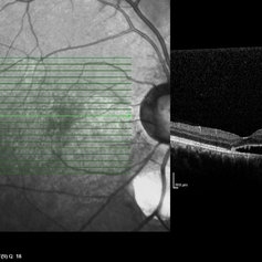 Coloboma of Disc & Choroid
Oct 6 2012 by Hamid Ahmadieh, MD
OCT image of a 25-year-old woman with serous retinal detachment secondary to coloboma of disc associated with coloboma of choroid.
Photographer: Hamid Ahmadieh, MD, Ophthalmic Research Center, Labbafinejad Medical Center, Shahid Beheshti University of Medical Sciences
Imaging device: Heidelberg Spectralis
Condition/keywords: coloboma of choroid, coloboma of optic disc, optical coherence tomography (OCT), serous retinal detachment
-
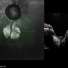 Coloboma of Disc & Choroid
Oct 6 2012 by Hamid Ahmadieh, MD
OCT image of a 25-year-old woman with a small coloboma of choroid associated with coloboma of disc.
Photographer: Hamid Ahmadieh, MD, Ophthalmic Research Center, Labbafinejad Medical Center, Shahid Beheshti University of Medical Sciences
Imaging device: Heidelberg Spectralis
Condition/keywords: coloboma of choroid, coloboma of optic disc, optical coherence tomography (OCT)
-
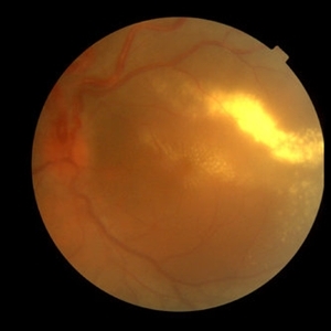 Von Hippel-Lindau 1
Oct 13 2012 by Hamid Ahmadieh, MD
Color fundus photograph of the left eye of a 25-year-old woman with exudative retinal detachment secondary to retinal angiomatosis (Von Hippel-Lindau).
Photographer: Hamid Ahmadieh, MD, Ophthalmic Research Center, Labbafinejad Medical Center, Shahid Beheshti University of Medical Sciences
Imaging device: Topcon Fundus Camera
Condition/keywords: exudative retinal detachment, retinal angiomatous proliferation (RAP), Von Hippel-Lindau
-
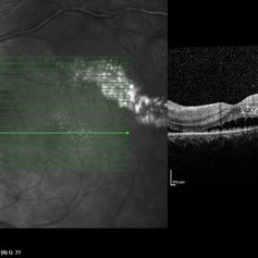 Von Hippel-Lindau 2
Oct 13 2012 by Hamid Ahmadieh, MD
OCT image of the left eye of a 25-year-old woman with exudative retinal detachment secondary to retinal angiomatosis (Von Hippel-Lindau).
Photographer: Hamid Ahmadieh, MD, Ophthalmic Research Center, Labbafinejad Medical Center, Shahid Beheshti University of Medical Sciences
Imaging device: Heidelberg Spectralis
-
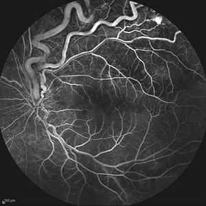 Von Hippel-Lindau 3
Oct 13 2012 by Hamid Ahmadieh, MD
Early FA image of the left eye of a 25-year-old woman with exudative retinal detachment secondary to retinal angiomatosis (Von Hippel-Lindau).
Photographer: Hamid Ahmadieh, MD, Ophthalmic Research Center, Labbafinejad Medical Center, Shahid Beheshti University of Medical Sciences
Imaging device: Heidelberg Spectralis
-
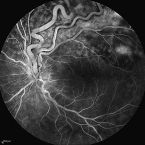 Von Hippel-Lindau
Oct 13 2012 by Hamid Ahmadieh, MD
Late FA image of the left eye of a 25-year-old woman with exudative retinal detachment secondary to retinal angiomatosis (Von Hippel-Lindau).
Photographer: Soodabeh Fooladin, Negah Eye Center, Tehran
Imaging device: Heidelberg Spectralis
Condition/keywords: exudative retinal detachment, retinal angiomatous proliferation (RAP)
-
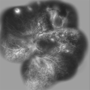 Von Hippel-Lindau
Oct 13 2012 by Hamid Ahmadieh, MD
Wide field FA image of the left eye of a 25-year-old woman with exudative retinal detachment secondary to retinal angiomatosis (Von Hippel-Lindau).
Photographer: Soodabeh Fooladin, Negah Eye Center, Tehran
Imaging device: Heidelberg Spectralis
Condition/keywords: exudative retinal detachment, retinal angiomatous proliferation (RAP), Von Hippel-Lindau
-
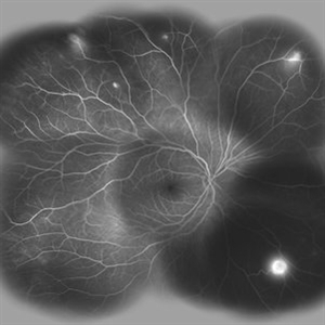 Von Hippel-Lindau
Oct 13 2012 by Hamid Ahmadieh, MD
Wide field FA image of the right eye of a 25-year-old woman with retinal angiomatosis (Von Hippel-Lindau). Fundus of the right eye seemed to be normal in ophthalmoscopy.
Photographer: Soodabeh Fooladin, Negah Eye Center, Tehran
Imaging device: Heidelberg Spectralis
Condition/keywords: exudative retinal detachment, retinal angiomatous proliferation (RAP), Von Hippel-Lindau
-
 Familial Exudative Vitreoretinopathy (FEVR)
Oct 13 2012 by Hamid Ahmadieh, MD
Color fundus photograph of the right eye of a 24-year-old woman with FEVR and visual acuity of 20/25.
Photographer: Hamid Ahmadieh, MD, Ophthalmic Research Center, Labbafinejad Medical Center, Shahid Beheshti University of Medical Sciences
Imaging device: Topcon Fundus Camera
Condition/keywords: familial exudative vitreoretinopathy (FEVR)
-
 Familial Exudative Vitreoretinopathy (FEVR)
Oct 13 2012 by Hamid Ahmadieh, MD
Color fundus photograph of the Left eye of a 24-year-old woman with FEVR and visual acuity of 20/30.
Photographer: Hamid Ahmadieh, MD, Ophthalmic Research Center, Labbafinejad Medical Center, Shahid Beheshti University of Medical Sciences
Imaging device: Topcon Fundus Camera
Condition/keywords: familial exudative vitreoretinopathy (FEVR)
-
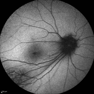 Familial Exudative Vitreoretinopathy (FEVR)
Oct 13 2012 by Hamid Ahmadieh, MD
Autofluorescence imaging of the right eye of a 24-year-old woman with FEVR and visual acuity of 20/25.
Photographer: Hamid Ahmadieh, MD, Ophthalmic Research Center, Labbafinejad Medical Center, Shahid Beheshti University of Medical Sciences
Imaging device: Heidelberg Spectralis
Condition/keywords: autofluorescence imaging
-
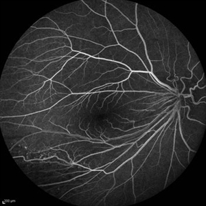 Familial Exudative Vitreoretinopathy (FEVR)
Oct 13 2012 by Hamid Ahmadieh, MD
Early FA image of the right eye of a 24-year-old woman with FEVR and visual acuity of 20/25.
Photographer: Hamid Ahmadieh, MD, Ophthalmic Research Center, Labbafinejad Medical Center, Shahid Beheshti University of Medical Sciences
Imaging device: Heidelberg Spectralis
Condition/keywords: familial exudative vitreoretinopathy (FEVR)
-
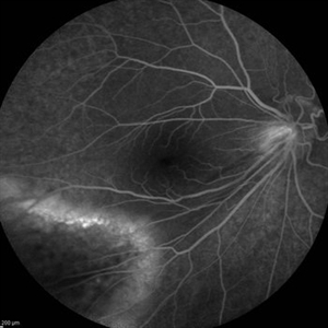 Familial Exudative Vitreoretinopathy (FEVR)
Oct 13 2012 by Hamid Ahmadieh, MD
Late FA image of the right eye of a 24-year-old woman with FEVR and visual acuity of 20/25.
Photographer: Hamid Ahmadieh, MD, Ophthalmic Research Center, Labbafinejad Medical Center, Shahid Beheshti University of Medical Sciences
Imaging device: Heidelberg Spectralis
Condition/keywords: familial exudative vitreoretinopathy (FEVR)
-
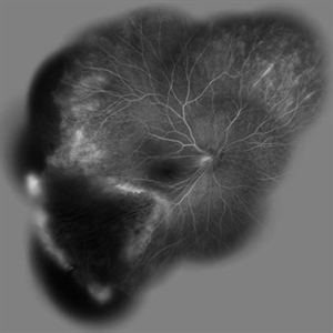 Familial Exudative Vitreoretinopathy (FEVR)
Oct 13 2012 by Hamid Ahmadieh, MD
Wide field FA image of the right eye of a 24-year-old woman with FEVR and visual acuity of 20/25.
Photographer: Hamid Ahmadieh, MD, Ophthalmic Research Center, Labbafinejad Medical Center, Shahid Beheshti University of Medical Sciences
Imaging device: Heidelberg Spectralis
Condition/keywords: familial exudative vitreoretinopathy (FEVR)
-
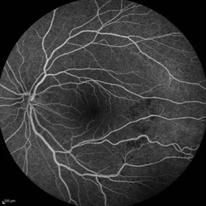 Familial Exudative Vitreoretinopathy (FEVR)
Oct 13 2012 by Hamid Ahmadieh, MD
FA image of the Left eye of a 24-year-old woman with FEVR and visual acuity of 20/30.
Photographer: Hamid Ahmadieh, MD, Ophthalmic Research Center, Labbafinejad Medical Center, Shahid Beheshti University of Medical Sciences
Imaging device: Heidelberg Spectralis
Condition/keywords: familial exudative vitreoretinopathy (FEVR)
-
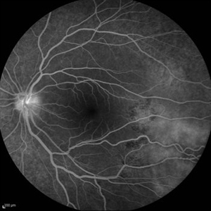 Familial Exudative Vitreoretinopathy (FEVR)
Oct 13 2012 by Hamid Ahmadieh, MD
Late FA image of the Left eye of a 24-year-old woman with FEVR and visual acuity of 20/30.
Photographer: Hamid Ahmadieh, MD, Ophthalmic Research Center, Labbafinejad Medical Center, Shahid Beheshti University of Medical Sciences
Imaging device: Heidelberg Spectralis
Condition/keywords: familial exudative vitreoretinopathy (FEVR)
-
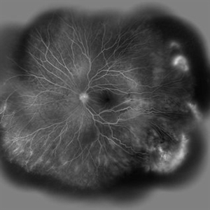 Familial Exudative Vitreoretinopathy (FEVR)
Oct 13 2012 by Hamid Ahmadieh, MD
Wide field FA image of the Left eye of a 24-year-old woman with FEVR and visual acuity of 20/30.
Photographer: Hamid Ahmadieh, MD, Ophthalmic Research Center, Labbafinejad Medical Center, Shahid Beheshti University of Medical Sciences
Imaging device: Heidelberg Spectralis
Condition/keywords: familial exudative vitreoretinopathy (FEVR)
-
---thumb.jpg/image-square;max$300,300.ImageHandler) Familial Exudative Vitreoretinopathy (FEVR)
Familial Exudative Vitreoretinopathy (FEVR)
Oct 13 2012 by Hamid Ahmadieh, MD
Color fundus photograph of the right eye of a 48-year-old man with advanced FEVR and visual acuity of 20/100.
Photographer: Hamid Ahmadieh, MD, Ophthalmic Research Center, Labbafinejad Medical Center, Shahid Beheshti University of Medical Sciences
Imaging device: Topcon Fundus Camera
Condition/keywords: familial exudative vitreoretinopathy (FEVR)
-
---thumb.jpg/image-square;max$300,300.ImageHandler) Familial Exudative Vitreoretinopathy (FEVR)
Familial Exudative Vitreoretinopathy (FEVR)
Oct 13 2012 by Hamid Ahmadieh, MD
Color fundus photograph of the right eye of a 48-year-old man with advanced FEVR and visual acuity of 20/100.
Photographer: Hamid Ahmadieh, MD, Ophthalmic Research Center, Labbafinejad Medical Center, Shahid Beheshti University of Medical Sciences
Imaging device: Topcon Fundus Camera
-
---thumb.jpg/image-square;max$300,300.ImageHandler) Familial Exudative Vitreoretinopathy (FEVR)
Familial Exudative Vitreoretinopathy (FEVR)
Oct 13 2012 by Hamid Ahmadieh, MD
Color fundus photograph & OCT image of the right eye of a 48-year-old man with advanced FEVR and visual acuity of 20/100. Traction retinoschisis is visible in OCT.
Photographer: Hamid Ahmadieh, MD, Ophthalmic Research Center, Labbafinejad Medical Center, Shahid Beheshti University of Medical Sciences
Imaging device: Topcon OCT
Condition/keywords: optical coherence tomography (OCT), traction retinoschisis
-
---thumb.jpg/image-square;max$300,300.ImageHandler) Familial Exudative Vitreoretinopathy (FEVR)
Familial Exudative Vitreoretinopathy (FEVR)
Oct 13 2012 by Hamid Ahmadieh, MD
OCT image of the right eye of a 48-year-old man with advanced FEVR, traction retinoschisis and visual acuity of 20/100.
Photographer: Hamid Ahmadieh, MD, Ophthalmic Research Center, Labbafinejad Medical Center, Shahid Beheshti University of Medical Sciences
Imaging device: Topcon OCT
Condition/keywords: optical coherence tomography (OCT), traction retinoschisis
-
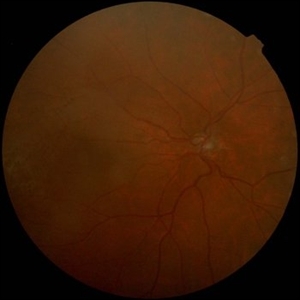 FEVR+ Optic Disc Hypoplasia 1
Oct 13 2012 by Hamid Ahmadieh, MD
Color fundus photograph of the right eye of a 26-year-old man with FEVR and optic disc hypoplasia; visual acuity of 20/50.
Photographer: Hamid Ahmadieh, MD, Ophthalmic Research Center, Labbafinejad Medical Center, Shahid Beheshti University of Medical Sciences
Imaging device: Topcon Fundus Camera
Condition/keywords: optic disc hypoplasia
-
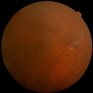 FEVR+ Optic Disc Hypoplasia 2
Oct 13 2012 by Hamid Ahmadieh, MD
Color fundus photograph of the right eye of a 24-year-old woman with FEVR and optic disc hypoplasia; visual acuity of 20/50. Her brother and sister were also involved with the same ocular disorder.
Photographer: Hamid Ahmadieh, MD, Ophthalmic Research Center, Labbafinejad Medical Center, Shahid Beheshti University of Medical Sciences
Imaging device: Topcon Fundus Camera
Condition/keywords: familial exudative vitreoretinopathy (FEVR), optic disc hypoplasia
-
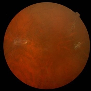 FEVR+ Optic Disc Hypoplasia 3
Oct 13 2012 by Hamid Ahmadieh, MD
Color fundus photograph of the left eye of a 24-year-old woman with FEVR and optic disc hypoplasia; visual acuity of 20/50. Her brother and sister were also involved with the same ocular disorder.
Photographer: Hamid Ahmadieh, MD, Ophthalmic Research Center, Labbafinejad Medical Center, Shahid Beheshti University of Medical Sciences
Imaging device: Topcon Fundus Camera
Condition/keywords: familial exudative vitreoretinopathy (FEVR), optic disc hypoplasia
-
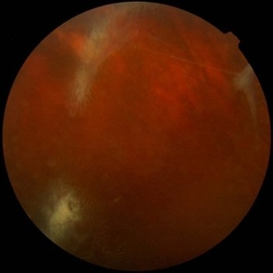 FEVR+ Optic Disc Hypoplasia 4
Oct 13 2012 by Hamid Ahmadieh, MD
Color fundus photograph of the right eye of a 24-year-old woman with FEVR and optic disc hypoplasia; visual acuity of 20/50. Her brother and sister were also involved with the same ocular disorder.
Photographer: Hamid Ahmadieh, MD, Ophthalmic Research Center, Labbafinejad Medical Center, Shahid Beheshti University of Medical Sciences
Imaging device: Topcon Fundus Camera
Condition/keywords: familial exudative vitreoretinopathy (FEVR), optic disc hypoplasia
-
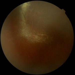 FEVR+ Optic Disc Hypoplasia 5
Oct 13 2012 by Hamid Ahmadieh, MD
Color fundus photograph of the right eye of a 24-year-old woman with FEVR and optic disc hypoplasia; visual acuity of 20/50. Her brother and sister were also involved with the same ocular disorder.
Photographer: Hamid Ahmadieh, MD, Ophthalmic Research Center, Labbafinejad Medical Center, Shahid Beheshti University of Medical Sciences
Imaging device: Topcon Fundus Camera
Condition/keywords: familial exudative vitreoretinopathy (FEVR), optic disc hypoplasia
-
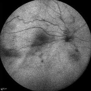 FEVR+ Optic Disc Hypoplasia 6
Oct 13 2012 by Hamid Ahmadieh, MD
Autofluorescence imaging of the right eye of a 24-year-old woman with FEVR and optic disc hypoplasia; visual acuity of 20/50. Her brother and sister were also involved with the same ocular disorder.
Photographer: Hamid Ahmadieh, MD, Ophthalmic Research Center, Labbafinejad Medical Center, Shahid Beheshti University of Medical Sciences
Imaging device: Heidelberg Spectralis
Condition/keywords: autofluorescence imaging, familial exudative vitreoretinopathy (FEVR), optic disc hypoplasia
-
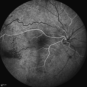 FEVR+ Optic Disc Hypoplasia 7
Oct 13 2012 by Hamid Ahmadieh, MD
Early FA image of the right eye of a 24-year-old woman with FEVR and optic disc hypoplasia; visual acuity of 20/50. Her brother and sister were also involved with the same ocular disorder.
Photographer: Hamid Ahmadieh, MD, Ophthalmic Research Center, Labbafinejad Medical Center, Shahid Beheshti University of Medical Sciences
Imaging device: Heidelberg Spectralis
Condition/keywords: familial exudative vitreoretinopathy (FEVR), optic disc hypoplasia
-
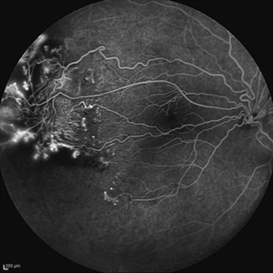 FEVR+ Optic Disc Hypoplasia
Oct 13 2012 by Hamid Ahmadieh, MD
FA image of the right eye of a 24-year-old woman with FEVR and optic disc hypoplasia; visual acuity of 20/50. Her brother and sister were also involved with the same ocular disorder.
Photographer: Hamid Ahmadieh, MD, Ophthalmic Research Center, Labbafinejad Medical Center, Shahid Beheshti University of Medical Sciences
Imaging device: Heidelberg Spectralis
Condition/keywords: familial exudative vitreoretinopathy (FEVR), optic disc hypoplasia
-
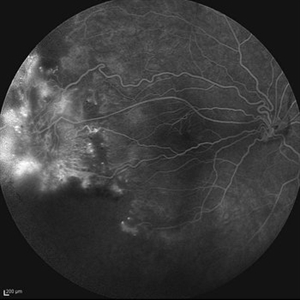 Peripheral Nonperfusion and Optic Disc Hypoplasia
Oct 13 2012 by Hamid Ahmadieh, MD
Late FA image of the right eye of a 24-year-old woman with peripheral retinal nonperfusion and fibrovascular proliferation associated with optic disc hypoplasia; visual acuity of 20/50. Her brother and sister were also involved with the same ocular disorder.
Photographer: Hamid Ahmadieh, MD, Ophthalmic Research Center, Labbafinejad Medical Center, Shahid Beheshti University of Medical Sciences
Imaging device: Heidelberg Spectralis
Condition/keywords: fibrovascular proliferation, optic disc hypoplasia, peripheral retinal nonperfusion
-
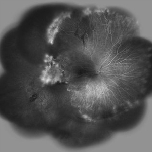 FEVR+ Optic Disc Hypoplasia
Oct 13 2012 by Hamid Ahmadieh, MD
Wide field FA image of the right eye of a 24-year-old woman with FEVR and optic disc hypoplasia; visual acuity of 20/50. Her brother and sister were also involved with the same ocular disorder.
Photographer: Hamid Ahmadieh, MD, Ophthalmic Research Center, Labbafinejad Medical Center, Shahid Beheshti University of Medical Sciences
Imaging device: Heidelberg Spectralis
Condition/keywords: familial exudative vitreoretinopathy (FEVR), optic disc hypoplasia
-
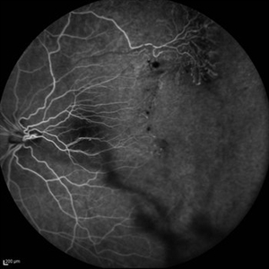 FEVR+ Optic Disc Hypoplasia 11
Oct 13 2012 by Hamid Ahmadieh, MD
Early FA image of the left eye of a 24-year-old woman with FEVR and optic disc hypoplasia; visual acuity of 20/50. Her brother and sister were also involved with the same ocular disorder.
Photographer: Hamid Ahmadieh, MD, Ophthalmic Research Center, Labbafinejad Medical Center, Shahid Beheshti University of Medical Sciences
Imaging device: Heidelberg Spectralis
Condition/keywords: familial exudative vitreoretinopathy (FEVR), optic disc hypoplasia
-
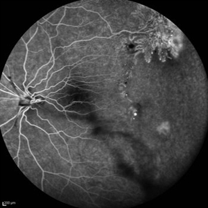 Peripheral Nonperfusion and Optic Disc Hypoplasia
Oct 13 2012 by Hamid Ahmadieh, MD
FA image of the left eye of a 24-year-old woman with peripheral retinal nonperfusion and fibrovascular proliferation associated with optic disc hypoplasia; visual acuity of 20/50. Her brother and sister were also involved with the same ocular disorder.
Photographer: Hamid Ahmadieh, MD, Ophthalmic Research Center, Labbafinejad Medical Center, Shahid Beheshti University of Medical Sciences
Imaging device: Heidelberg Spectralis
Condition/keywords: fibrovascular proliferation, optic disc hypoplasia, peripheral retinal nonperfusion
-
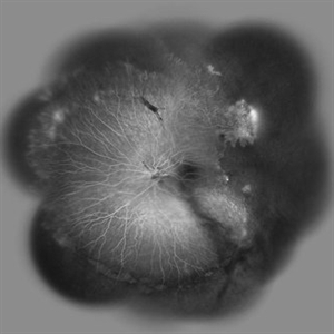 FEVR+ Optic Disc Hypoplasia 13
Oct 13 2012 by Hamid Ahmadieh, MD
Wide field FA image of the left eye of a 24-year-old woman with FEVR and optic disc hypoplasia; visual acuity of 20/50. Her brother and sister were also involved with the same ocular disorder.
Photographer: Hamid Ahmadieh, MD, Ophthalmic Research Center, Labbafinejad Medical Center, Shahid Beheshti University of Medical Sciences
Imaging device: Heidelberg Spectralis
Condition/keywords: familial exudative vitreoretinopathy (FEVR), optic disc hypoplasia
-
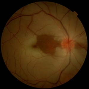 Central Retinal Artery Occlusion & Cilioretinal Artery Sparing
Dec 22 2012 by Hamid Ahmadieh, MD
Color fundus photograph of the right eye of a 34-year-old man with sudden drop of vision due to CRAO. Please notice cherry red spot despite cilioretinal artery sparing .
Photographer: Zohre Salimi; Labbafinejad Medical Center, Shahid Beheshti University of Medical Sciences, Tehran
Imaging device: Topcon Fundus Camera
Condition/keywords: central retinal artery occlusion (CRAO), cherry red spot, cilioretinal sparing
-
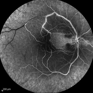 Central Retinal Artery Occlusion & Cilioretinal Artery Sparing
Dec 22 2012 by Hamid Ahmadieh, MD
Late phase FA image of the right eye of a 34-year-old man with sudden drop of vision due to CRAO. The macula is involved despite cilioretinal artery sparing .
Photographer: Zohre Salimi; Labbafinejad Medical Center, Shahid Beheshti University of Medical Sciences , Tehran
Imaging device: Heidelberg HRA
Condition/keywords: central retinal artery occlusion (CRAO), cilioretinal sparing
-
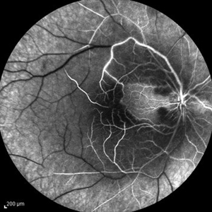 Central Retinal Artery Occlusion & Cilioretinal Artery Sparing
Dec 22 2012 by Hamid Ahmadieh, MD
Late phase FA image of the right eye of a 34-year-old man with sudden drop of vision due to CRAO. The macula is involved despite cilioretinal artery sparing .
Photographer: Zohre Salimi; Labbafinejad Medical Center, Shahid Beheshti University of Medical Sciences
Imaging device: Heidelberg HRA
Condition/keywords: central retinal artery occlusion (CRAO), cilioretinal sparing
-
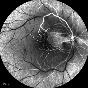 Central Retinal Artery Occlusion & Cilioretinal Artery Sparing
Dec 22 2012 by Hamid Ahmadieh, MD
FA image of the right eye of a 34-year-old man with sudden drop of vision due to CRAO. The macula is involved despite cilioretinal artery sparing .
Photographer: Zohre Salimi; Labbafinejad Medical Center, Shahid Beheshti University of Medical Sciences , Tehran
Imaging device: Heidelberg HRA
Condition/keywords: central retinal artery occlusion (CRAO), cilioretinal sparing
-
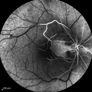 Central Retinal Artery Occlusion & Cilioretinal Artery Sparing
Dec 22 2012 by Hamid Ahmadieh, MD
Mid arteriovenous phase FA image of the right eye of a 34-year-old man with sudden drop of vision due to CRAO. The macula is involved despite cilioretinal artery sparing .
Photographer: Zohre Salimi; Labbafinejad Medical Center, Shahid Beheshti University of Medical Sciences
Imaging device: Heidelberg HRA
Condition/keywords: central retinal artery occlusion (CRAO), cilioretinal sparing
-
 Central Retinal Artery Occlusion & Cilioretinal Artery Sparing
Dec 22 2012 by Hamid Ahmadieh, MD
Early phase FA image of the right eye of a 34-year-old man with sudden drop of vision due to CRAO. The macula is involved despite cilioretinal artery sparing .
Photographer: Zohre Salimi; Labbafinejad Medical Center, Shahid Beheshti University of Medical Sciences , Tehran
Imaging device: Heidelberg HRA
Condition/keywords: central retinal artery occlusion (CRAO), cilioretinal sparing
-
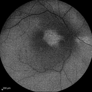 Central Retinal Artery Occlusion & Cilioretinal Artery Sparing
Dec 22 2012 by Hamid Ahmadieh, MD
Autofluorescence imaging of the right eye of a 34-year-old man with sudden drop of vision due to CRAO. The macula is involved despite cilioretinal artery sparing .
Photographer: Zohre Salimi; Labbafinejad Medical Center, Shahid Beheshti University of Medical Sciences
Imaging device: Heidelberg HRA
Condition/keywords: autofluorescence imaging, central retinal artery occlusion (CRAO), cilioretinal sparing
A project from the American Society of Retina Specialists