 Initializing download.
-
 By Hamid Ahmadieh, MD
Labbafinejad Medical Center
Co-author(s): Dr Masoome Valipur - Uploaded on Oct 10, 2015.
- Last modified by Hamid Ahmadieh, MD on Dec 29, 2015.
- Rating
- Appears in
- Miscellaneous
- Condition/keywords
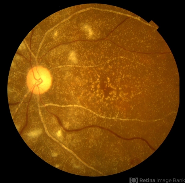
- primary hyperoxaluria, oxalosis, color fundus photograph
- Photographer
- Shabnam Pooreh, Negah Eye Center, Tehran, Iran
- Imaging device
- Fundus camera
- Description
- Color fundus photograph of the left eye of a 55-year-old woman with primary hyperoxaluria and oxalosis leading to intraretinal and subretinal deposition of calcium oxalate crystals . In addition, deposition of these crystals in the retinal vessels has led to the occlusion of retinal arterioles and venules leading to multiple cotton wools and dot and blot retinal hemorrhages.

---thumb.jpg/image-square;max$79,0.ImageHandler "Primary Hyperoxaluria and Oxalosis")
---thumb.jpg/image-square;max$79,0.ImageHandler "Primary Hyperoxaluria and Oxalosis")
---thumb.jpg/image-square;max$79,0.ImageHandler "Primary Hyperoxaluria and Oxalosis")
---thumb.jpg/image-square;max$79,0.ImageHandler "Primary Hyperoxaluria and Oxalosis")
---thumb.jpg/image-square;max$79,0.ImageHandler "Primary Hyperoxaluria and Oxalosis")
---thumb.jpg/image-square;max$79,0.ImageHandler "Primary Hyperoxaluria and Oxalosis")
---thumb.jpg/image-square;max$79,0.ImageHandler "Primary Hyperoxaluria and Oxalosis")
---thumb.jpg/image-square;max$79,0.ImageHandler "Primary Hyperoxaluria and Oxalosis")
---thumb.jpg/image-square;max$79,0.ImageHandler "Primary Hyperoxaluria and Oxalosis")
---thumb.jpg/image-square;max$79,0.ImageHandler "Primary Hyperoxaluria and Oxalosis")
