-
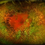
 Wyburn Mason Racemose Angiomatosis
May 22 2016 by Olivia Rainey
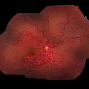
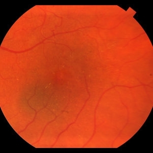
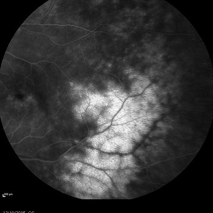
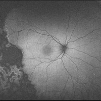
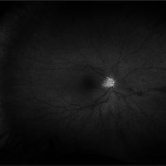
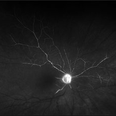
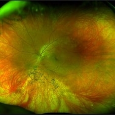
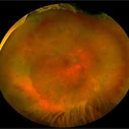
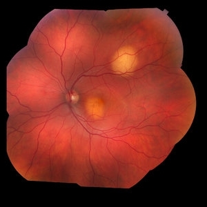
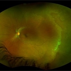
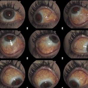
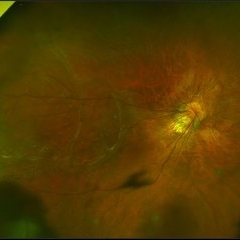
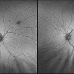
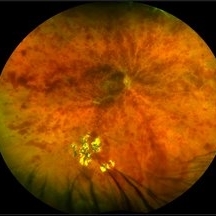
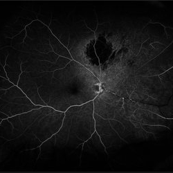
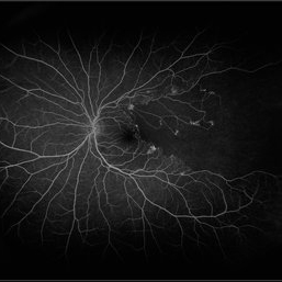
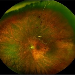
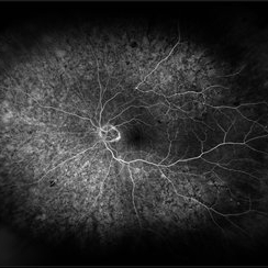
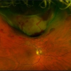
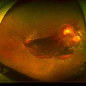
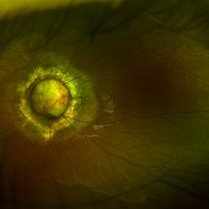
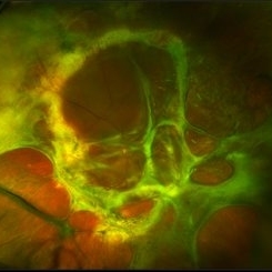
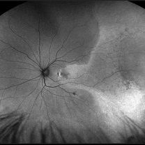
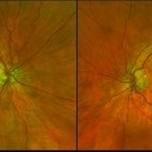
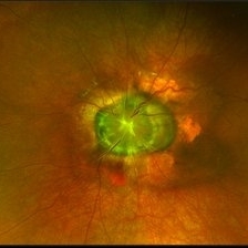
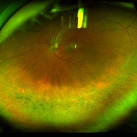
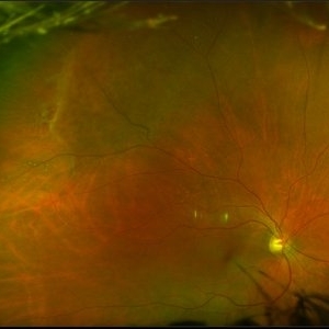
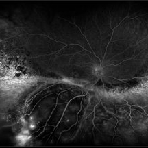
Color fundus montage of an 13-year-old female with arteriovenous malformation (Wyburn Mason Racemose Angiomatosis) affecting her right eye. The retinal arteriovenous malformation appears to be stable. She presented with NLP in the eye, strabismus, and peripheral retinal ischemia. She is at risk for neovascular complications; however, she is currently being treated with Sirolimus. Since she is on this systemically, there is no need to perform intraocular anti-VEGF injections or PRP laser. She also presented with optic atrophy affecting her left eye, secondary to chiasmal involvement of arteriovenous malformation. She has had a potential progressive visual field loss involving the temporal aspect of her visual field from the left eye. There is sector optic atrophy. Presumably, this is due to a compressive effect of her arteriovenous malformation on the nasal nerve fiber layer (corresponding to the temporal visual field) crossing to the right occipital cortex at the chiasm.
Photographer: Olivia Rainey
Imaging device: Topcon 50dx
Condition/keywords: arteriovenous malformation, color fundus photograph, color photo, montage, peripheral ischemia, Sirolimus
-
 Ocular Parasitosis
May 22 2016 by Olivia Rainey
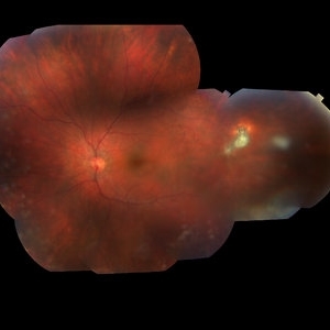
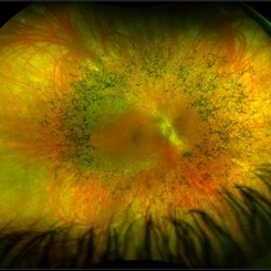
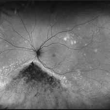
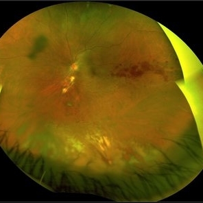
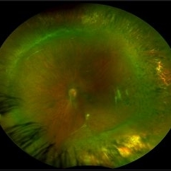
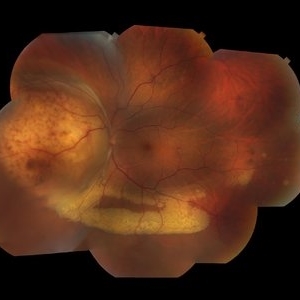
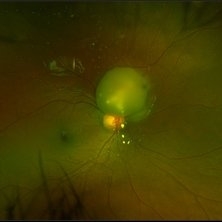
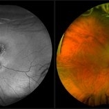
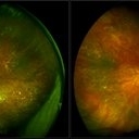
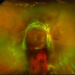
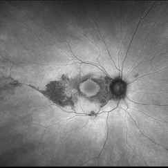
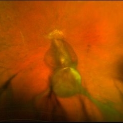
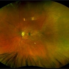
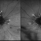
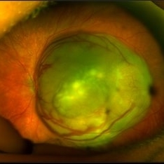
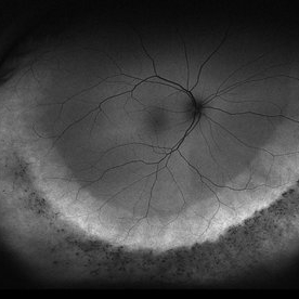
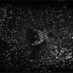
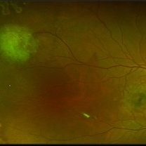
Color fundus montage of an 12-year-old boy with ocular parasitosis affecting his left eye. Patient presented with decreased vision and recent travel to Florida. The specimen was lost in the lab and was never recovered.
Photographer: Olivia Rainey
Imaging device: Topcon 50dx
Condition/keywords: color fundus photograph, color photo, intraocular foreign body, left eye, montage, parasite
-
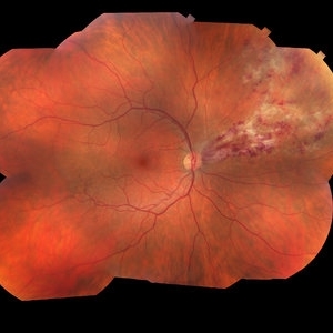
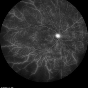
 Central Retinal Vein Occlusion with Severe Ischemia
May 22 2016 by Olivia Rainey
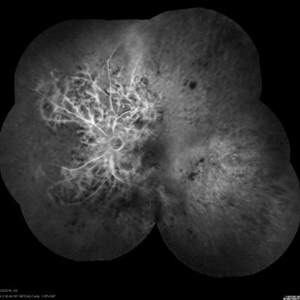
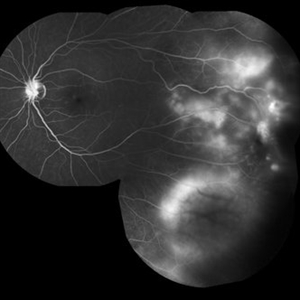
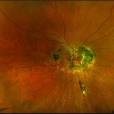
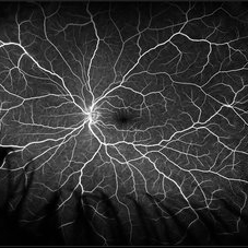
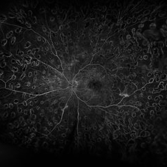
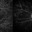
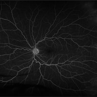
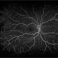
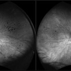
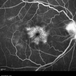
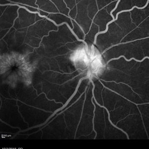
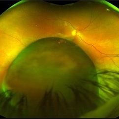
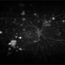
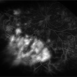
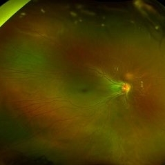
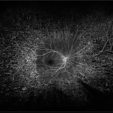
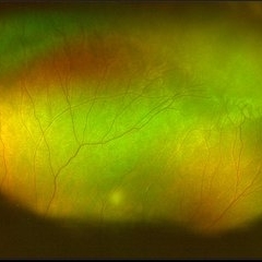
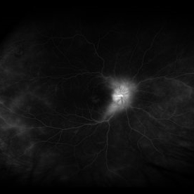
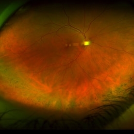
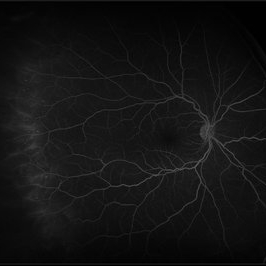
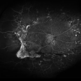
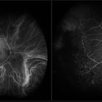
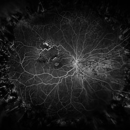
Composite fluorescein angiogram of the left eye of a male with a Central Retinal Vein Occlusion with severe ischemia.
Photographer: Olivia Rainey
Imaging device: Heidelberg Spectralis
Condition/keywords: central retinal vein occlusion (CRVO), composite, fluorescein leakage, ischemic CRVO
-
 Retinitis Pigmentosa
May 27 2016 by Olivia Rainey
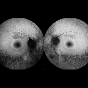
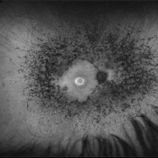
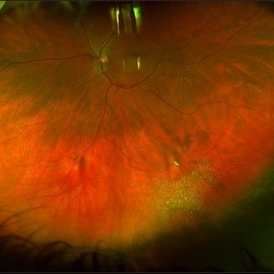
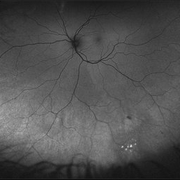
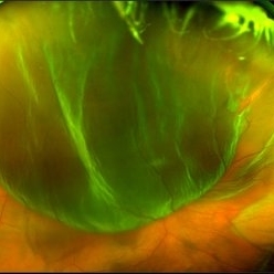
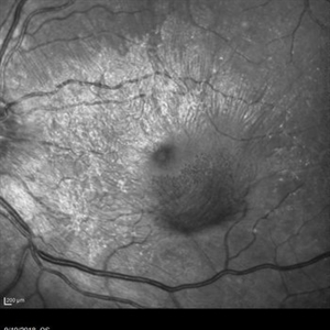
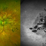
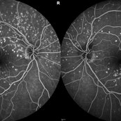
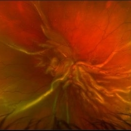
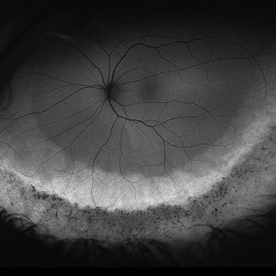
Bilateral fundus autofluorescence images of retinitis pigmentosa.
Photographer: Olivia Rainey
Imaging device: Heidelberg Spectralis
Condition/keywords: 50 degrees, bilateral, fundus autofluorescence (FAF), hereditary retinal dystrophy, retinitis pigmentosa
-
 Choroidal Nevus
May 27 2016 by Olivia Rainey
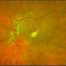
Color fundus image of a small choroidal nevus near the macula.
Photographer: Olivia Rainey
Imaging device: Topcon50dx
Condition/keywords: 20 degrees, choroidal nevus, color fundus photograph, color photo, macula
-
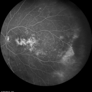
 Coats Disease
May 27 2016 by Olivia Rainey
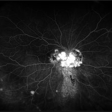
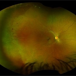
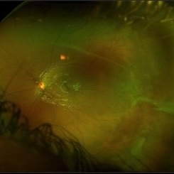
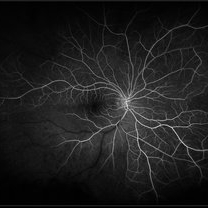
Composite fluorescein angiogram of the left eye of a man with Coats Disease.
Photographer: Olivia Rainey
Imaging device: Heidelberg Spectralis
Condition/keywords: Coats' disease, composite, fluorescein angiogram (FA), fluorescein leakage, Heidelburg Spectralis
-
 Choroidal Melanoma through the Pupil
May 28 2016 by Olivia Rainey
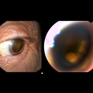
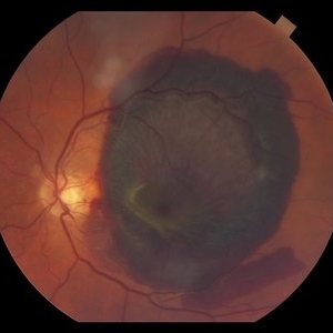
External image of the left eye of a man with metastatic choroidal melanoma, secondary to lung cancer. There was an obstruction of view to the inferior retina, and this prompted the photographer to pull back to see what the problem was.
Photographer: Olivia Rainey
Imaging device: Topcon 50dx
Condition/keywords: choroidal metastasis, color photo, external photography
-
 Diabetic Macular Edema
May 28 2016 by Olivia Rainey
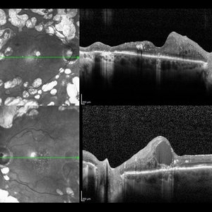
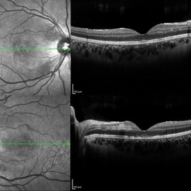
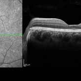
Optical coherence tomography of an 54-year-old female with diabetic macular edema affecting both eyes. Patient has a history of proliferative diabetic retinopathy s/p PRP/PPV/MP/EL, and glaucoma s/p tube shunt in both eyes. There has been a persistence of her macular edema and limited response to antiVEGF therapy, which puts into question whether there is another cause for her edema. Leading the possible causes is her renal insufficiency and fluid retention. Patient was seeing 20/50 in the right eye and 20/80 in the left eye.
Photographer: Olivia Rainey
Imaging device: Heidelberg Spectralis
Condition/keywords: anti-VEGF, diabetic macular edema, edema, glaucoma, optical coherence tomography (OCT), pan-retinal photocoagulation (PRP), proliferative diabetic retinopathy (PDR)
-
 Retinoschisis
May 28 2016 by Olivia Rainey
Late image from a fluorescein angiogram of a patient's left eye with retinoschisis.
Photographer: Olivia Rainey
Imaging device: Heidelberg Spectralis
Condition/keywords: fluorescein angiogram (FA), retinoschisis, temporal retina
-
 Proliferative Diabetic Retinopathy
May 28 2016 by Olivia Rainey
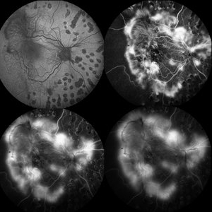
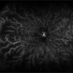
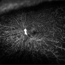
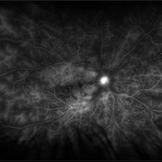
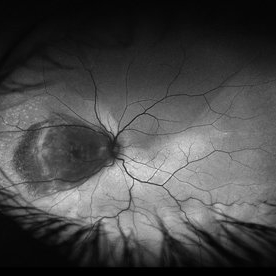
Fluorescein angiogram series of an 30-year-old male with proliferative diabetic retinopathy affecting his right eye. The patient presented with worsening neovascularization and scar tissue contracting in macula in the right eye. He experienced a decline in vision secondary to macula ischemia. Patient was seeing 20/400 and with PH 20/200 in the right eye and HM in the left eye.
Photographer: Olivia Rainey
Imaging device: Heidelberg Spectralis
Condition/keywords: diabetes, FA early phase, FA late phase, FA mid phase, fluorescein leakage, fundus autofluorescence (FAF), neovascularization (NV), proliferative diabetic retinopathy (PDR)
-
 Central Retinal Artery Occlusion
May 16 2017 by Olivia Rainey
Fluorescein angiogram of an 66-year-old female with a central retinal artery occlusion affecting her left eye.
Photographer: Olivia Rainey
Imaging device: Heidelberg Spectralis
Condition/keywords: 50 degrees, central retinal artery occlusion (CRAO), fluorescein angiogram (FA), left eye, mid phase, retinal ischemia
-
 Central Serous Retinopathy
May 16 2017 by Olivia Rainey
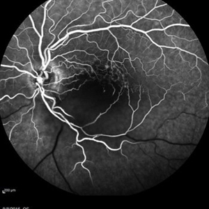
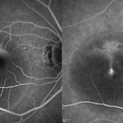
Simultaneous fluorescein and indocyanine green angiography of an 37-year-old male with central serous retinopathy affecting his right eye. Patient's vision declined from 20/25 to 20/80 in the right eye. He elected for treatment with photodynamic therapy.
Photographer: Olivia Rainey
Imaging device: Heidelberg Spectralis
Condition/keywords: 30 degrees, central serous retinopathy (CSR), fluorescein angiogram (FA), fluorescein leakage, Heidelburg Spectralis, indocyanine green (ICG) angiography, late phase, mushroom cloud
-
 Familial Exudative Vitreoretinopathy
May 16 2017 by Olivia Rainey
Ultra-wide-field autofluorescence image of an 10-year-old male with familial exudative vitreoretinopathy s/p laser.
Photographer: Olivia Rainey
Imaging device: Optos California
Condition/keywords: autofluorescence imaging, familial exudative vitreoretinopathy (FEVR), laser scarring, Optos, ultra-wide field imaging
-
 Chorioretinal Scar
May 16 2017 by Olivia Rainey
Fundus photograph of an 17-year-old male with a macular scar affecting his right eye secondary to exudation from Coats disease.
Photographer: Olivia Rainey
Imaging device: Topcon 50dx
Condition/keywords: 20 degrees, chorioretinal scar, Coats' disease, color fundus photograph, color photo, fundus photograph
-
 Central Retinal Artery Occlusion
May 25 2017 by Olivia Rainey
Ultra-wide field fluorescein angiography, taken at 42 seconds, of an 73-year-old female with a central retinal artery occlusion in her right eye.
Photographer: Olivia Rainey
Imaging device: Optos California
Condition/keywords: central retinal artery occlusion (CRAO), early phase, fluorescein angiogram (FA), ischemia, non-perfusion, Optos, ultra-wide field imaging
-
 Central Retinal Artery Occlusion
May 25 2017 by Olivia Rainey
UItra-widefield fluorescein angiography, taken at 6 minutes and 22 seconds, of an 73-year-old woman with a central retinal artery occlusion in her right eye.
Photographer: Olivia Rainey
Imaging device: Optos California
Condition/keywords: central retinal artery occlusion (CRAO), fluorescein angiogram (FA), ischemia, late phase, non-perfusion, Optos, ultra-wide field imaging
-
 Retinitis Pigmentosa
May 26 2017 by Olivia Rainey
Ultra-wide-field pseudocolor image of the right eye of an 39-year-old female with Retinitis Pigmentosa. She had slightly atypical appearance due to asymmetry: sectoral atrophy in left eye, compared to 360 degree bone spicule formation in right eye. Ddx: Pigmentary degeneration vs infection vs X-linked RP carrier due to asymmetry. Recommended genetic testing through My Retina Tracker, as well as visual field and ERG testing. Patient's vision was sc20/100 PH 20/70 in the right eye and sc20/80 PH 20/40 in the left.
Photographer: Olivia Rainey
Imaging device: Optos California
Condition/keywords: bone spicule, fundus photograph, Optos, peripheral bone spicules, pseudocolor, retinitis pigmentosa, ultra-wide field imaging
-
 Retinitis Pigmentosa
May 26 2017 by Olivia Rainey
Ultra-wide-field pseudocolor image of the left eye of an 39-year-old female with Retinitis Pigmentosa. She had slightly atypical appearance due to asymmetry: sectoral atrophy in left eye, compared to 360 degree bone spicule formation in right eye. Ddx: Pigmentary degeneration vs infection vs X-linked RP carrier due to asymmetry. Recommended genetic testing through My Retina Tracker, as well as visual field and ERG testing. Patient's vision was sc20/100 PH 20/70 in the right eye and sc20/80 PH 20/40 in the left eye.
Photographer: Olivia Rainey
Imaging device: Optos California
Condition/keywords: bone spicule, fundus photograph, left eye, Optos, peripheral bone spicules, pseudocolor, retinitis pigmentosa, ultra-wide field imaging
-
 Retinitis Pigmentosa
May 26 2017 by Olivia Rainey
Ultra-wide-field fundus autofluorescence image of the left eye of an 39-year-old female with Retinitis Pigmentosa. She had slightly atypical appearance due to asymmetry: sectoral atrophy in left eye, compared to 360 degree bone spicule formation in right eye. Ddx: Pigmentary degeneration vs infection vs X-linked RP carrier due to asymmetry. Recommended genetic testing through My Retina Tracker, as well as visual field and ERG testing. Patient's vision was sc20/100 PH 20/70 in the right eye and sc20/80 PH 20/40 in the left eye.
Photographer: Olivia Rainey
Imaging device: Optos
Condition/keywords: autofluorescence imaging, hyperautofluorescence, hypoautofluorescence, left eye, Optos, peripheral bone spicules, retinitis pigmentosa, ultra-wide field imaging
-
 Retinitis Pigmentosa
May 26 2017 by Olivia Rainey
Ultra-wide-field pseudocolor image of the left eye of an 39-year-old female with Retinitis Pigmentosa. She had slightly atypical appearance due to asymmetry: sectoral atrophy in left eye, compared to 360 degree bone spicule formation in right eye. Ddx: Pigmentary degeneration vs infection vs X-linked RP carrier due to asymmetry. Recommended genetic testing through My Retina Tracker, as well as visual field and ERG testing. Patient's vision was sc20/100 PH 20/70 in the right eye and sc20/80 PH 20/40 in the left eye.
Photographer: Olivia Rainey
Imaging device: Optos California
Condition/keywords: autofluorescence imaging, bone spicule, hyperautofluorescent ring, hypoautofluorescence, Optos, peripheral bone spicules, retinitis pigmentosa, ultra-wide field imaging
-
 Optic Nerve Hemangioblastoma
May 30 2017 by Olivia Rainey
Ultra-wide-field color fundus photograph of the right eye of an 29-year-old female with an optic nerve hemangioblastoma secondary to Von Hippel-Lindau Syndrome.
Photographer: Olivia Rainey
Imaging device: Optos California
Condition/keywords: color fundus photograph, optic nerve, Optos, retinal hemangioblastoma, ultra-wide field imaging, Von Hippel-Lindau
-
 Von Hippel-Lindau Syndrome with Retinal Hemangiomas
May 30 2017 by Olivia Rainey
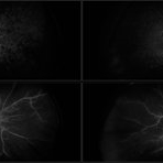
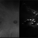
Ultra-wide-field fluorescein angiogram of the left eye of an 29-year-old female with multiple retinal hemangiomas secondary to Von Hippel-Lindau Syndrome.
Photographer: Olivia Rainey
Imaging device: Optos California
Condition/keywords: fluorescein angiogram (FA), fluorescein leakage, left eye, Optos, retinal hemangioma, ultra-wide field imaging, Von Hippel-Lindau
-
 Optic Disc Hemangioblastoma
May 30 2017 by Olivia Rainey
Ultra-wide-field fluorescein angiogram of the right eye of an 29-year-old female with an optic nerve hemangioblastoma secondary to Von Hippel-Lindau Syndrome.
Photographer: Olivia Rainey
Imaging device: Optos California
Condition/keywords: fluorescein angiogram (FA), fluorescein leakage, optic disc, Optos, retinal hemangioblastoma, ultra-wide field imaging, Von Hippel-Lindau
-
 Acute Retinal Necrosis secondary to Herpes Zoster Ophthalmicus
Jan 9 2018 by Olivia Rainey
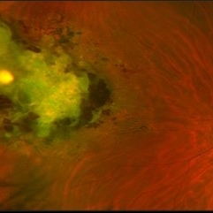
Ultra-wide field Optos pseudocolor montage of an 40-year-old female presenting with acute retinal necrosis secondary to herpes zoster ophthalmicus affecting her right eye.
Photographer: Olivia Rainey
Imaging device: Optos California
Condition/keywords: acute retinal necrosis, color fundus photograph, Herpes zoster, montage, Optos, ultra-wide field imaging
-
 Endogenous Endophthalmitis With Suspected Systemic Candidiasis
Jan 9 2018 by Olivia Rainey
Ultra-wide field Optos pseudocolor image of an 45-year-old male presenting with endogenous endophthalmitis affecting his left eye. Candidiasis was at high considerations due to intravenous drug abuse and recent history of dental abscess. Patient developed a subretinal infiltrate resulting in central scotoma and responded well to anti-fungal treatment.
Photographer: Olivia Rainey
Imaging device: Optos California
Condition/keywords: candida endophthalmitis, drug abuse, endogenous endophthalmitis, left eye, Optos, pseudocolor, retinal infiltrates, scotoma, ultra-wide field imaging
-
 Proliferative Diabetic Retinopathy with Pre-retinal Hemorrhage
Jan 16 2018 by Olivia Rainey
Ultra-wide field pseudo-color image of an 57-year-old male with a large pre-retinal hemorrhage secondary to proliferative diabetic retinopathy affecting his left eye.
Photographer: Olivia Rainey
Imaging device: Optos California
Condition/keywords: color fundus photograph, diabetic mellitus, hemorrhage, left eye, neovascularization (NV), Optos, proliferative diabetic retinopathy (PDR), pseudocolor, ultra-wide field imaging
-
 Retinitis Pigmentosa with Asteroid Hyalosis
Jan 16 2018 by Olivia Rainey
Ultra-wide field pseudocolor fundus photograph of an 75-year-old female with Retinitis Pigmentosa with Asteroid Hyalosis affecting her right eye.
Photographer: Olivia Rainey
Imaging device: Optos California
Condition/keywords: asteroid hyalosis, color fundus photograph, Optos, pseudocolor, retinitis pigmentosa, ultra-wide field imaging
-
 Cytomegalovirus Retinitis
Jan 16 2018 by Olivia Rainey
Color fundus montage of an 37-year-old, HIV positive male with CMV retinitis affecting his right eye. Patient's vision was sc20/20-1. He received an intravitreal Ganciclovir injection as well. The referring physcian suspects his condition is secondary to his chemotherapy for large B cell lymphoma or stomach cancer. The patient had not started taking oral Valgancyclovir.
Photographer: Olivia Rainey
Imaging device: Topcon 50dx
Condition/keywords: CMV retinitis, color fundus photograph, cytomegalovirus (CMV), HIV, montage
-
 Plateau Iris from Aqueous Misdirection
Jan 18 2018 by Olivia Rainey
Anterior segment OCT of a 90-year-old female with plateau iris from aqueous misdirection affecting her right eye.
Photographer: Olivia Rainey
Imaging device: Heidelberg Spectralis
Condition/keywords: anterior segment, aqueous misdirection, Heidelburg Spectralis, optical coherence tomography (OCT), plateau iris
-
 Proliferative Diabetic Retinopathy
Jan 18 2018 by Olivia Rainey
Ultra-wide field fluorescein angiogram of a 57-year-old male with proliferative diabetic retinopathy affecting his right eye.
Photographer: Olivia Rainey
Imaging device: Heidelberg Spectralis
Condition/keywords: diabetes, fluorescein leakage, Heidelburg Spectralis, neovascularization of the disc (NVD), peripheral retinal nonperfusion, proliferative diabetic retinopathy (PDR), ultra-wide field imaging
-
 Posterior Uveitis with Cystoid Macular Edema
Jan 18 2018 by Olivia Rainey
Ultra-wide field fluorescein angiogram of a 59-year-old female with posterior uveitis and chronic cystoid macular edema affecting her left eye. Interestingly, she has peripheral capillary nonperfusion inferotemporal, which could be driving CME.
Photographer: Olivia Rainey
Imaging device: Heidelberg Spectralis
Condition/keywords: 102 degrees, cystoid macular edema (CME), fluorescein leakage, Heidelburg Spectralis, left eye, peripheral retinal nonperfusion, posterior uveitis, ultra-wide field imaging
-
 Choroidal Metastasis
Jan 24 2018 by Olivia Rainey
Color fundus montage of a 35-year-old male with choroidal metastasis from the lung. Before the diagnosis was confirmed, the patient had multiple CT scans revealing only pneumonia, with no signs of cancer.
Photographer: Olivia Rainey
Imaging device: Topcon 50DX
Condition/keywords: choroidal metastasis, color fundus photograph, lung cancer metastasis, montage
-
 Proliferative Diabetic Retinopathy with Diabetic Macular Edema
Jan 24 2018 by Olivia Rainey
Ultra-wide field fluorescein angiogram of a 59-year-old male with proliferative diabetic retinopathy and diabetic macular edema affecting his left eye.
Photographer: Olivia Rainey
Imaging device: Optos
Condition/keywords: diabetes, diabetic macular edema, fluorescein leakage, ischemia, laser scarring, left eye, proliferative diabetic retinopathy (PDR), ultra-wide field imaging
-
 Penetrating Trauma of an Inadvertent Sub-Tenon's Kenalog Injection
Jan 31 2018 by Olivia Rainey
Ultra-wide field pseudocolor photograph of a 38-year-old female with penetrating trauma after an inadvertent sub-tenon's kenalog injection affecting her left eye. Patient has a large dehemoglobinized vitreous hemorrhage settling inferior near the entry wound. The exit wound has developed chorioretinal scarring and the disruption of several veins near the optic nerve, resulting in a branch retinal vein occlusion.
Photographer: Olivia Rainey
Imaging device: Optos
Condition/keywords: branch retinal vein occlusion (BRVO), chorioretinal scar, color fundus photograph, dehemoglobinized hemorrhage, kenalog, left eye, montage, Optos, penetrating trauma, sub-tenon's, ultra-wide field imaging
-
 White Without Pressure/Dot Blot Hemorrhages
Jan 31 2018 by Olivia Rainey
Ultra-wide field pseudocolor photograph of a 57-year-old female with white without pressure affecting her right eye. Patient will be having bloodwork done to rule out possible sarcoidosis or sickle cell.
Photographer: Olivia Rainey
Imaging device: Optos
Condition/keywords: blot hemorrhages, Optos, ultra-wide field imaging
-
 White Without Pressure
Jan 31 2018 by Olivia Rainey
Ultra-wide field pseudocolor photograph of a 57-year-old female with white without pressure affecting her left eye. Patient will be having bloodwork done to rule out possible sarcoidosis or sickle cell.
Photographer: Olivia Rainey
Imaging device: Optos
Condition/keywords: blot hemorrhages, color fundus photograph, left eye, Optos, ultra-wide field imaging, white without pressure
-
 Familial Exudative Vitreoretinopathy
Feb 2 2018 by Olivia Rainey
Ultra-wide field montage of a 37-year-old female with familial exudative vitreoretinopathy affecting her left eye. Cryotherapy, laser destruction of retinopathy, and a scleral buckle was performed to stabilize the retina in 2017.
Photographer: Olivia Rainey
Imaging device: Optos
Condition/keywords: familial exudative vitreoretinopathy (FEVR), fibrotic neovascularization, laser scarring, left eye, montage, Optos, scleral buckle, tractional retinal detachment, ultra-wide field imaging
-
 Massive Subretinal Hemorrhage Due to Exudative Macular Degeneration
Feb 2 2018 by Olivia Rainey
Color fundus photograph of a 74-year-old male with a massive subretinal hemorrhage due to exudative macular degeneration.
Photographer: Olivia Rainey
Imaging device: Topcon 50dx
Condition/keywords: 50 degrees, age-related macular degeneration (AMD), color fundus photograph, left eye, subretinal hemorrhage
-
 Disciform Scar Due to Exudative Macular Degeneration
Feb 2 2018 by Olivia Rainey
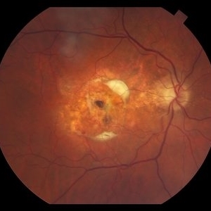
Color fundus photograph of a 74-year-old male with a central disciform scar due to exudative macular degeneration.
Photographer: Olivia Rainey
Imaging device: Topcon 50dx
Condition/keywords: 50 degrees, age-related macular degeneration (AMD), central disciform scar, color fundus photograph
-
 External Photography of Likely Xanthogranuloma
Feb 2 2018 by Olivia Rainey
An external photography series of a 51-year-old female with conjunctival lesions affecting both eyes. Patient is going through testing to help with diagnosis.
Photographer: Olivia Rainey
Imaging device: Topcon 50dx
Condition/keywords: external photography, xanthogranuloma
-
 Choroidal Melanoma
Feb 2 2018 by Olivia Rainey
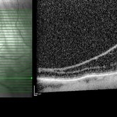
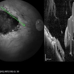
Optical coherence tomography with enhanced depth imaging of a 78-year-old female with choroidal melanoma with subretinal fluid affecting her right eye.
Photographer: Olivia Rainey
Imaging device: Heidelberg Spectralis
Condition/keywords: enhanced depth imaging, infrared image, optical coherence tomography (OCT), subretinal fluid, superior retina
-
 Optic Nerve Head Drusen with OCT
Feb 2 2018 by Olivia Rainey
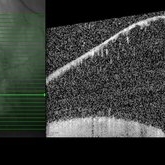
Optical coherence tomography with enhanced depth imaging of a 86-year-old male with optic nerve head drusen affecting his right eye. This patient has also been diagnosed with pseudoxanthoma elasticum and macular degeneration.
Photographer: Olivia Rainey
Imaging device: Heidelberg Spectralis
Condition/keywords: enhanced depth imaging, infrared image, macular degeneration, optic disc drusen, optic nerve, optical coherence tomography (OCT), pseudoxanthoma elasticum (PXE)
-
 Optic Nerve Head Drusen
Feb 9 2018 by Olivia Rainey
Fundus autofluorescence of a 49-year-old female with optic nerve head drusen affecting her left eye. The patient has pseudoxanthoma elasticum with choroidal neovascularization and has been receiving anti-VEGF treatment for many years.
Photographer: Olivia Rainey
Imaging device: Heidelberg Spectralis
Condition/keywords: 30 degrees, anti-VEGF, choroidal neovascularization (CNV), fundus autofluorescence (FAF), Heidelburg Spectralis, left eye, optic disc, optic nerve drusen, pseudoxanthoma elasticum (PXE)
-
 Macula Sparring Tractional Retinal Detachment
Feb 9 2018 by Olivia Rainey
Ultra-wide field pseudocolor image of a 22-year-old male with a macula sparring tractional retinal detachment relating to retinopathy of prematuritiy affecting his right eye.
Photographer: Olivia Rainey
Imaging device: Optos
Condition/keywords: color fundus photograph, demarcation line, macula sparring, Optos, retinopathy of prematurity (ROP), tractional retinal detachment, ultra-wide field imaging
-
 Proliferative Diabetic Retinopathy
Mar 7 2018 by Olivia Rainey
Ultra-wide field fluorescein angiogram of a patient with proliferative diabetic retinopathy affecting the left eye.
Photographer: Olivia Rainey
Imaging device: Optos
Condition/keywords: diabetes, fluorescein leakage, ischemia, left eye, neovascularization (NV), neovascularization of the disc (NVD), Optos, proliferative diabetic retinopathy (PDR), ultra-wide field imaging
-
 Ocular Metastasis of Breast Cancer
Mar 13 2018 by Olivia Rainey
Color fundus montage of a 45-year-old female presenting with ocular metastasis affecting her left eye. She had been treated for pneumonia, had progressive lumbar back pain, and a 29 pound weight loss recently. She reported that she had a breast lump and a mammogram, but had not been provided the results. After she was sent to oncology, it was confirmed that she has widely metastatic breast cancer and tumors throughout the brain. The oncology team felt she could not wait weeks for her chemotherapy to start and consequently decided to do whole brain radiation and treat the affected eye just posterior to the lens.
Photographer: Olivia Rainey
Imaging device: Topcon DX50
Condition/keywords: choroidal metastasis, color fundus photograph, exudative detachment, left eye, lipid exudation, montage
-
 Retinal Detachment with Giant Tear
Mar 13 2018 by Olivia Rainey
Ultra-wide field pseduocolor image of a 36-year-old male with an giant inferior tear, causing a retinal detachment.
Photographer: Olivia Rainey
Imaging device: Optos
Condition/keywords: color fundus photograph, giant retinal tear, inferior retina, macular splitting, Optos, ultra-wide field imaging
-
 Congenital Retinal Pigment Epithelial Hypertrophy (CHRPE) Associated with Gardner's Syndrome
Mar 13 2018 by Olivia Rainey
Ultra-wide field fundus autofluorescence images of a 14-year-old patient with congenital retinal pigment epithelial hypertrophy affecting both eyes as a manifestation of Gardner's Syndrome.
Photographer: Olivia Rainey
Imaging device: Optos
Condition/keywords: bilateral, familial adenomatous polyposis, fundus autofluorescence (FAF), Gardner Syndrome, hypofluorescent lesions, Optos, ultra-wide field imaging
-
 Purtscher's Retinopathy
Mar 30 2018 by Olivia Rainey
Bilateral OCTS of a 21-year-old female with Purtscher's Retinopathy affecting both eyes. Patient developed acute pancreatitis triggered by hypercalcemia and likely increased alcohol consumption.
Photographer: Olivia Rainey
Imaging device: Spectralis
Condition/keywords: acute pancreatitis, bilateral, optical coherence tomography (OCT), Purtscher's retinopathy
-
 Peripheral Retinal Ischemia
Apr 26 2018 by Olivia Rainey
Ultra-wide field fluorescein angiogram of a 55-year-old female with peripheral retinal ischemia affecting her left eye. CTA head and neck performed on 11/16/15 and showed calcified atherosclerotic plaque involving the intracranial internal carotid arteries with resulting luminal narrowing. Intracranial vertebral arteries have smooth luminal contours. CTA neck normal. Likely from internal carotid plaques. Sickle cell disease came back negative.
Photographer: Olivia Rainey
Imaging device: Optos California
Condition/keywords: fluorescein angiogram (FA), fluorescein leakage, left eye, Optos, retinal ischemia, ultra-wide field imaging
-
 Central Retinal Vein Occlusion
Jul 13 2018 by Olivia Rainey
Ultra-wide field fluorescein angiogram of a patient presenting with a central retinal vein occlusion their right eye.
Photographer: Olivia Rainey
Imaging device: Optos
Condition/keywords: central retinal vein occlusion (CRVO), fluorescein angiogram (FA), fluorescein leakage, hemorrhage, Optos, ultra-wide field imaging
-
 Central Retinal Vein Occlusion
Jul 13 2018 by Olivia Rainey
Ultra-wide field, pseudocolor montage of a patient presenting with a central retinal vein occlusion, as well as, an inferior chorioretinal scar in their right eye.
Photographer: Olivia Rainey
Imaging device: Optos
Condition/keywords: central retinal vein occlusion (CRVO), chorioretinal scar, montage, Optos, pseudocolor, ultra-wide field imaging
-
 Retinocytoma
Jul 13 2018 by Olivia Rainey
Ultra-wide field pseudocolor image of a 5-year-old male with a retinocytoma affecting his right eye. The retinal tumor has associated calcium which looks suspicious for retinoblastoma. However, there are a number of atypical features which raise the possibility of a masquerade tumor.
Photographer: Olivia Rainey
Imaging device: Optos
Condition/keywords: Optos, pseudocolor, retinocytoma, ultra-wide field imaging
-
 Pseudocolor Fundus of Peripheral Retinoschisis
Jul 26 2018 by Olivia Rainey
Ultra-wide field pseudocolor image of a 49-year-old male with non-progressive peripheral retinoschisis of his left eye. Patient was asymptomatic and had no prior trauma or surgery to his eye. Recommended observation at this time.
Photographer: Olivia Rainey
Imaging device: Optos
Condition/keywords: fundus photograph, left eye, Optos, pseudocolor, retinoschisis, ultra-wide field imaging
-
 Autofluorescence of Peripheral Retinoschisis
Jul 26 2018 by Olivia Rainey
Ultra-wide field autofluorescence image of a 49-year-old male with non-progressive peripheral retinoschisis of his left eye. Patient was asymptomatic and had no prior trauma or surgery to his eye. Recommended observation at this time.
Photographer: Olivia Rainey
Imaging device: Optos
Condition/keywords: autofluorescence imaging, left eye, Optos, retinoschisis, ultra-wide field imaging
-
 Peripheral Retinoschisis
Jul 26 2018 by Olivia Rainey
Optical coherence tomography of a 49-year-old male with non-progressive peripheral retinoschisis of his left eye. Patient was asymptomatic and had no prior trauma or surgery to his eye. Recommended observation at this time.
Photographer: Olivia Rainey
Imaging device: Heidelberg Spectralis
Condition/keywords: Heidelburg Spectralis, left eye, optical coherence tomography (OCT), retinoschisis
-
 Multiple Astrocytic Hamartomas
Jul 26 2018 by Olivia Rainey
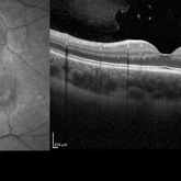
Optical coherence tomography of a 7-year-old female with multiple astrocytic harmartomas as a retinal manifestation of tuberous sclerosis. Patient came to our office to rule out possible drug toxicity from Sabril, a an anticonvulsant. There were no signs of retinal toxicity by extended ophthalmoscopy or imaging, yet she will be monitored every 6 months.
Photographer: Olivia Rainey
Imaging device: Heidelberg Spectralis
Condition/keywords: astrocytic hamartoma, Heidelburg Spectralis, infrared image, left eye, optical coherence tomography (OCT), tuberous sclerosis
-
 Retinal Ischemia Secondary to Diabetic Retinopathy
Aug 29 2018 by Olivia Rainey
Fluorescein angiogram series of a 57-year-old male patient with proliferative diabetic retinopathy of the right eye. Patient has delayed AV transit with significant retinal ischemia and retinal capillary nonperfusion. The ischemia is extensive resulting in neovascularization of the iris and consequently neovascular glaucoma.
Photographer: Olivia Rainey
Imaging device: Optos
Condition/keywords: diabetes, disc hyperfluorescene, fluorescein angiogram (FA), non-perfusion, Optos, proliferative diabetic retinopathy (PDR), retinal ischemia, ultra-wide field imaging, vitreous hemorrhage
-
 Central Retinal Vein Occlusion With Macular Edema
Aug 29 2018 by Olivia Rainey
Ultra-widefield fluorescein angiogram of an 58-year-old male with a central retinal vein occlusion with macular edema affecting his right eye. Fluorescein showed delayed transit with late leakage.
Photographer: Olivia Rainey
Imaging device: Optos
Condition/keywords: central retinal vein occlusion (CRVO), FA late phase leakage, fluorescein angiogram (FA), Optos, tortuous vessels, ultra-wide field imaging
-
 Malignant Melanoma
Sep 11 2018 by Olivia Rainey
Ultra-wide field autofluorescence and pseudocolor montage of a 57-year-old male s/p I-125 brachytherapy for malignant melanoma affecting his right eye. The patient’s radiation retinopathy has resulted in retinal vascular occlusive disease and optic nerve edema.
Photographer: Olivia Rainey
Imaging device: Optos
Condition/keywords: branch retinal vein occlusion (BRVO), fundus autofluorescence (FAF), I-125 brachytherapy, malignant melanoma, montage, Optos, pseudocolor, radiation retinopathy, ultra-wide field imaging
-
 Radiation Retinopathy resulting in Retinal Vascular Occlusive Disease
Sep 11 2018 by Olivia Rainey
Ultra-wide field fluorescein angiography of a 57-year-old male s/p I-125 brachytherapy for malignant melanoma affecting his right eye. The patient’s radiation retinopathy has resulted in retinal vascular occlusive disease and optic nerve edema.
Photographer: Olivia Rainey
Imaging device: Optos
Condition/keywords: branch retinal vein occlusion (BRVO), fluorescein angiogram (FA), I-125 brachytherapy, malignant melanoma, optic disc edema, Optos, radiation retinopathy, ultra-wide field imaging
-
 Branch Retinal Artery Occlusion
Sep 11 2018 by Olivia Rainey
Ultra-wide field fluorescein angiogram of a 46-year-old male with a branch retinal artery occlusion affecting his left eye. The longstanding occlusion and has resulted in peripheral nonperfusion and neovascularization.
Photographer: Olivia Rainey
Imaging device: Optos
Condition/keywords: branch retinal artery occlusion (BRAO), fluorescein angiogram (FA), left eye, neovascularization (NV), non-perfusion, Optos
-
 Branch Retinal Vein Occlusion
Sep 11 2018 by Olivia Rainey
Ultra-wide field pseudocolor montage of an 84-year-old female with a branch retinal vein occlusion affecting her left eye. Patient recently had a PPV for a epiretinal membrane in her left eye and shortly after developed an occlusion.
Photographer: Olivia Rainey
Imaging device: Optos
Condition/keywords: branch retinal artery occlusion (BRAO), hemorrhage, left eye, montage, Optos, pseudocolor, ultra-wide field imaging
-
 Bilateral Lymphoma Metastasis after Resolution with IVM
Sep 19 2018 by Olivia Rainey
Ultra-wide field, pseudocolor fundus images of an 86-year-old female treated with intravitreal methothrexate as a management of subretinal infiltrate in the macula of the right eye, as a manifestation of leukemia. Her last intravitreal methotrexate injection was 5/1/18.
Photographer: Olivia Rainey
Imaging device: Optos
Condition/keywords: bilateral, leukemia, methotrexate, Optos, pseudocolor, ultra-wide field imaging, uveitis
-
 Bilateral Lymphoma Metastasis after Resolution with IVM
Sep 19 2018 by Olivia Rainey
Ultra-wide field, autofluorescence images of an 86-year-old female treated with intravitreal methothrexate as a management of subretinal infiltrate in the macula of the right eye, as a manifestation of leukemia. Her last intravitreal methotrexate injection was 5/1/18.
Photographer: Olivia Rainey
Imaging device: Optos
Condition/keywords: bilateral, fundus autofluorescence (FAF), lymphoma, Optos, ultra-wide field imaging, uveitis
-
 Retinal Detachment with Retinal Tears
Dec 11 2018 by Olivia Rainey
Ultra-wide field pseudocolor image of a 56-year-old male with a large superior retinal detachment with retinal tears affecting his right eye.
Photographer: Olivia Rainey
Imaging device: Optos
Condition/keywords: Optos, pseudocolor, retinal tear with detachment
-
 Branch Retinal Vein Occlusion With Peripheral Pigmentary Change
Jan 15 2019 by Olivia Rainey
Ultra-wide field fluorescein angiogram of an 85-year-old female with a branch retinal vein occlusion with peripheral pigmentary changes. Patient developed a BRVO after a PPV for an epiretinal membrane.
Photographer: Olivia Rainey
Imaging device: Optos
Condition/keywords: branch retinal vein occlusion (BRVO), epiretinal membrane (ERM), fluorescein angiogram (FA), left eye, Optos, pigmentary retinal dystrophy
-
 Cystoid Macular Edema Secondary to Panuveitis
Jan 15 2019 by Olivia Rainey
Fluorescein angiogram of a 55-year-old female with cystoid macular edema secondary to uveitis affecting her right eye. Patient was diagnosed with sarcoidosis.
Photographer: Olivia Rainey
Imaging device: Heidelberg Spectralis
Condition/keywords: 30 degrees, cystoid macular edema (CME), fluorescein angiogram (FA), fluorescein leakage, Heidelburg Spectralis, sarcoidosis, uveitis
-
 Weiss Ring
Jan 15 2019 by Olivia Rainey
Fluorescein angiogram of a 55-year-old female with a Weiss ring affecting her right eye. Patient was diagnosed with sarcoidosis. She has cystoid macular edema secondary to panuveitis.
Photographer: Olivia Rainey
Imaging device: Heidelberg Spectralis
Condition/keywords: 30 degrees, cystoid macular edema (CME), fluorescein angiogram (FA), fluorescein leakage, Heidelburg Spectralis, optic nerve, sarcoidosis, uveitis, Weiss ring
-
 Epiretinal Membrane With Traction
Jan 15 2019 by Olivia Rainey
Red-free image of a 72-year-old female with an epiretinal membrane with inferior traction affecting her left eye. Patient had seen distortion for one month prior to her appointment and stated that the change happened suddenly. Patient deferred surgery.
Photographer: Olivia Rainey
Imaging device: Heidelberg Spectralis
Condition/keywords: 30 degrees, epiretinal membrane (ERM), Heidelburg Spectralis, left eye, macular traction, red-free
-
 Ciliary Body Melanoma (Class 2 PRAME Positive)
Jan 17 2019 by Olivia Rainey
Pseudocolor optos fundus image of an 61-year-old male with ciliary body melanoma affecting his right eye, which tested PRAME positive placing him in class 2. The melanoma had caused a hemorrhagic retinal detachment. Patient was referred for a retinal detachment and was experiencing cloudy vision affecting his inferior field. He had been seeing flashes inferiorly 1-2x a daily for about 6-8 months.
Photographer: Olivia Rainey
Imaging device: Optos
Condition/keywords: ciliary body melanoma, class 2, Optos, PRAME positive
-
 Diabetic Tractional Retinal Detachment
Jan 23 2019 by Olivia Rainey
Ultra-wide field pseudocolor image of an 43-year-old female with a diabetic tractional retinal detachment and a vitreous hemorrhage affecting her right eye.
Photographer: Olivia Rainey
Imaging device: Optos
Condition/keywords: diabetes, diabetic traction detachment, Optos, pan-retinal photocoagulation (PRP), proliferative diabetic retinopathy (PDR), pseudocolor, ultra-wide field imaging, vitreous hemorrhage
-
 Large Choroidal Melanoma
Mar 29 2019 by Olivia Rainey
Ultra-wide field pseudocolor image of a 59-year-old male with a large choroidal melanoma affecting his right eye. Patient presented with a superior visual field defect 3 months prior to examination, but reports that the vision has been "bad" in the right eye since he was 3 years old. The risks, benefits and alternatives for treating the melanoma were thoroughly discussed and the patient has elected to proceed with enucleation, which we will schedule for the near future. Patient reports a brother with "blood cancer" and a father with throat cancer.
Photographer: Olivia Rainey
Imaging device: Optos
Condition/keywords: Optos, ultra-wide field imaging
-
 Proliferative Diabetic Retinopathy
Mar 29 2019 by Olivia Rainey
Ultra-wide field fluorescrein angiogram of a 48-year-old male with proliferative diabetic retinopathy affecting his right eye. Patient was treated with pan-retinal laser follow his fluorescein angiogram and will likely need intravitreal injections in the near future.
Photographer: Olivia Rainey
Imaging device: Optos
Condition/keywords: diabetes, diabetic macular edema, fluorescein angiogram (FA), fluorescein leakage, late phase, Optos, proliferative diabetic retinopathy (PDR), ultra-wide field imaging
-
 Proliferative Diabetic Retinopathy with Retinal Ischemia
Mar 29 2019 by Olivia Rainey
Ultra-wide field fluorescrein angiogram of a 42-year-old female with proliferative diabetic retinopathy with retinal ischemia affecting her left. Patient had been noticing a vision decline and floaters over the past few months. She was treated with Avastin in her right eye and then her left one week later.
Photographer: Olivia Rainey
Imaging device: Optos
Condition/keywords: diabetes, diabetic macular edema, early phase, fluorescein angiogram (FA), fluorescein leakage, neovascularization of the disc (NVD), Optos, proliferative diabetic retinopathy (PDR), retinal ischemia, ultra-wide field imaging
-
 Proliferative Diabetic Retinopathy with Retinal Ischemia
Mar 29 2019 by Olivia Rainey
Ultra-wide field fluorescrein angiogram of a 42-year-old female with proliferative diabetic retinopathy with retinal ischemia affecting her right eye. Patient had been noticing a vision decline and floaters over the past few months. She was treated with Avastin in her right eye and then her left one week later.
Photographer: Olivia Rainey
Imaging device: Optos
Condition/keywords: diabetes, diabetic macular edema, fluorescein angiogram (FA), fluorescein leakage, late phase, neovascularization of the disc (NVD), Optos, proliferative diabetic retinopathy (PDR), retinal ischemia, ultra-wide field imaging
-
 Penetrating Trauma with Retinal Detachment
Apr 30 2019 by Olivia Rainey
Ultra-wide field pseudocolor image of a 39-year-old female with penetrating trauma resulting in a retinal detachment with an intraretinal hemorrhage affecting the left eye. Patient was struck with a champagne glass in October of 2018, which lacerated the eyelid and globe. Patient was "seeing red" when she first came to the office and after multiple surgeries she was seeing 20/20 at her last check in April 2019.
Photographer: Olivia Rainey
Imaging device: Optos
Condition/keywords: hemorrhage, left eye, Optos, penetrating trauma, ruptured globe, ultra-wide field imaging
-
 Choroidal Melanoma with a Serous Retinal Detachment
Apr 30 2019 by Olivia Rainey
Ultra-wide field fundus autofluorescence of a 67-year-old male with serous retinal detachment, secondary to a large choroidal melanoma affecting the left eye. Patient reported a curtain affecting his superior field for about 2-3 months prior to examination. Patient elects radiation bracytherapy with a guarded visual expectation secondary to the location of the tumor, touching the optic disc.
Photographer: Olivia Rainey
Imaging device: Optos
Condition/keywords: autofluorescence imaging, left eye, Optos, ultra-wide field imaging
-
 Morning Glory Syndrome
Jun 19 2019 by Olivia Rainey
Ultra-wide field pseudocolor image of an 10-year-old girl with Morning Glory Syndrome affecting her left eye. Patient is able to count fingers at 4 feet.
Photographer: Olivia Rainey
Imaging device: Optos
Condition/keywords: left eye, Morning Glory Syndrome, Optos, pseudocolor, ultra-wide field imaging
-
 Plaquenil Toxicity
Jun 20 2019 by Olivia Rainey
Ultra-wide field fundus autofluorescence photograph of a 60-year-old female with plaquenil toxicity affecting both eyes. Patient was taking plaquenil for management of Lupus, but discontinued use in 2011, but continues to be affected with severe progression of toxicity. Patient developed macular edema affecting her right eye and has received 5 Avastin injections.
Photographer: Olivia Rainey
Imaging device: Optos
Condition/keywords: autofluorescence imaging, focal pigmentary changes, fundus autofluorescence (FAF), macular edema, Optos, plaquenil toxicity, ultra-wide field imaging
-
 Severe macular atrophy secondary to Pseudoxanthoma Elasticum and CNV
Jul 26 2019 by Olivia Rainey
Ultra-wide field color/autofluorescence comparison of a 54-year-old female with severe macular atrophy secondary to pseudoxanthoma elasticum. Patient developed choroidal neovascularization that does not warrant treatment due to the patient's poor visual acuity. Patient has significantly attenuated ERG. Genetic testing is recommended to rule out retinitis pigmentosa. The mechanism is yet to be determined, but there is a thought that ABCC6 mutation causes alteration of plasma lipid composition may be implicated in the systemic changes observed in PXE.
Photographer: Olivia Rainey
Imaging device: Optos
Condition/keywords: angioid streaks, atrophic central lesion, autofluorescence imaging, choroidal neovascularization (CNV), hypofluorescent lesions, macular atrophy, Optos, pigmentary retinal dystrophy, pseudoxanthoma elasticum (PXE), ultra-wide field imaging
-
 Proliferative Diabetic Retinopathy with Active Neovascularization
Jul 30 2019 by Olivia Rainey
Ultra-wide field fluorescein angiogram of a 36-year-old male with proliferative diabetic retinopathy with active neovascularization affecting his left eye. Patient presented with seeing flashing lights and trouble seeing to drive at night. His vision was sc20/50-1 PH20/40-2 in the left eye. There are suspicious vessels within the inferonasal retina of the patient's left eye. Labs ordered and are negative for sickle cell.
Photographer: Olivia Rainey
Imaging device: Optos
Condition/keywords: diabetes, diabetic macular edema, fluorescein angiogram (FA), fluorescein leakage, ischemia, late phase, left eye, neovascularization (NV), Optos, proliferative diabetic retinopathy (PDR), ultra-wide field imaging
-
 Chronic Total Retinal Detachment
Oct 8 2019 by Olivia Rainey
Ultra-wide field pseudocolor image of a 57-year-old male with a chronic total retinal detachment affecting his right eye. Patient presented with klebsiella endophthalmitis in UK, and was in medically induced coma with tracheostomy. He awoke after sedation with loss of vision in both eyes, later developing a retinal detachment in both eyes. He had his first repair with gas then with silicone oil. Although he is developing band K and corneal decompensation due to oil in the AC, he has a chronic cicatricial retinal detachment with subretinal and preretinal PVR with large stretch breaks. His vision may worsen with surgery and his eye is hypotenous and if repair is possible, oil will need to be replaced and thus may still migrate to the AC thus would defer intervention until absolutely necessary given risks of vision loss in his only seeing eye. Given this is his only eye, observation is recommended at this time.
Photographer: Olivia Rainey and Amber Poss
Imaging device: Optos
Condition/keywords: chronic retinal detachment, klebsiella endopthalmitis, laser scarring, Optos, proliferative vitreoretinopathy (PVR), retinectomy, silicone oil
-
 Closed Funnel Retinal Detachment
Oct 8 2019 by Olivia Rainey
Ultra-wide field pseudocolor image of a 57-year-old male with a closed funnel retinal detachment with anterior and posterior napkin rings affecting his left eye. Patient presented with klebsiella endophthalmitis in UK, and was in medically induced coma with tracheostomy. He awoke after sedation with loss of vision in both eyes, later developing a retinal detachment in both eyes. Prior inflammation attributable to prephthisical state and chronic funnel retinal detachment. The eye is inoperable and observation is recommended.
Photographer: Olivia Rainey and Amber Poss
Imaging device: Optos
Condition/keywords: blind eye, funnel, hypotony, klebsiella endopthalmitis, left eye, Optos
-
 Retinopathy of Prematurity
Oct 8 2019 by Olivia Rainey
Ultra-wide field pseudocolor image of a 15-year-old male with retinopathy of prematurity affecting both of his eyes. Patient was born at 22 weeks and had a birth weight of 434g. He presented with esotropia, nystagmus, and severe macular/vascular dragging in the right eye.
Photographer: Olivia Rainey
Imaging device: Optos
Condition/keywords: esotropia, macular dragging, myopia, nystagmus, Optos, retinopathy of prematurity (ROP), ultra-wide field imaging, vascular arrest, vascular dragging
-
 Retinopathy of Prematurity
Oct 8 2019 by Olivia Rainey
Ultra-wide field pseudocolor image of a 15-year-old male with retinopathy of prematurity affecting both of his eyes. Patient was born at 22 weeks and had a birth weight of 434g. Although his posterior vasculature and macula appears normal on exam, he has vascular arrest to anterior zone 2 or posterior zone. He has no NV or RD on exam however his left eye peripheral avascular area is concerning moving forward. Recommend close follow-up, however may elect laser ablation in the future.
Photographer: Olivia Rainey
Imaging device: Optos
Condition/keywords: failure to vascularize, left eye, myopia, nystagmus, Optos, retinopathy of prematurity (ROP), ultra-wide field imaging
-
 Macula Off Retinal Detachment with CNV
Nov 11 2019 by Olivia Rainey
Ultra-wide field pseudocolor photograph of a 42-year-old female with a long-standing, macula-off retinal detachment affecting her left eye. Patient was unaware of vision loss until testing her visual acuity and she denied seeing flashing lights. Patient decided to proceed with scleral buckling. The CNV is potentially secondary the retinal detachment, but may be myopic related or idiopathic. The CNV appears fibrotic and inactive. The patient was warned that this will absolutely limit how much vision she recovers once the retina is reattached.
Photographer: Olivia Rainey
Imaging device: Optos California
Condition/keywords: choroidal neovascularization (CNV), chronic retinal detachment, fundus autofluorescence (FAF), left eye, montage, Optos, retinal detachment of the macula, ultra-wide field imaging
-
 Macula Off Retinal Detachment with CNV
Nov 11 2019 by Olivia Rainey
Ultra-wide field pseudocolor photograph of a 42-year-old female with a long-standing, macula-off retinal detachment affecting her left eye. Patient was unaware of vision loss until testing her visual acuity and she denied seeing flashing lights. Patient decided to proceed with scleral buckling. The CNV is potentially secondary the retinal detachment, but may be myopic related or idiopathic. The CNV appears fibrotic and inactive. The patient was warned that this will absolutely limit how much vision she recovers once the retina is reattached.
Photographer: Olivia Rainey
Imaging device: Optos California
Condition/keywords: choroidal neovascularization (CNV), left eye, montage, Optos, pseudocolor, retinal detachment of the macula, ultra-wide field imaging
-
 Proliferative Diabetic Retinopathy
Nov 13 2019 by Olivia Rainey
Ultra-wide field fluorescein angiogram at 29 seconds of a 52-year-old male with proliferative diabetic retinopathy affecting his right eye. Patient is receiving Eylea intravitreal injections and has had panretinal photocoagulation in the past. Patient's vision tested 20/40 and with pinholes to 20/30.
Photographer: Olivia Rainey
Imaging device: Optos California
Condition/keywords: diabetes, diabetic macular edema, early phase, FA early phase, fluorescein angiogram (FA), intravitreal injection, ischemia, pan-retinal photocoagulation (PRP), proliferative diabetic retinopathy (PDR)
-
 Familial Drusen with Extrafoveal Pigment Epithelial Detachment
Nov 21 2019 by Olivia Rainey
Late images from a fluorescein angiogram of a 66-year-old female with familial drusen with extrafoveal pigment epithelial detachments affecting both eyes. The physician is presuming that the small multifocal PED's result from her familial drusen. These remain asymptomatic and clinically stable on her exam 11-20-19. VA OD: Dcc20/25-1+1 and VA OS: Dcc20/25-2.
Photographer: Olivia Rainey
Imaging device: Heidelberg Spectralis
Condition/keywords: drusen, drusenoid PED, familial drusen, fluorescein angiogram (FA), hyperfluorescence, late phase, pigment epithelial detachment (PED)
-
 Macular Pattern Dystrophy Associated with MELAS
Dec 19 2019 by Olivia Rainey
Bilateral wide field pseudocolor images of a 54-year-old female with macular pattern dystrophy associated with MELAS. The patient is positive for m.3243A>G in MT-TL1. She had stroke in her 40s, hearing loss in her 30s, and has early onset diabetes. MyRetinaTracker shows VUS in RP1L1. Mutation in RP1L1 have been describe in other families with occult macular dystrophy. Farnsworth D15 is showing mild tritan abnormality, which is most commonly seen with acquired maculopathies. 12/17/19 patient's Optos and OCT show mild progression of atrophy.
Photographer: Olivia Rainey
Imaging device: Optos California
Condition/keywords: advanced geographic atrophy, bilateral, fundus photograph, MELAS, Optos, pattern macular dystrophy, pseudocolor, wide angle imaging
-
 Macular Pattern Dystrophy Associated with MELAS
Dec 19 2019 by Olivia Rainey
Bilateral wide field fundus autofluorescence images of a 54-year-old female with macular pattern dystrophy associated with MELAS. The patient is positive for m.3243A>G in MT-TL1. She had stroke in her 40s, hearing loss in her 30s, and has early onset diabetes. MyRetinaTracker shows VUS in RP1L1. Mutation in RP1L1 have been describe in other families with occult macular dystrophy. Farnsworth D15 is showing mild tritan abnormality, which is most commonly seen with acquired maculopathies. 12/17/19 patient's Optos and OCT show mild progression of atrophy.
Photographer: Olivia Rainey
Imaging device: Optos California
Condition/keywords: advanced geographic atrophy, bilateral, fundus autofluorescence (FAF), MELAS, Optos, pattern macular dystrophy, wide angle imaging
-
 Retinopathy of Prematurity
Dec 26 2019 by Olivia Rainey
Ultra-wide field pseudocolor image of a 7 month old, corrected gestational age at 3 months, with retinopathy of prematurity, stage 1-2 zone 3 affecting her left eye. Patient was born at 23 weeks and her birth weight was 459 g (1.01192 lbs). She was born 3 days after her twin sister. She was in the NICU for over 5 months. She had patent ductus arteriosus (PDA) corrected by ligation. No current motor or social delay. Patient could fix and follow up at her appointment.
Photographer: Olivia Rainey
Imaging device: Optos California
Condition/keywords: flat neovascularization, foveal avascular zone (FAZ), left eye, Optos, pseudocolor, retinopathy of prematurity (ROP), retinopathy of prematurity stage 1, retinopathy of prematurity stage2, zone III
-
 Chronic Rhegmatogenous Retinal Detachment
Dec 27 2019 by Olivia Rainey
Ultra-wide field pseudocolor fundus photography of a 22-year-old male with a rhegmatogenous retinal detachment affecting his right eye. Patient states that he has been seeing flickering lights in his inferotemporal field when transitioning from light to dark areas over a year beginning June 2018. Patient has limited visual potential due to longstanding nature of detachment. He elected for surgery.
Photographer: Olivia Rainey
Imaging device: Optos California
Condition/keywords: chronic retinal detachment, light perception, macula-off, Optos, proliferative vitreoretinopathy (PVR), ultra-wide field imaging, vitreous haze
-
 Morning Glory Syndrome
Jan 6 2020 by Olivia Rainey
Ultra-wide field pseudocolor image of a 23-month-old male with morning glory syndrome affecting his left eye. Patient presented with esotropia affecting his left eye and strabismic amblyopia affecting both eyes. He could fix and follow on exam and his medical history was unremarkable.
Photographer: Olivia Rainey
Imaging device: Optos California
Condition/keywords: esotropia, left eye, macular, Morning Glory Syndrome, Optos, strabismic amblyopia, ultra-wide field imaging
-
 Retinoblastoma Without Vitreous Seeding
Jan 27 2020 by Olivia Rainey
Ultra-wide field pseudocolor image of a 5-week-old infant's left eye with retinoblastoma without vitreous seeding. The right eye was also affected to a similar degree. There is suspect for new germline mutation. There was calcium on B scan OS>OD as well. Referred to Kellogg Eye Center for treatments.
Photographer: Olivia Rainey
Imaging device: Optos California
Condition/keywords: infant, left eye, Optos, pseudocolor, retinoblastoma, speculum, tumor, ultra-wide field imaging
-
 Syphilitic Uveitis
Apr 2 2020 by Olivia Rainey
Ultrawide-field fluorescein angiogram of a 42-year-old male with syphilitic uveitis affecting his right eye more than his left. Patient is HIV positive. He developed hearing loss and palm/leg/scalp rash prompting diagnosis of neurosyphilis, s/p IM and full IV course of 2.4 Mil PCN G, and finished this course 3/9/20. He admits to recent rectal bleeding with ongoing plan for colonoscopy 3/16/20. He has a history of extensive travel including London, Hong Kong, and Bangkok. His husband has also been treated with IV PCN G, however per chart review he has multiple sexual partners. Patient's vision was 20/20 in each eye.
Photographer: Olivia Rainey
Imaging device: Optos California
Condition/keywords: disc hyperfluorescence, FA late phase leakage, fluorescein angiogram (FA), fluorescein leakage, HIV, late phase, optic nerve edema, Optos, phelbitis, syphilis neuroretinopathy, ultra-wide field imaging, uveitis
-
 Pigmentary Retinal Dystrophy
May 5 2020 by Olivia Rainey
Ultra-widefield fundus autofluorescence image of an 44-year-old male with pigmentary retinal dystrophy affecting both eyes. He presented with decreased night vision for 6 months prior to his appointment. He stated that his recovery time from transitioning from dark to light areas is reduced. He stated that his peripheral vision has never been very good for most of his life. He admits to environmental hearing loss. Patient denies family history of blin. His vision was 20/20 in both eyes. His full field ERG, visual fields were not consistent with RP. Genetic testing with ID Your IRD and annual follow up has been recommended.
Photographer: Olivia Rainey, OCT-C, COA
Imaging device: Optos California
Condition/keywords: fundus autofluorescence (FAF), hyperautofluorescence, hypoautofluorescence, inferior retina, left eye, Optos, ultra-wide field imaging
-
 Pigmentary Retinal Dystrophy
May 5 2020 by Olivia Rainey
Ultra-widefield pseudocolor image of an 44-year-old male with pigmentary retinal dystrophy affecting both eyes. He presented with decreased night vision for 6 months prior to his appointment. He stated that his recovery time from transitioning from dark to light areas is reduced. He stated that his peripheral vision has never been very good for most of his life. He admits to environmental hearing loss. Patient denies family history of blin. His vision was 20/20 in both eyes. His full field ERG, visual fields were not consistent with RP. Genetic testing with ID Your IRD and annual follow up has been recommended.
Photographer: Olivia Rainey, OCT-C, COA
Imaging device: Optos California
Condition/keywords: inferior retina, left eye, Optos, pigment, pseudocolor, ultra-wide field imaging
-
 Pigmentary Retinal Dystrophy
May 5 2020 by Olivia Rainey
Ultra-widefield fundus autofluorescence image of an 44-year-old male with pigmentary retinal dystrophy affecting both eyes. He presented with decreased night vision for 6 months prior to his appointment. He stated that his recovery time from transitioning from dark to light areas is reduced. He stated that his peripheral vision has never been very good for most of his life. He admits to environmental hearing loss. Patient denies family history of blin. His vision was 20/20 in both eyes. His full field ERG, visual fields were not consistent with RP. Genetic testing with ID Your IRD and annual follow up has been recommended.
Photographer: Olivia Rainey, OCT-C, COA
Imaging device: Optos California
Condition/keywords: fundus autofluorescence (FAF), hyperautofluorescence, hypoautofluorescence, inferior retina, Optos, pigment, ultra-wide field imaging
-
 Pigmentary Retinal Dystrophy
May 5 2020 by Olivia Rainey
Ultra-widefield pseudocolor image of an 44-year-old male with pigmentary retinal dystrophy affecting both eyes. He presented with decreased night vision for 6 months prior to his appointment. He stated that his recovery time from transitioning from dark to light areas is reduced. He stated that his peripheral vision has never been very good for most of his life. He admits to environmental hearing loss. Patient denies family history of blin. His vision was 20/20 in both eyes. His full field ERG, visual fields were not consistent with RP. Genetic testing with ID Your IRD and annual follow up has been recommended.
Photographer: Olivia Rainey, OCT-C, COA
Imaging device: Optos California
Condition/keywords: inferior retina, Optos, pigmentary retinal dystrophy, pseudocolor, ultra-wide field imaging
-
 Peripheral Retinal Vasculitis
May 27 2020 by Olivia Rainey
Ultra-widefield fluorescein angiogram of a 58-year-old female with possible peripheral vasculitis. There was no venous access for this patient, so the fluorescein was administered orally. The image was taken at 7:33 after oral administration. The physician stated that the peripheral nonperfusion could be a sign of previous vasculitis, although could also be a result of uncontrolled diabetes. She was asked to obtain additional bloodwork in order to rule out sarcoidosis, as well as sickle cell. It does not appear the nonperfusion has progressed since her last evaluation. Her vision was 20/40 in the right eye at the time the image was taken.
Photographer: Olivia Rainey, OCT-C, COA
Imaging device: Optos California
Condition/keywords: diabetes, fluorescein angiogram (FA), hypertensive retinopathy, non-perfusion, Optos, oral fluorescein, peripheral retinal vasculitis, ultra-wide field imaging
-
 Plateau Fovea with Inner Retinal Thinning
May 27 2020 by Olivia Rainey
Optical coherence tomography of the left eye of a 20-year-old male with Alport Syndrome. The patient did not present with any ocular or visual symptoms, yet the distinct "plateau contour" of his fovea was noted on OCT during his visit. The patient presented with 20/25 vision at the time of his visit. There was myelinated nerve fiber layer noted in both eyes, but these features had remained stable from his appointment three years prior. The physician noted that myelinated nerve fiber was a congenital change, and had not affected his vision or health of the eye, nor is a feature of Alport Syndrome.
Photographer: Olivia Rainey, OCT-C, COA
Imaging device: Heidelberg Spectralis
Condition/keywords: Alports disease, Heidelburg Spectralis, inner retinal thinning, left eye, optical coherence tomography (OCT), plateau fovea
-
 Severe Retinal Ischemia Secondary to Diabetic Retinopathy
Jun 2 2020 by Olivia Rainey
Ultra-widefield fluorescein angiography of a 77-year-old female with severe retinal ischemia secondary to diabetic retinopathy affecting her right eye. Patient has received ten Eylea injections in her right eye. The efficacy of continuing injections in the right eye, based largely on her severe retinal ischemia, was discussed at her appointment. The retina is essentially non-perfused and based on these findings, the physician recommended to discontinue treatment in the right eye. With a systemic workup, it has been determined that her retinal ischemia is due to diabetic small vessel disease. Her vision has remained at hand motion since October of 2019.
Photographer: Olivia Rainey, OCT-C, COA
Imaging device: Optos California
Condition/keywords: diabetes, diabetic blindness, diabetic macular edema, diabetic retinopathy, early phase, fluorescein angiogram (FA), fluorescein leakage, macular scar, Optos, pan-retinal photocoagulation (PRP), retinal ischemia, ultra-wide field imaging
-
 Bullous Retinoschisis with Outer Retinal Holes
Jun 15 2020 by Olivia Rainey
Ultra-widefield pseudocolor fundus photograph of a 56-year-old female with bullous retinoschisis with outer retinal holes affecting her right eye. The physician noted superotemporal retinoschisis in her monoculcar functioning eye. There was no demarcation line and no inner or outer layer breaks at her first appointment in February of 2020. On 6/15/20 she had a new onset outer holes and SRF tracking inferiorly. The physician recommended observation, however if this continues to progress we have discussed indications for barrier laser.
Photographer: Olivia Rainey, OCT-C, COA
Imaging device: Optos California
Condition/keywords: bullous retinoschisis, Optos, outer layer breaks, outer layer hole, pseudocolor, subretinal fluid, superior retina, ultra-wide field imaging
-
 Severe Proliferative Diabetic Retinopathy with Asteroid Hyalosis
Aug 25 2020 by Olivia Rainey
Ultra-widefield fluorescein angiogram of a 47-year-old male with severe prolifterative diabetic retinopathy with very extensive neovascularization with fibrosis and traction affecting his right eye. The patient also has asteroid hyalosis affecting the eye. He was diagnosed with Type 1 diabetes in the late 1970s. The patient's vision sc20/100 PH20/70-2. He received treatment of panretinal photocoagulation following the angiogram.
Photographer: Olivia Rainey, OCT-C, COA
Imaging device: Optos California
Condition/keywords: asteroid hyalosis, FA early phase, fibrotic neovascularization, fluorescein angiogram (FA), hyperfluorescence, neovascularization (NV), ultra-wide field imaging
-
 Peripheral Exudative Hemorrhagic Chorioretinopathy
Oct 7 2020 by Olivia Rainey
Fluorecein and ICG angiography of a 80-year-old male with peripheral exudative hemorrhagic chorioretinopathy affecting his right eye. Patient noted only mild floaters for a couple weeks OD on 10/5/2020. The physician strongly suspects that the lesion is subretinal blood (likely from PEHCR) rather than choroidal melanoma. There is blocking on FA and ICG with a definite lack of intrinsic vessels within the mass lesion. He will monitor closely, as the patient is monocular with a history of multiple surgeries (which the family believes PPV for "scar tissue") OS mostly in 2013. His family also reports remembering possibly being told there was a "small mass" in the left eye at one point in their surgical course. The physician believes that it's possible this was a bleed related to PEHCR as it typically exists as a bilateral condition.
Photographer: Olivia Rainey, OCT-C, COA
Imaging device: Optos California
Condition/keywords: fluorescein angiogram (FA), indocyanine green (ICG) angiography, monocular, Optos, peripheral exudative hemorrhagic chorioretinopathy (PEHCR), periphery, subretinal hemorrhage, ultra-wide field imaging
-
 Retinal Cavernous Hemangioma
Oct 21 2020 by Olivia Rainey
Ultra-widefield image of a 31-year-old male presenting with a Retinal Cavernous Hemangioma affecting his left eye. Patient was 18-years-old when he was diagnosed with a retinal cavernous hemangioma. He has had a few episodes of vitreous hemorrhages since then. His vision was 20/20-1 in both eyes.
Photographer: Olivia Rainey, OCT-C, COA
Imaging device: Optos California
Condition/keywords: cavernous hemangioma of the retina, color fundus photograph, fundus photograph, left eye, Optos, pseudocolor, ultra-wide field imaging
-
 Retinal Cavernous Hemangioma
Oct 22 2020 by Olivia Rainey
Widefield OCT of a 31-year-old male presenting with a retinal cavernous hemangioma affecting his left eye. Patient was 18-years-old when he was diagnosed with a retinal cavernous hemangioma. He has had a few episodes of vitreous hemorrhages since then. His vision was 20/20-1 in both eyes.
Photographer: Becca Harris
Imaging device: Heidelberg Spectralis
Condition/keywords: 50 degrees, cavernous hemangioma of the retina, Heidelburg Spectralis, left eye, optical coherence tomography (OCT), wide angle imaging
-
 Retinal Cavernous Hemangioma
Oct 22 2020 by Olivia Rainey
Ultra-widefield fluorescein and ICG angiogram of a 31-year-old male presenting with a retinal cavernous hemangioma affecting his left eye. Patient was 18-years-old when he was diagnosed with a retinal cavernous hemangioma. He has had a few episodes of vitreous hemorrhages since then. His vision was 20/20-1 in both eyes.
Photographer: Becca Harris
Imaging device: Optos California
Condition/keywords: cavernous hemangioma of the retina, fluorescein angiogram (FA), indocyanine green (ICG) angiography, late phase, left eye, Optos, ultra-wide field imaging
-
 Exudative Retinal Detachment and Branch Retinal Vein Occulsion
Oct 29 2020 by Olivia Rainey
Ultra-widefield fluorescein anigogram of a 51-year-old female with an exudative retinal detachment and branch retinal vein occlusion with retinal neovascularization affecting her right eye. The physician stated that the multiple aneurysmal dilations noted in the inferior periphery are responsible for the exudative RD seen on exam. He is considering Coat's vs FEVR given family history of aneurysms/congenital heart pathology per patient. He encouraged the patient to control their blood pressure, cholesterol, blood sugar, and co-morbidities which may have promoted the BRVO. He recommended antiVEGF injections to control the vascular leakage. Given the severe presentation and imminent threat to her vision, he recommended Eylea as first line therapy.
Photographer: Olivia Rainey, OCT-C, COA
Imaging device: Optos California
Condition/keywords: branch retinal vein occlusion (BRVO), chronic retinal detachment, fluorescein angiogram (FA), fluorescein leakage, inferior retina, inferior retinal detachment, Optos, ultra-wide field imaging
-
 Branch Retinal Vein Occlusion
Dec 9 2020 by Olivia Rainey
Ultra-widefield angiogram of a 78-year-old male with a branch retinal vein occlusion affecting his right eye. The patient was diagnosed on 12/17/12 at another practice. The physician noted that there wasn't NVE noted, however areas of micoaneurysmal dilation is present. She noted retinal ischemia secondary to BRVO. 12/8/20 leakage on FA noted to be worsening compared to his previous angiography. She has concern for progressing NVE and recommends sector PRP after injection of antiVEGF series of 3 for the health of the eye.
Photographer: Olivia Rainey, OCT-C, COA
Imaging device: Optos California
Condition/keywords: branch retinal vein occlusion (BRVO), macular branch retinal vein occlusion (BRVO), non-perfusion, scleral buckle, vitreoretinal surgery
-
 Multiple Branch Retinal Artery Occlusions
Jan 14 2021 by Olivia Rainey
Ultra-widefield fluorescein angiogram of a 54-year-old male with multiple branch retinal artery occlusions affecting his right eye. In October 2020, the patient reported a 5 min episode of half of vision OD going black then returning. Then on 1/2/21 he noted superior half of vision going black OD then went urgently to the hospital. He was given tPA in hospital and was admitted for 1 day and had MRI brain, which showed a few lesion in the brain. He has had carotid stent placed on left side and reports right side carotid 100% blocked based on prior imaging. There were intra-arteriolar plaques appreciated on exam and FA and no neovascularization.
Photographer: Olivia Rainey, OCT-C, COA
Imaging device: Optos California
Condition/keywords: branch retinal artery occlusion (BRAO), fluorescein angiogram (FA), non-perfusion, Optos, ultra-wide field imaging
-
 Proliferative Sickle Cell Retinopathy
Jan 29 2021 by Olivia Rainey
Ultra-widefield fundus photograph of a 24-year-old female with proliferative sickle cell retinopathy affecting her right eye. He performed scatter PRP OD on 12/2/2020 to nonperfusion in temporal far periphery. The patient's 12/2020 Hb electrophoresis came back showing Hb SC (rather than sickle cell trait). Patient was born at full term, but she reports that her mother used drugs while pregnant with the patient. The patient also mentioned that her niece has full sickle cell disease and her grandmother, mother, and sibling all have condition on the sickle cell spectrum.
Photographer: Olivia Rainey, OCT-C, COA
Imaging device: Optos California
Condition/keywords: fundus photograph, laser photocoagulation, neovascularization (NV), neovascularization elsewhere (NVE), Optos, pseudocolor, sea fan, sickle cell retinopathy
-
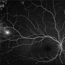 Proliferative Sickle Cell Retinopathy
Jan 29 2021 by Olivia Rainey
Ultra-widefield fluorescein angiogram of a 24-year-old female with proliferative sickle cell retinopathy affecting her right eye. The physician's interpretation of the fluorescein shows seafan neovascularization superotemporally, AV anastomeses, and good peripheral laser. He performed scatter PRP OD on 12/2/2020 to nonperfusion in temporal far periphery. The patient's 12/2020 Hb electrophoresis came back showing Hb SC (rather than sickle cell trait). Patient was born at full term, but she reports that her mother used drugs while pregnant with the patient. The patient also mentioned that her niece has full sickle cell disease and her grandmother, mother, and sibling all have condition on the sickle cell spectrum.
Photographer: Olivia Rainey, OCT-C, COA
Imaging device: Optos California
Condition/keywords: fluorescein angiogram (FA), fluorescein leakage, neovascularization (NV), neovascularization elsewhere (NVE), Optos, sea fan, sickle cell retinopathy
-
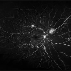 Proliferative Diabetic Retinopathy
Jan 29 2021 by Olivia Rainey
Ultra-widefield fluorescein angiogram of a 65-year-old male with proliferative diabetic retinopathy affecting his right eye. The patient's diabetic retinopathy has progressed significantly since he was last seen in 2014. It was recommended to begin antiVEGF to control DME followed by laser treatment OU.
Photographer: Olivia Rainey, OCT-C, COA
Imaging device: Optos California
Condition/keywords: anti-VEGF, diabetes, diabetic macular edema, fluorescein angiogram (FA), fluorescein leakage, neovascularization (NV), neovascularization elsewhere (NVE), non-perfusion, Optos, proliferative diabetic retinopathy (PDR), ultra-wide field imaging
-
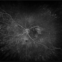 Retinal Vasculitis
Jan 29 2021 by Olivia Rainey
Ultra-widefield fluorescein angiogram of a 71-year-old female with retinal vascultitis affecting her left eye. The patient had a bilateral TAB performed in 5/2019 and 7/2019 which showed heterozygous for prothrombin mutation, but this would correlate with venous thromboembolism rather than arterial embolism. She has a history of elevated ESR and CRP. She experienced a new onset of vitreous debris on 6-1-2020 combined with findings of severe vascular occlusion/vasculitis with diffuse leakage on angiography suggests underlying inflammatory component rather than prothrombotic occlusion secondary to prothrombin mutation. There is persistent significant vasculitis noted on angiography on 1/25/2021.
Photographer: Lauren Schuler
Imaging device: Optos California
Condition/keywords: fluorescein angiogram (FA), fluorescein leakage, left eye, non-perfusion, Optos, peripheral retinal vasculitis, retinal vasculitis, ultra-wide field imaging
-
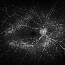 Peripheral Drusen
Jan 19 2022 by Olivia Rainey
Ultra-widefield fluorescein angiogram of an 83-year-old female with Peripheral Drusen affecting both eyes. The patient presented on 1/19/2022 with slightly decreased vision since her last appointment. Her vision was sc20/25-2 in the right eye. The physician did not believe that the peripheral drusen represents AMD and recommended monitoring. The patient also had mild diabetic retinopathy at the time of her visit.
Photographer: Olivia Rainey, OCT-C, COA
Imaging device: Optos California
Condition/keywords: background diabetic retinopathy (BDR), fluorescein angiogram (FA), Optos, Peripheral drusen, staining, ultra-wide field imaging
-
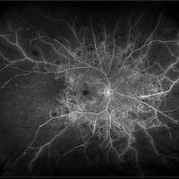 Central Retinal Vein Occlusion with Severe Retinal Ischemia
Jan 19 2022 by Olivia Rainey
Ultra-widefield fluorescein angiogram of a 56-year-old male with a Central Retinal Vein Occlusion with Severe Retinal Ischemia affecting his right eye. The patient presented on 1/19/2022, sc20/20-2 vision in the right eye. The patient has had a good response to Eylea with complete resolution of edema. The physician is considering PRP to ischemic periphery in the future and given the degree of ischemia in both eyes, she recommends that the patient's PCP check carotid Doppler US.
Photographer: Olivia Rainey, OCT-C, COA
Imaging device: Optos California
Condition/keywords: central retinal vein occlusion (CRVO), FA late phase, fluorescein angiogram (FA), ischemic CRVO, Optos, retinal ischemia, ultra-wide field imaging
-
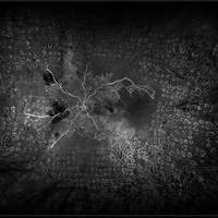 Central Retinal Vein Occlusion with Retinal Neovascularization
Jan 19 2022 by Olivia Rainey
Ultra-widefield fluorescein angiogram of a 56-year-old male with a Central Retinal Vein Occlusion with Retinal Neovascularization affecting his left eye. The patient presented on 1/19/2022 with scNLP vision in the left eye. The patient has good PRP, however areas of ischemia still remain untreated by laser. He also has severe neovascular glaucoma contributing to his poor vision.
Photographer: Olivia Rainey, OCT-C, COA
Imaging device: Optos California
Condition/keywords: central retinal vein occlusion (CRVO), FA early phase, fluorescein angiogram (FA), hemorrhage, ischemic CRVO, left eye, neovascular glaucoma, Optos, pan-retinal photocoagulation (PRP), retinal ischemia, retinal neovascularization, ultra-wide field imaging
-
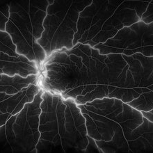 Central Retinal Vein Occlusion
Jan 21 2022 by Olivia Rainey
Ultra-widefield fluorescein angiogram of a 23-year-old female with a Central Retinal Vein Occlusion affecting her left eye. The patient presented on 12/22/2021 cc20/40-2 vision in the left eye. The patient reported recent trauma of being hit with a fist on both sides of face followed by vision loss. The patient has history of Hashimoto's thyroid disease. The following labs have been ordered, PT, PTT, CBC, antithrombin III activity, protein C, protein S, Factor V Leiden mutation, Prothrombin (G20210A), lipid panel, HbA1c, quantiferon gold, RPR, and CXR.
Photographer: Olivia Rainey, OCT-C, COA
Imaging device: Optos California
Condition/keywords: central retinal vein occlusion (CRVO), disc leakage, fluorescein angiogram (FA), fluorescein leakage, left eye, non-ischemic central retinal vein occlusion (CRVO), Optos, trauma, ultra-wide field imaging
-
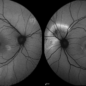 Central Serous Chorioretinopathy
Jan 25 2022 by Olivia Rainey
Widefield fundus autofluorescence of a 60-year-old male with Central Serous Chorioretinopathy affecting both eyes. Chronic history of CSR followed with observation without treatment prior to presenting at our office. The physician noted significant findings on exam and imaging with multifocal areas of inactive and active changes in the right eye and subfoveal subretinal fluid with recent visual decline in the left eye. There are hyper and hypoautofluorescent changes, consistent with CSR.
Photographer: Olivia Rainey, OCT-C, COA
Imaging device: Heidelberg Spectralis
Condition/keywords: 55-degrees, central serous chorioretinopathy (CSCR), central serous retinopathy (CSR), chronic central serous chorioretinopathy (CSCR), fundus autofluorescence (FAF), heidelberg spectralis, left eye
-
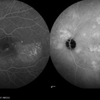 Central Serous Chorioretinopathy
Jan 25 2022 by Olivia Rainey
Late phase widefield fluorescein angiography of a 60-year-old male with Central Serous Chorioretinopathy. Chronic history of CSR followed with observation without treatment prior to presenting at our office. The physician noted subfoveal subretinal fluid with recent visual decline. FA shows multifocal leakage and ICG shows hypercyanescence. OCTA, ICG, and FA consistent with CSR, and without concern for CNVM thus will observe without anti-VEGF at this time. PDT therapy recommended.
Photographer: Olivia Rainey, OCT-C, COA
Imaging device: Heidelberg Spectralis
Condition/keywords: 55-degrees, central serous chorioretinopathy (CSCR), central serous retinopathy (CSR), chronic central serous chorioretinopathy (CSCR), fluorescein angiogram (FA), heidelberg spectralis, indocyanine green (ICG) angiography, left eye
-
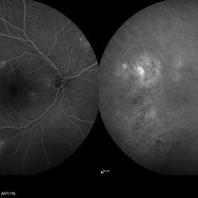 Central Serous Chorioretinopathy
Jan 25 2022 by Olivia Rainey
Late phase widefield fluorescein angiography of a 60-year-old male with Central Serous Chorioretinopathy. Chronic history of CSR followed with observation without treatment prior to presenting at our office. The physician noted significant findings on exam and imaging with multifocal areas of inactive and active changes OD. FA shows superotemporal macular leakage, subtle inferonasal macular leakage and staining as well as multifocal hypercyanescence on ICG. Fortunately foveal sparing and thus observation is recommended at this time OD.
Photographer: Olivia Rainey, OCT-C, COA
Imaging device: Heidelberg Spectralis
Condition/keywords: 55-degrees, central serous chorioretinopathy (CSCR), central serous retinopathy (CSR), chronic central serous chorioretinopathy (CSCR), fluorescein angiogram (FA), fluorescein leakage, heidelberg spectralis, indocyanine green (ICG) angiography, late phase
-
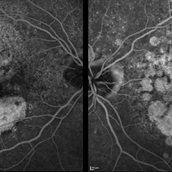 Macular Degeneration with Extensive Geographic Atrophy
Jan 26 2022 by Olivia Rainey
Heidelberg Spectralis fluorescein angiography of a 94-year-old woman with Macular Degeneration affecting both eyes. The FA reveals transmission defects consistent with RPE changes and geographic atrophy of RPE of both eyes, as well as window defects consistent with peripheral scarring in the right eye. The patient's vision was Dcc20/70 in both eyes at the visit the images were taken.
Photographer: Olivia Rainey, OCT-C, COA
Imaging device: Heidelberg Spectralis
Condition/keywords: 30-degrees, choroidal neovascularization (CNV), dry age-related macular degeneration (dry AMD), early phase, fluorescein angiogram (FA), geographic atrophy, heidelberg spectralis, macular degeneration, neovascular age-related macular degeneration (AMD)
-
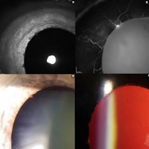 Iris Vascular Tuft
Jul 5 2022 by Olivia Rainey
Anterior segment imaging of a 66-year-old male with Vascular Disorders of Iris and Ciliary Body affecting his right eye. The physician stated that the findings are most consistent with a benign vascular tuft at the pupillary margin. The patient presented at the office with 20/20 vision in both eyes and had no ocular complaints at the time of his appointment.
Photographer: Olivia Rainey, OCT-C, COA
Imaging device: Heidelberg Spectralis, Slit Lamp with Samsung Galaxy 7
Condition/keywords: anterior segment, fluorescein angiogram (FA), heidelberg spectralis, infrared image, near infrared autofluorescence (NIRAF), slit lamp photo, vascular anomaly, vascular disorders of iris and ciliary body, vascular tuft
-
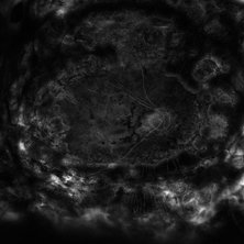 Combined Tractional and Rhegmatogenous Retinal Detachment
Jan 30 2023 by Olivia Rainey
Ultra-widefield fluorescein angiography of a combined tractional and rhegmatogenous retinal detachment repair affecting the left eye. The retina is attached following silicone oil placement during most recent surgery. The patient was seeing CF at the time the image was taken.
Photographer: Olivia Rainey, OCT-C, COA
Imaging device: Optos California
Condition/keywords: diabetes, diabetic macular edema, diabetic retinopathy, fluorescein angiogram (FA), hyperfluorescence, right eye, scleral buckle, silicone oil, tractional retinal detachment, ultra-wide field imaging, ultra-widefield image
-
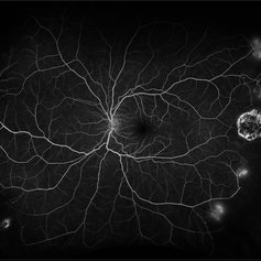 Proliferative Sickle Cell Retinopathy
Feb 1 2023 by Olivia Rainey
Ultra-widefield fluorescein angiography of a 25-year old male with Proliferative Sickle Cell Retinopathy affecting his left eye. Patient stated that he was born with Sickle disease (SC), and has yearly eye exams. He noted no vision concerns over the last year but has typically experienced sickle attacks about 1-2 per year. The physician noted that the fluorescein obtained showed peripheral nonperfusion affecting the patient's nasal and temporal retina as well as neovascularization affecting his left eye more than his right. He recommended pan retinal photocoagulation in his left eye for his temporal and nasal retina, as as well as his right eye following.
Photographer: Olivia Rainey, OCT-C, COA
Imaging device: Optos California
Condition/keywords: early phase, fluorescein angiogram (FA), fluorescein leakage, left eye, neovascularization (NV), proliferative retinopathy, sickle cell retinopathy, ultra-wide field imaging, ultra-widefield image
-
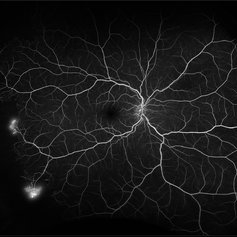 Proliferative Sickle Cell Retinopathy
Feb 1 2023 by Olivia Rainey
Ultra-widefield fluorescein angiography of a 25-year old male with Proliferative Sickle Cell Retinopathy affecting his right eye. Patient stated that he was born with Sickle disease (SC), and has yearly eye exams. He noted no vision concerns over the last year but has typically experienced sickle attacks about 1-2 per year. The physician noted that the fluorescein obtained showed peripheral nonperfusion affecting the patient's nasal and temporal retina as well as neovascularization affecting his left eye more than his right. He recommended pan retinal photocoagulation in his left eye for his temporal and nasal retina, as as well as his right eye following.
Photographer: Olivia Rainey, OCT-C, COA
Imaging device: Optos California
Condition/keywords: early phase, fluorescein angiogram (FA), fluorescein leakage, neovascularization (NV), non-perfusion, proliferative retinopathy, right eye, sickle cell retinopathy, ultra-wide field imaging, ultra-widefield image
A project from the American Society of Retina Specialists