 Initializing download.
-
 By Olivia Rainey
Retina Specialists of Michigan
Co-author(s): Thomas Aaaberg, MD - Uploaded on Apr 30, 2019.
- Last modified by Olivia Rainey on Apr 3, 2020.
- Rating
- Appears in
- Miscellaneous
- Condition/keywords
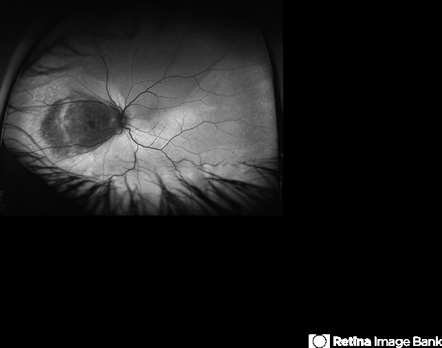
- autofluorescence imaging, Optos, ultra-wide field imaging, left eye
- Photographer
- Olivia Rainey
- Imaging device
-
Fundus camera
Optos - Description
- Ultra-wide field fundus autofluorescence of a 67-year-old male with serous retinal detachment, secondary to a large choroidal melanoma affecting the left eye. Patient reported a curtain affecting his superior field for about 2-3 months prior to examination. Patient elects radiation bracytherapy with a guarded visual expectation secondary to the location of the tumor, touching the optic disc.


")


