 Initializing download.
-
 By Olivia Rainey
Retina Specialists of Michigan
Co-author(s): Greg Bever, MD and Olivia Rainey, OCT-C, COA - Uploaded on Oct 22, 2020.
- Last modified by Caroline Bozell on Oct 23, 2020.
- Rating
- Appears in
- Miscellaneous
- Condition/keywords
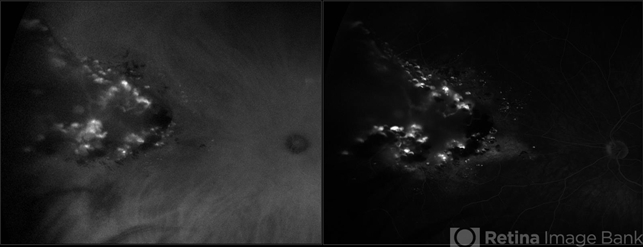
- ultra-wide field imaging, cavernous hemangioma of the retina, Optos, fluorescein angiogram (FA), indocyanine green (ICG) angiography, late phase, left eye
- Photographer
- Becca Harris
- Imaging device
-
Fundus camera
Optos California - Description
- Ultra-widefield fluorescein and ICG angiogram of a 31-year-old male presenting with a retinal cavernous hemangioma affecting his left eye. Patient was 18-years-old when he was diagnosed with a retinal cavernous hemangioma. He has had a few episodes of vitreous hemorrhages since then. His vision was 20/20-1 in both eyes.
")


")
")
")
")

")