 Initializing download.
-
 By Olivia Rainey
Retina Specialists of Michigan
Co-author(s): Thomas Aaberg, MD - Uploaded on May 27, 2020.
- Last modified by Olivia Rainey on Jun 3, 2020.
- Rating
- Appears in
- Miscellaneous
- Condition/keywords
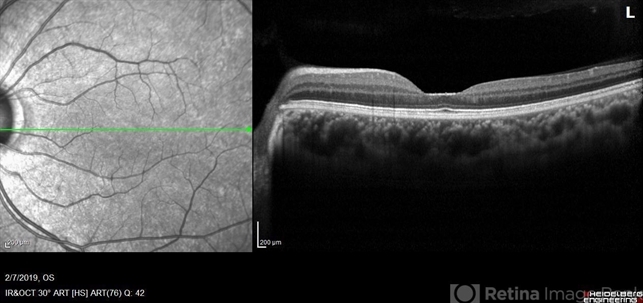
- plateau fovea, inner retinal thinning, Alports disease, optical coherence tomography (OCT), Heidelburg Spectralis, left eye
- Photographer
- Olivia Rainey, OCT-C, COA
- Imaging device
-
Optical coherence tomography system
Heidelberg Spectralis - Description
- Optical coherence tomography of the left eye of a 20-year-old male with Alport Syndrome. The patient did not present with any ocular or visual symptoms, yet the distinct "plateau contour" of his fovea was noted on OCT during his visit. The patient presented with 20/25 vision at the time of his visit. There was myelinated nerve fiber layer noted in both eyes, but these features had remained stable from his appointment three years prior. The physician noted that myelinated nerve fiber was a congenital change, and had not affected his vision or health of the eye, nor is a feature of Alport Syndrome.

")


")
")
")


