 Initializing download.
-
 By Olivia Rainey
Retina Specialists of Michigan
Co-author(s): Thomas Aaaberg, MD - Uploaded on Nov 11, 2019.
- Last modified by Olivia Rainey on Apr 3, 2020.
- Rating
- Appears in
- Miscellaneous
- Condition/keywords
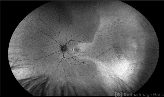
- fundus autofluorescence (FAF), ultra-wide field imaging, Optos, left eye, choroidal neovascularization (CNV), retinal detachment of the macula, chronic retinal detachment, montage
- Photographer
- Olivia Rainey
- Imaging device
-
Fundus camera
Optos California - Description
- Ultra-wide field pseudocolor photograph of a 42-year-old female with a long-standing, macula-off retinal detachment affecting her left eye. Patient was unaware of vision loss until testing her visual acuity and she denied seeing flashing lights. Patient decided to proceed with scleral buckling. The CNV is potentially secondary the retinal detachment, but may be myopic related or idiopathic. The CNV appears fibrotic and inactive. The patient was warned that this will absolutely limit how much vision she recovers once the retina is reattached.


")

")

