File number: 28420
Comments
-
 Suber S. Huang, MD, MBA, FASRS (September 21 2018)
Excellent capture in the young patient. Consider adding clinical course and classifying under tumors/neoplasia. Thank you for sharing!
Sign in to comment.
 Initializing download.
-
 By Olivia Rainey
Retina Specialists of Michigan
Co-author(s): Thomas Aaaberg, MD - Uploaded on Jul 13, 2018.
- Last modified by Olivia Rainey on Apr 3, 2020.
- Rating
- Appears in
- Miscellaneous
- Condition/keywords
- retinocytoma, ultra-wide field imaging, pseudocolor, Optos
- Photographer
- Olivia Rainey
- Imaging device
-
Fundus camera
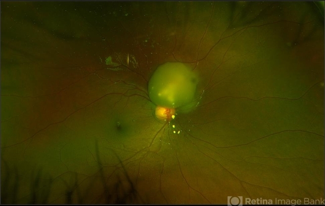
Optos - Description
- Ultra-wide field pseudocolor image of a 5-year-old male with a retinocytoma affecting his right eye. The retinal tumor has associated calcium which looks suspicious for retinoblastoma. However, there are a number of atypical features which raise the possibility of a masquerade tumor.


![Choroidal Nevus[004]](/Content/imagebank/Choroidal-Nevus-004---thumb.jpg/image-square;max$79,0.ImageHandler "Choroidal Nevus[004]")


