 Initializing download.
-
 By Olivia Rainey
Retina Specialists of Michigan
Co-author(s): Liliya Sutherland, DO - Uploaded on Jul 26, 2019.
- Last modified by Olivia Rainey on Apr 3, 2020.
- Rating
- Appears in
- Miscellaneous
- Condition/keywords
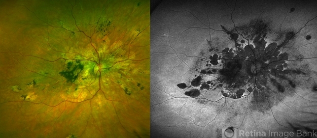
- pseudoxanthoma elasticum (PXE), choroidal neovascularization (CNV), angioid streaks, atrophic central lesion, autofluorescence imaging, ultra-wide field imaging, Optos, pigmentary retinal dystrophy, hypofluorescent lesions, macular atrophy
- Photographer
- Olivia Rainey
- Imaging device
-
Fundus camera
Optos - Description
- Ultra-wide field color/autofluorescence comparison of a 54-year-old female with severe macular atrophy secondary to pseudoxanthoma elasticum. Patient developed choroidal neovascularization that does not warrant treatment due to the patient's poor visual acuity. Patient has significantly attenuated ERG. Genetic testing is recommended to rule out retinitis pigmentosa. The mechanism is yet to be determined, but there is a thought that ABCC6 mutation causes alteration of plasma lipid composition may be implicated in the systemic changes observed in PXE.


")


")