Search results (35 results)
-
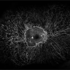
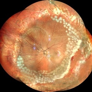
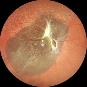 The Bullet Ridden Retina
Feb 17 2024 by SHISHIR VERGHESE, MS, FVRS, FAICO (Retina)
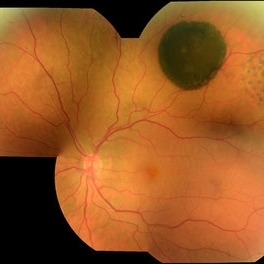
Fundus image obtained of a case of lasered branch retinal vein occlusion (BRVO) with fibrovascular proliferation (FVP) where the laser marks have given way to multiple small retinal holes due to traction from the same.
Photographer: DIVYA SHAJI
Imaging device: NIDEK MIRANTE
Condition/keywords: BRVO, chronic retinal detachment
-
 New Retinal Detachment 6w s/p RD repair
Nov 16 2023 by Virginia Gebhart
13 year old male presented with new blind spot 6 weeks s/p RD repair with cryo/scleral buckle/prophylaxis laser with gas bubble. New RD involving the macula, posterior to scleral buckle, secondary to PVD. Small gas bubble remaining. Pt was brought back to OR for repeat PPV and silicone oil repair
Photographer: Virginia Gebhart
Imaging device: Optos
Condition/keywords: gas bubble, Retinal Detachment, retinal detachment of the macula, scleral buckle
-
 Macula Off Retinal Detachment
Mar 22 2023 by Zach Seim
An ultra-widefield fundus image of a 65 year old male with a Macula Off Retinal Detachment. Patient's vision at the time of the image was CF at 6 Feet and surgical options were discussed. Fluid-gas exchange was performed without complications.
Photographer: Zach Seim
Imaging device: Optos California
Condition/keywords: left eye, macula off retinal detachment, OPTOS CALIFORNIA, scanning laser ophthalmoscope, ultra-widefield image
-
 High risk Proliferative Diabetic Retinopathy treated with Pan Retinal Photocoagulation
Nov 5 2022 by Somnath Chakraborty, MD
A Fundus Photo Montage of 43 year old Asian Male with Type 2 Diabetes Mellitus since 7 years who presented with sudden onset diminition of vision in his Left eye. BCVA OS was 20/200. He was diagnosed to have Pre retinal bleed due to Proliferative Diabetic Retinopathy and was treated with Pan Retinal Photocoagulation. This image shows a large neo-cascular frond at the disc and superior to it with Pre-retinal bleed and Fresh laser marks along
Photographer: Pulak Roy
Condition/keywords: diabetic blindness, diabetic retinopathy vitrectomy study (DRVS), fresh laser burns, laser photocoagulation, preretinal hemorrhage, proliferative diabetic retinopathy (PDR)
-
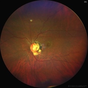 Optic disc pit
Mar 21 2022 by T. P . VIGNESH, MBBS,MS
Fundus photo of Left eye of a 55 year male patient revealing optic disc pit with temporal barrage laser marks and foveal schisis with RPE atrophic changes.
Photographer: Bharathi Singaravel
Imaging device: Zeiss Clarus
Condition/keywords: Optic disc pit
-
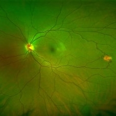 Isolated Retinal Capillary Hemangioblastoma
Mar 11 2022 by Bryon R McKay, MD, PhD, FRCSC, DRCPSC - Retina
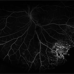
Optos widefield fundus photograph and IVFA of a 23-year-old female with asymptomatic isolated retinal capillary hemangioblastoma without exudation. IVFA demonstrates some mild late leakage. The tumor measures 1.5mm and was effectively ablated with laser photocoagulation.
Imaging device: Optos
Condition/keywords: retina capillary hemangioblastoma
-
 Coloboma involving the Optic nerve, Retina, and Choroid
Dec 6 2021 by Jesus Lozano, MD
78-year-old woman after prophylactic laser photocoagulation (PLP) for her RE Coloboma involving the optic nerve, retina, and choroid. At 6 month follow up, patient preserved her FC vision as it was before the procedure. Retina attached.
Photographer: Yair Bet Yosef, Hadassah Medical Center. Israel
Imaging device: Optos Silverstone fundus image
Condition/keywords: coloboma, coloboma of choroid, coloboma of macula, coloboma of optic disc, PLP, prophylactic photocoagulation
-
 Diabetic Retinopathy Treated with PRP Laser
Jun 8 2021 by Ronald Coriasso
Diabetic retina treated with complete 360 PRP laser, taken during fluorescein angiogram.
Photographer: Ronald Coriasso
Imaging device: OPTOS
Condition/keywords: pan-retinal photocoagulation (PRP)
-
 Attached Retina in a Silicon Oil Filled Buckled Eye with Retinectomy
Apr 17 2021 by Navneet Mehrotra, DNB
Fundus photograph of a 12-year-old boy operated for re retinal detachment with PVR showing attached retina with fresh and old laser marks, silicon oil filled and relaxing retinectomy.
Photographer: Dr Nivesh Gupta, Retina Foundation
Imaging device: Nidek mirante
Condition/keywords: proliferative vitreoretinopathy (PVR), retinectomy, scleral buckle
-
 Sickle Cell Retinopathy
Feb 15 2021 by Kim Barrett
24-year-old female with Sickle Cell Retinopathy, stage 3. She confirms she has the trait as well as her grandmother, mother and a sibling. She has seafan neovascularization superotemporal OD. Current VA is 20/20. Photo is pre-PRP laser with areas of non-profusion temporally.
Photographer: Kim Barrett C.O.A. Retina Specialist of Michigan, Grand Rapids, MI
Imaging device: Optos California
Condition/keywords: neovascularization (NV), pan-retinal photocoagulation (PRP), sickle cell retinopathy, stage 3, trait
-
 Retinopathy of Prematurity S/P Laser Complications
Apr 30 2020 by Giselle DeOliveira
Gonio photograph of 13-year-old female with retinopathy of prematurity, s/p laser complications.
Photographer: Giselle DeOliveira
Imaging device: Retcam III
Condition/keywords: laser, retinopathy of prematurity (ROP)
-
 Retinal Detachment with Giant Retinal Tear and Macular Hole
Jan 6 2020 by MATTEO FORLINI, MD
A 61-year-old-male patient presented with sudden diminution of vision in the right eye due to retinal detachment with giant retinal tear and macular hole. Best corrected visual acuity (BCVA) at presentation was 20/200. A 23 G vitrectomy was performed. The edges of the tear were unrolled and complete retinal re-attachment under PFCL was achieved. A 360 degree intraoperative endolaser was performed on the peripheral retina as well as around the edges of the tears. PFCL was exchanged with silicone oil 5000cs as final tamponade. At six-months follow-up retina was attached and macular hole was repaired. Best-corrected visual acuity is 20/125 at present.
Photographer: Matteo Forlini MD, San Marino Hospital, Republic of San Marino
Condition/keywords: full thickness macular hole, giant retinal tear, silicone oil
-
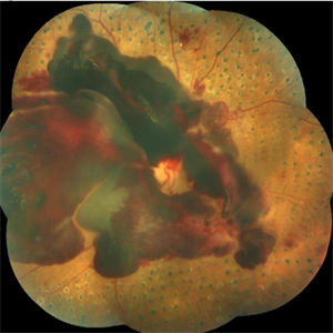
 Retinoblastoma Stage 5 After One Cycle of Systemic Chemotherapy and Laser Ablation
Sep 17 2019 by Sophia El Hamichi, MD
A 1-year-old patient with stage 5B retinoblastoma, fundus after one cycle of systemic chemotherapy and laser ablation.
Photographer: Abby Orcutt-Hayes, Murray Ocular Oncology and Retina
Condition/keywords: chemoreduction, laser photocoagulation, montage, retinoblastoma, stage 5
-
 Coloboma
Oct 2 2019 by John S. King, MD
27-year-old white female with bilateral, isolated, inferior, chorioretinal colobomas; she has a history of retinal laser anterior to the edge of the coloboma OD secondary to a limited RD. This is the right eye.
Photographer: Shelly Blair
Imaging device: Optos CA
Condition/keywords: coloboma of choroid
-
 CHRPE
CHRPE
Oct 8 2019 by DIEGO TOLENTINO
CHRPE plus laser barricade around retinal break
Photographer: Diego Tolentino
Condition/keywords: congenital hypertrophy of the retinal pigment epithelium (CHRPE)
-
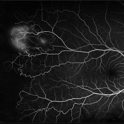
 Coats' Disease
Jul 16 2019 by Kim Barrett
Ultra-wide field fluorescein angiogram of a 23-year-old male with Coats' disease, presented with distorted vision affecting his left eye. He reported seeing flashes and floaters since January of 2019, but the flashes had resolved. He was treated with Intravitreal Preservative Free Triamcinolone in the office and scheduled for PRP laser in the near future.
Photographer: Kim Barrett
Imaging device: Optos
Condition/keywords: Coats' disease, fluorescein angiogram (FA), fluorescein leakage, inferior retina, ischemia, left eye, Optos, ultra-wide field imaging
-
 Laser Induced BRAO in IRVAN Syndrome
May 3 2019 by Deependra Vikram Singh, MD FASRS
Fundus photograph of a 26-year-old man with IRVAN syndrome referred for vitreous surgery in OS for secondary rhegmatogenous retinal detachment. OD has received laser photocoagulation for capillary nonperfusion areas and retinal artery macroaneurysm associated with retinal vasculitis. Fundus photograph of OD shows laser induced nasal BRAO. Case re-emphasizes why laser for macroaneurysm should be avoided in cases with IRVAN.
Photographer: Deependra V Singh, Eye-Q Superspecialty Eye Hospitals. Gurugram, India
Imaging device: Zeiss Visucam 500
Condition/keywords: arteriolar macroaneurysm, branch retinal artery occlusion (BRAO), laser photocoagulation
-
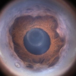
 Optos Picture With Speculum: Dislocated Natural Lens
Oct 9 2018 by John S. King, MD
55-year-old white female with history of pathologic myopia+, lattice (laser), SB OU (1990s), and dislocated natural lenses OU that had been watched for years. In the fellow eye she developed phacolytic glaucoma and a PPV, PPL was performed. Plan for both eyes are monitoring. I wanted to get a good picture of her lens today with the optos machine, as the other pics had artifact from the lower lid. It worked out well to use a speculum in the left eye. Vision cc is 20/400 J1+ OD and 20/40 J2 OS; aphakic OU; vitreous clear OD; dislocated lens OS (see pic); retinas attached.
Photographer: Maisee Yang
Imaging device: Optos California
Condition/keywords: dislocated crystalline lens, pathologic myopia, scleral buckle, staphyloma
-
 Laser Pointer Maculopathy
Jul 1 2018 by John S. King, MD
9-year-old with two month history of decreased vision that has improved some over time. 20/20 OD and 20/30 OS. Further prompting, he admitted to playing with "toy lasers" with his friends recently. He also looked at the eclipse, but used protective eye wear. Photos show small RPE defects characterized by a hyperpigmented center with hypopigmented halo.
Photographer: Karin Aletter
Imaging device: Topcon
Condition/keywords: laser pointer maculopathy, laser pointer retinopathy, maculopathy
-
 Subhyaloid Hemorrhage, Proliferative Diabetic Retinopathy
May 31 2018 by awaneesh m upadhyay, MBBS, DNB
Right eye fundus photography of a 63-year-old male came with sudden onset defective vision with history of laser photocoagulation done for proliferative diabetic retinopathy.
Photographer: Dr Awaneesh Upadhyay
Condition/keywords: laser photocoagulation, proliferative diabetic retinopathy (PDR), subhyaloid hemorrhage
-
 Giant Retinal Tear
May 1 2018 by Talia R Kaden, MD
Fundus Photograph of the right eye of a 51-year-old taxi driver with a newly lasered giant retinal tear. He had a history of a retinal detachment from a giant retinal tear in his left eye.
Photographer: Maria Pei, Bellevue Hospital, New York University, NY
Imaging device: Topcon TRC 501x
Condition/keywords: barrier laser, fresh laser burns, giant retinal tear
-
 Valsalva Retinopathy
Apr 6 2018 by Jun Dong Dong
A 45-year-old male with no definite history of activities which can suddenly increase the intrathoracic pressure refused the laser treatment and chose the observation.
Photographer: Jun Dong, Shanghai Aier Eye Hospital
Condition/keywords: subhyaloid hemorrhage
-
 Capillary Nonperfusion
Apr 12 2018 by SUSHIL BHATT
OPTOS ultra wide field angiogram of an 45 years old diabetic male patient shows capillary nonperfusion areas with inadequate laser.
Photographer: Bhatt Sushil PGIMER chandigarh INDIA
Imaging device: OPTOS Ultra wide Field
Condition/keywords: capillary nonperfusion
-
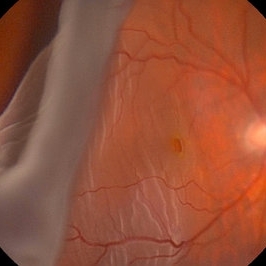
 Horse Shoe Tear
Sep 16 2017 by Purva Patwari
Asymptomatic horse shoe tear found on preoperative cataract assessment of a 54-year-old male patient. Laser barrage was done and he underwent Phacoemulsification surgery a month later.
Photographer: Dr Purva Patwari,Patwari Retina Center,Ahmedabad,India
Imaging device: Zeiss Visucam 500
-
 Macula-Sparing GRT RRD
Jul 6 2017 by Andrew A. Moshfeghi, MD, MBA, FASRS
Wide-field fundus photograph of a 43-year-old myopic man with a history of lattice retinal degeneration status posterior barrier laser performed elsewhere who presented with a giant-retinal tear associated retinal detachment of the right eye.
Photographer: Jay Jiang, University of Southern California Roski Eye Institute
Imaging device: Optos California
Condition/keywords: acute retinal detachment, giant retinal tear, lattice degeneration
 Loading…