Search results (832 results)
-
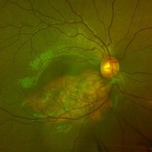
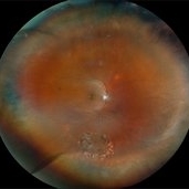
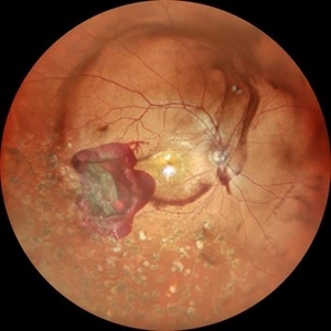 Subhyaloid Hemorrhage With Dispersed Vitreous Hemorrhage in a Case of Old Lasered Branch Retinal Vein Occlusion
Jul 12 2025 by Akansha Sharma
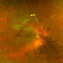
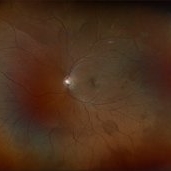
Color fundus photograph of a 32 year old hypertensive and diabetic male with subhyaloid hemorrhage with dispersed vitreous hemorrhage in a case of old lasered branch retinal vein occlusion.
Photographer: DR. AKANSHA SHARMA
Condition/keywords: branch retinal vein occlusion (BRVO), laser photocoagulation, SHH, subhyaloid hemorrhage, VH, vitreous hemorrhage
-
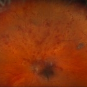
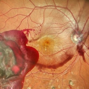 Subhyaloid Hemorrhage With Dispersed Vitreous Hemorrhage in a Case of Old Lasered Branch Retinal Vein Occlusion
Jul 12 2025 by Akansha Sharma
Color fundus photograph of a 32 year old hypertensive and diabetic male with subhyaloid hemorrhage with dispersed vitreous hemorrhage in a case of old lasered branch retinal vein occlusion.
Photographer: DR. AKANSHA SHARMA
Condition/keywords: branch retinal vein occlusion (BRVO), laser photocoagulation, SHH, subhyaloid hemorrhage, VH, vitreous hemorrhage
-
 Repaired Retinal Detachment
Jun 24 2025 by Kimberly Wakester
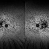
Optomap RGB of an 45-year-old woman with a repaired retinal detachment in the right eye. The operative eye is doing well three-month s/p surgery. Retina is attached 360 on SB. There is resolving residual SRF at 6:00. Discussed the possible need for added laser. Will continue to observe and will return in 3 months for follow up exam.
Photographer: Kimberly Wakester, COA, OCT-C
Imaging device: Optos California
Condition/keywords: repaired RD, scleral buckle
-
 Laser Marks
Jun 4 2025 by Paulina Araujo
The 55-degree central fundus photograph of the right eye shows evidence of old laser marks along the inferior temporal arcade.
Photographer: Paulina D.Araujo Martínez, Asociación para Evitar la Ceguera en México I.A.P., Hospital Dr Luis Sánchez Bulnes.
Condition/keywords: Attached retina with Endolaser marks, laser
-
 The Dread of the Crimson Red
Jun 2 2025 by Thirumalesh Mochi Basavaraj, MD
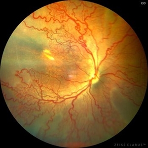
Fundus photograph of a 64 year man post laser depicting a regressed NVD in the superior aspect and a Persistent Neo vascularization in the inferior aspect
Photographer: Vivek
Condition/keywords: Neovascularisation at the Disc (NVD), proliferative diabetic retinopathy (PDR)
-
 Aggressive Posterior Retinopathy of Prematurity (APROP)
May 16 2025 by KANWALJEET HARJOT MADAN, M.S. (Ophthalmology); FAICO (Vitreous - Retina)
This is the fundus picture of right eye of a premature neonate depicting Aggressive Posterior Retinopathy of Prematurity (APROP). It is a severe rapidly progressing form of retinopathy that can lead to vision loss and blindness. It requires prompt diagnosis and treatment in the form of anti-VEGF agents and laser photocoagulation.
Photographer: Dr. Kanwaljeet Harjot Madan, Thind Eye Hospital, Jalandhar City (Punjab) INDIA.
Imaging device: Zeiss Clarus
Condition/keywords: Oxygen Exposure, retinopathy of prematurity (ROP)
-
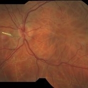
 High Risk Proliferative Diabetic Retinopathy with Sub-hyaloid Hemorrhage
May 13 2025 by Anupama Kiran Kumar
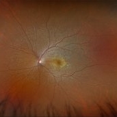
This image shows a case of high risk proliferative diabetic retinopathy. The retina is unlasered with a taut posterior hyaloid and a sub-hyaloid hemorrhage at the macula and along the arcades ,sparing the fovea.
Photographer: Mr Pratap
Imaging device: Mirante SLO/OCT (Nidek Co., Gamagori, Japan)
Condition/keywords: Diabetes, Diabetic Retinopathy, proliferative diabetic retinopathy (PDR), subhyaloid hemorrhage
-
 Bot Fly Larvae
Apr 29 2025 by Daniela Bogenschutz
57 year-old male referred for decreased vision from optometrist. His only complaint was floaters and the letters were moving on the screen. He had never been out of the country, but is a farmer. Upon examination, our retina specialist found a bot fly larvae with numerous tracks made in this patient's retina. Patient was treated with laser to kill the larvae which was successful and he has been monitored yearly.
Photographer: Daniela Bogenschutz, OSC; Retina Consultants of Carolina, P.A.
Imaging device: Topcon
Condition/keywords: Bot Fly Larvae
-
 Hourglass in an Eye
Apr 22 2025 by KRISHNENDU NANDI, MS
A twenty-five-year-young male presented with a decrease in vision in the right eye following a blunt trauma with a football. On examination the BCVA in the right eye was CFCF and the left eye was 6/6, N6. The anterior segment was within normal limits. AT was 12 and 10 mm of Hg in the right and left eyes, respectively. Fundus examination reveals subhyaloid haemorrhage in the right eye with an attached retina. The fundus of the left eye was within normal limits. YAG laser hyaloidotomy was done with an energy of 2 mJ in the right eye. After 3 weeks the BCVA in the right eye improved to 6/9, N6.
Photographer: Dr. Krishnendu Nandi
Imaging device: Topcon
Condition/keywords: Trauma, YAG HYALOIDOTOMY, Young Male
-
 Choroidal Osteoma
Apr 17 2025 by Gustavo Uriel Fonseca Aguirre
Scanning laser ophthalmoscopy reveals a well-circumscribed, yellowish-white choroidal osteoma overlying the macular region and extending into the inferior temporal vascular arcade. Retinal vessels course normally over the tumor surface, with no evidence of subretinal fluid or hemorrhage. The surrounding retina shows preserved architecture without secondary degenerative changes.
Photographer: Gustavo U. Fonseca Aguirre, Hospital Conde de Valenciana, Ciudad de México
Condition/keywords: choroidal osteoma, macular choroidal osteoma
-
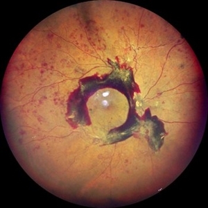
 Superior Rhegmatogenous Retinal Detachment (RRD) in the Right Eye, With a Retinal Tear Located Between the 1 and 2 O'clock Positions
Apr 4 2025 by Cesar Orlando Oviedo Vera
A 45-year-old male patient presented with a sudden onset of decreased visual acuity in the right eye, with a 24-hour progression. Upon examination, Image 1 revealed a superior rhegmatogenous retinal detachment in the right eye, with a retinal tear located between the 1 and 2 o'clock positions. Image 2: Pneumatic retinopexy by intravitreal injection of Sulfur Hexafluoride gas (SF6) at the time of diagnosis with subsequent application of 532 nm laser around the retinal tear.
Photographer: Cesar Orlando Oviedo Vera, Hospital Militar de Especialidades Oftalmológicas
Imaging device: Optos
Condition/keywords: Pneumatic Retinopexy, Retinal tear, Rhegmatogenous retinal detachment, SF6, Superior rhegmatogenous retinal detachment
-
 Pneumatic Retinopexy by Intravitreal Injection of Sulfur Hexafluoride Gas (SF6) at the Time of Diagnosis With Subsequent Application of 532 Nm Laser Around the Retinal Tear
Apr 4 2025 by Cesar Orlando Oviedo Vera
A 45-year-old male patient presented with a sudden onset of decreased visual acuity in the right eye, with a 24-hour progression. Upon examination, Image 1 revealed a superior rhegmatogenous retinal detachment in the right eye, with a retinal tear located between the 1 and 2 o'clock positions. Image 2: Pneumatic retinopexy by intravitreal injection of Sulfur Hexafluoride gas (SF6) at the time of diagnosis with subsequent application of 532 nm laser around the retinal tear.
Photographer: Cesar Orlando Oviedo Vera, Hospital Militar de Especialidades Oftalmológicas
Imaging device: Optos
Condition/keywords: Pneumatic Retinopexy, Retinal tear, Rhegmatogenous retinal detachment, SF6, Superior rhegmatogenous retinal detachment
-
 Intravitreal Ozurdex Implant
Apr 3 2025 by Tejaswita Verma
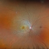
Fundus photo of a middle-aged diabetic male showing Ozurdex implant in situ with laser marks.
Photographer: Tejaswita Verma
Imaging device: MIRANTE
Condition/keywords: dexamethasone implant, ozurdex
-
 Sickle Cell Retinopathy
Feb 24 2025 by Kimberly Wakester
Optomap RGB image of an 24-year-old woman with sickle cell retinopathy in both eyes. There is overall progression of the ischemic vessels and vascular drops out compared to previous images completed in 2021. Oral FA was completed and shows possible progression of peripheral non-perfusion but difficult to determine due to drinking FA dye and images not being as bright. On Clinical exam there is no evidence of NV, RD, or RT in either eye. Patient understands the need for continued follow up care and the likely need for PRP laser in both eyes.
Photographer: Kimberly Wakester, COA
Imaging device: Optos California
Condition/keywords: sickle cell retinopathy
-
 Mac-on Retinal Detachment (Barely!)
Feb 6 2025 by Virginia Gebhart
FAF of 46 year old male with a mac-on retinal detachment from 1:00 to 6:00 with a single break at 3:00. Pt scheduled for emergent PPV/Laser/GFE
Photographer: Virginia Gebhart, Retina Consultants of Carolina
Imaging device: Optos California
Condition/keywords: autofluorescence imaging, retinal detachment
-
 FEVR
FEVR
Feb 6 2025 by Vishal Agrawal, MD, FRCS,FACS,FASRS
A 22- year male, one eyed patient came for routine examination. Fundus showed temporal straightening of Vessels. FA revealed peripheral avascular area and leakage. 3 siblings had the same findings with no history of prematurity. All the siblings underwent laser treatment.
Photographer: Dr Ayushi Gupta
Imaging device: Clarus 700
Condition/keywords: familial exudative vitreoretinopathy (FEVR)
-
 FEVR-FFA
Feb 5 2025 by Vishal Agrawal, MD, FRCS,FACS,FASRS
A 22- year male, one eyed patient came for routine examination. Fundus showed temporal straightening of Vessels. FA revealed peripheral avascular area and leakage. 3 siblings had the same findings with no history of prematurity. All the siblings underwent laser treatment.
Photographer: Dr Ayushi Gupta
Imaging device: Clarus 700
Condition/keywords: familial exudative vitreoretinopathy (FEVR)
-
 Multifocal Pattern Dystrophy
Feb 5 2025 by Kimberly Wakester
Optomap RGB and AF photograph of an 37-year-old woman with multifocal pattern dystrophy in both eyes. Previously believed to be Stargardts, but genetic testing returned positive for PRPH2 mutation. Likely Multifocal Pattern Dystrophy given phenotypical appearance of SGD. There is stable NVE in the left eye. Will continue to monitor both eyes and consider treatment with PRP laser if needed for NVE in the left eye.
Photographer: Kimberly Wakester, COA
Imaging device: Optos California
Condition/keywords: multifocal pattern dystrophy, NVE, PRPH2 Positive
-
 Retinal Detachment with Single Break
Feb 5 2025 by Virginia Gebhart
61 year old male with mac-off retinal detachment with single horseshoe tear. Macula has been off for several days and has developed associated cystic edema. Visual prognosis guarded. Pt schedule for PPV/Laser/GFE
Photographer: Virginia Gebhart, Retina Consultants of Carolina
Imaging device: Optos California
Condition/keywords: horseshoe tear, PVD, retinal detachment
-
 Eales Disease
Jan 31 2025 by Thirumalesh Mochi Basavaraj, MD
Ultra wide field image of a 24 year-old young healthy adult male with a visible sea fan neovascularization with partial PVD secondary to Scatter LASER photocoagulation with Vitreous and subhyaloid hemorrhage.
Photographer: Puttaswamy N K
Condition/keywords: Eales disease, Neovascularisation elsewhere (NVE), sea fan
-
 Lattice Degeneration With Atrophic Retinal Holes
Jan 30 2025 by Kimberly Wakester
Ultra-wide field montage fundus photograph of a 56-year-old woman with lattice degeneration with atrophic holes statues post laser. Patient also has a small CHRPE temporal to macula and trace ERM that is not visually significant. Will continue follow up care to monitor and treat as needed.
Photographer: Kimberly Wakester, COA
Imaging device: Optos California
Condition/keywords: atrophic retinal hole, CHRPE, epiretinal membrane (ERM), lattice degeneration, montage photo
-
 Central Retinal Vein Occlusion with Macular Edema
Jan 29 2025 by Kimberly Wakester
Fundus photograph of a 62-year-old man with central retinal vein occlusion with macular edema and a new PVD with an operculated retinal tear in the left eye. Laser to retinal tear was completed. Patient will return in 2-3 weeks for follow up exam with possible intravitreal injection for the CRVO with edema and to follow up on the operculated retinal tear s/p retinal tear laser.
Photographer: Kimberly Wakester, COA
Imaging device: Optos California
Condition/keywords: central retinal vein occlusion (CRVO), operculated tear, PVD
 Loading…