Search results (832 results)
-
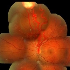
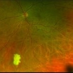
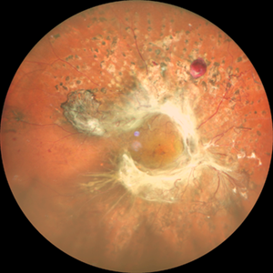
 Valsalva Retinopathy
Jan 26 2017 by JEFFERSON R SOUSA, Tecg.º (Biomedical Systems Technology)
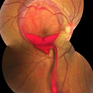
Male patient, 23-years-old, with low visual acuity in the right eye. In the ocular examination of the retinography, intense subhyaloidal hemorrhage. 2 minutes after laser application.
Photographer: JEFFERSON R SOUSA - Suel Abujamra Institute - São Paulo - Brazil
Imaging device: Topcon TRC-50 DX, Imaginet, 35 degree field. Flash 36 / Mosaic with four images.
Condition/keywords: subhyaloid hemorrhage, valsalva retinopathy
-
 Bot Fly Larvae
Apr 29 2025 by Daniela Bogenschutz
57 year-old male referred for decreased vision from optometrist. His only complaint was floaters and the letters were moving on the screen. He had never been out of the country, but is a farmer. Upon examination, our retina specialist found a bot fly larvae with numerous tracks made in this patient's retina. Patient was treated with laser to kill the larvae which was successful and he has been monitored yearly.
Photographer: Daniela Bogenschutz, OSC; Retina Consultants of Carolina, P.A.
Imaging device: Topcon
Condition/keywords: Bot Fly Larvae
-
 Hourglass in an Eye
Apr 22 2025 by KRISHNENDU NANDI, MS
A twenty-five-year-young male presented with a decrease in vision in the right eye following a blunt trauma with a football. On examination the BCVA in the right eye was CFCF and the left eye was 6/6, N6. The anterior segment was within normal limits. AT was 12 and 10 mm of Hg in the right and left eyes, respectively. Fundus examination reveals subhyaloid haemorrhage in the right eye with an attached retina. The fundus of the left eye was within normal limits. YAG laser hyaloidotomy was done with an energy of 2 mJ in the right eye. After 3 weeks the BCVA in the right eye improved to 6/9, N6.
Photographer: Dr. Krishnendu Nandi
Imaging device: Topcon
Condition/keywords: Trauma, YAG HYALOIDOTOMY, Young Male
-
 Iris
Iris
Apr 29 2019 by Stephanie Moolman
Multi-color images after Yag PI of iris.
Photographer: Stephanie Moolman, Dr Marissa Willemse, Pretoria, South Africa
Imaging device: Heidelberg Spectralis
Condition/keywords: glaucoma, iris, multicolor, NdYAG laser, peripheral iridotomy
-
 PRP laser
Mar 29 2013 by Henry J. Kaplan, MD
Right after PRP laser in PDR.
Condition/keywords: laser photocoagulation, pan-retinal photocoagulation (PRP)
-
 Stage IVB
Oct 9 2012 by Audina M. Berrocal, MD FASRS
Progression of ROP to Stage IVB despite laser treatment.
Photographer: Ditte Hess CRA, BPEI
Imaging device: RetCam Digital Imaging
Condition/keywords: retinopathy of prematurity (ROP)
-
 VHL With Capillary Hemangioma Pre-Rx
Dec 29 2016 by Manish Nagpal, MD, FRCS (UK), FASRS
VHL with hemangioma with feeder vessels.
Photographer: rakesh juneja
Condition/keywords: cryotherapy, hemangioma, laser, Von Hippel-Lindau
-
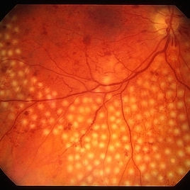
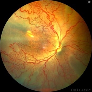 Aggressive Posterior Retinopathy of Prematurity (APROP)
May 16 2025 by KANWALJEET HARJOT MADAN, M.S. (Ophthalmology); FAICO (Vitreous - Retina)
This is the fundus picture of right eye of a premature neonate depicting Aggressive Posterior Retinopathy of Prematurity (APROP). It is a severe rapidly progressing form of retinopathy that can lead to vision loss and blindness. It requires prompt diagnosis and treatment in the form of anti-VEGF agents and laser photocoagulation.
Photographer: Dr. Kanwaljeet Harjot Madan, Thind Eye Hospital, Jalandhar City (Punjab) INDIA.
Imaging device: Zeiss Clarus
Condition/keywords: Oxygen Exposure, retinopathy of prematurity (ROP)
-
 Attached Retina With Lasered Retinectomy Status Post Multiple Surgeries for PVR With Presence of Silicon Oil and a PFCL Bubble
May 15 2014 by Manish Nagpal, MD, FRCS (UK), FASRS
Patient who underwent multiple surgeries due to recurrent PVR finally settled with a large retinectomy and a PFCL remnant bubble is seen.
Photographer: pooja barot, Optometrist, Retina Foundation, Ahmedabad
Condition/keywords: perfluorocarbon fluid
-
 Macula Off Retinal Detachment
Mar 22 2023 by Zach Seim
An ultra-widefield fundus image of a 65 year old male with a Macula Off Retinal Detachment. Patient's vision at the time of the image was CF at 6 Feet and surgical options were discussed. Fluid-gas exchange was performed without complications.
Photographer: Zach Seim
Imaging device: Optos California
Condition/keywords: left eye, macula off retinal detachment, OPTOS CALIFORNIA, scanning laser ophthalmoscope, ultra-widefield image
-
 Macular Tear
May 14 2014 by Avris Romario Diparaja Siahaan
Blue autofluorescence (BAF) a 40-year-old man with macular tear (had a photocoagulation laser).
Photographer: Avris Romario Diparaja Siahaan
Imaging device: Heidelberg HRA + OCT Spectralis
Condition/keywords: autofluorescence imaging, macular hole
-
 Melanocytoma of the Optic Nerve
Apr 6 2024 by Hector Gabriel Moreno Solano, MD, MHA
Optic Nerve laser scan image reconstruction of a 57-year-old male presented for an ophthalmological evaluation with a chief complaint of progressive visual loss. Indirect ophthalmoscopy revealed proliferative diabetic retinopathy, without macular edema, and a hyperpigmented lesion at the optic disc which corresponds to a melanocytoma.
Photographer: Héctor Gabriel Moreno-Solano, MD, MHA
Imaging device: Mirante
Condition/keywords: intraocular tumor, macular edema, melanocytoma, optic nerve
-
 Ozurdex Implant Related Tear
Jan 26 2022 by Tracey Grabowski
Ultra wide-field photograph of a 73-year-old female with an Ozurdex implant causing a retinal tear in the inferior retina. Prompt laser was added to prevent a retinal detachment and patient has been doing well since. Patient had no symptoms following the occurrence.
Photographer: Tracey Grabowski
Imaging device: Optos California
Condition/keywords: fundus photograph, inferior retina, optos, ozurdex, Ozurdex implant, retinal tear, treated retinal tear, ULTRA WIDE FIELD
-
 Posterior Ophthalmomyiasis Interna
Sep 20 2021 by Haley Tamanosky
Fundus photograph of 36-year-old woman after focal laser of a fly larva.
Photographer: Haley Tamanosky
Condition/keywords: focal laser, Posterior Ophthalmomyiasis Interna
-
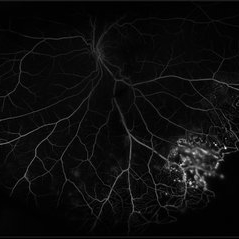
 Proliferative Sickle Cell Retinopathy
Jan 29 2021 by Olivia Rainey
Ultra-widefield fluorescein angiogram of a 24-year-old female with proliferative sickle cell retinopathy affecting her right eye. The physician's interpretation of the fluorescein shows seafan neovascularization superotemporally, AV anastomeses, and good peripheral laser. He performed scatter PRP OD on 12/2/2020 to nonperfusion in temporal far periphery. The patient's 12/2020 Hb electrophoresis came back showing Hb SC (rather than sickle cell trait). Patient was born at full term, but she reports that her mother used drugs while pregnant with the patient. The patient also mentioned that her niece has full sickle cell disease and her grandmother, mother, and sibling all have condition on the sickle cell spectrum.
Photographer: Olivia Rainey, OCT-C, COA
Imaging device: Optos California
Condition/keywords: fluorescein angiogram (FA), fluorescein leakage, neovascularization (NV), neovascularization elsewhere (NVE), Optos, sea fan, sickle cell retinopathy
-
 RAMA
RAMA
Jun 20 2016 by John S. King, MD
RAMA with 2 w co decreased vision; htn, afib using anticoag; light laser applied; 20/400.
Condition/keywords: ruptured macroaneurysm
-
 Retinopathy of Prematurity
Jul 12 2021 by Stefanie Palmer
Retinopathy of prematurity Stage 3 in a 5 month old baby. The flying baby technique was used to create this image.
Photographer: Stefanie Palmer CRA
Imaging device: scanning laser ophthalmoscope
Condition/keywords: retinopathy of prematurity (ROP), retinopathy of prematurity stage 3
-
 Sickle Cell Retinopathy
Feb 15 2021 by Kim Barrett
24-year-old female with Sickle Cell Retinopathy, stage 3. She confirms she has the trait as well as her grandmother, mother and a sibling. She has seafan neovascularization superotemporal OD. Current VA is 20/20. Photo is pre-PRP laser with areas of non-profusion temporally.
Photographer: Kim Barrett C.O.A. Retina Specialist of Michigan, Grand Rapids, MI
Imaging device: Optos California
Condition/keywords: neovascularization (NV), pan-retinal photocoagulation (PRP), sickle cell retinopathy, stage 3, trait
-
 Advanced Proliferative Diabetic Retinopathy
Nov 4 2017 by Hamid Ahmadieh, MD
Merged color fundus photograph of the left eye of a 30-year-old woman with type1 diabetes since childhood. Note laser scars, severe fibrous proliferation, traction RD and macular dragging.
Photographer: Shabnam Poureh, Negah Eye Center, Tehran, Iran
Condition/keywords: color fundus photograph, diabetes, fibrous proliferation, proliferative diabetic retinopathy (PDR), severe traction
-
 Annular Tractional Retinal Detachment
Jul 4 2024 by Hector Gabriel Moreno Solano, MD, MHA
52-year-old Hispanic female patient with a diagnosis of type II diabetes mellitus of 15 years of evolution, comes to the retina service for progressive visual loss in the right eye (single functional eye) with visual acuity of 20/100, Fundus examination reveals laser-modified proliferative diabetic retinopathy with activity + annular tractional retinal detachment with macular involvement.
Photographer: Hector Gabriel Moreno Solano, MD, MHA, HGZ #20 IMSS Puebla.
Imaging device: Mirante
Condition/keywords: macular detachment, proliferative diabetic retinopathy (PDR), tractional retinal detachment
-
 Branch Retinal Vein Occlusion
Jan 10 2014 by Manish Nagpal, MD, FRCS (UK), FASRS
Fundus photo of a 60 year old woman who had a ischemic BRVO and underwent Sectoral laser along with a Ozurdex implant sequentially in the same sitting.
Photographer: Pooja Rathod, Optometrist, Retina Foundation
Imaging device: Topcon
Condition/keywords: branch retinal vein occlusion (BRVO), Ozurdex implant, sectoral laser
-
 Central Retinal Artery Occlusion
Apr 20 2018 by Kim Barrett
64-year-old female woke with no vision in her right eye. This image was taken at 6:11 minutes and the vessels have not filled. Patient has been treated with PRP laser and anti-VEGF therapy. Current vision is CF @ 2 ft.
Photographer: Kim Barrett C.O.A.
Imaging device: Heidelberg
Condition/keywords: central retinal artery occlusion (CRAO), diabetes, hypertension, smoker, uncontrolled
-
 Central Retinal Vein Occlusion with Retinal Neovascularization
Jan 19 2022 by Olivia Rainey
Ultra-widefield fluorescein angiogram of a 56-year-old male with a Central Retinal Vein Occlusion with Retinal Neovascularization affecting his left eye. The patient presented on 1/19/2022 with scNLP vision in the left eye. The patient has good PRP, however areas of ischemia still remain untreated by laser. He also has severe neovascular glaucoma contributing to his poor vision.
Photographer: Olivia Rainey, OCT-C, COA
Imaging device: Optos California
Condition/keywords: central retinal vein occlusion (CRVO), FA early phase, fluorescein angiogram (FA), hemorrhage, ischemic CRVO, left eye, neovascular glaucoma, Optos, pan-retinal photocoagulation (PRP), retinal ischemia, retinal neovascularization, ultra-wide field imaging
-
 Coats' Disease
Jul 16 2019 by Kim Barrett
Ultra-wide field fluorescein angiogram of a 23-year-old male with Coats' disease, presented with distorted vision affecting his left eye. He reported seeing flashes and floaters since January of 2019, but the flashes had resolved. He was treated with Intravitreal Preservative Free Triamcinolone in the office and scheduled for PRP laser in the near future.
Photographer: Kim Barrett
Imaging device: Optos
Condition/keywords: Coats' disease, fluorescein angiogram (FA), fluorescein leakage, inferior retina, ischemia, left eye, Optos, ultra-wide field imaging
-
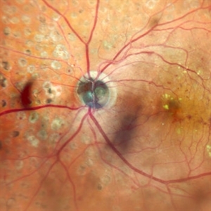
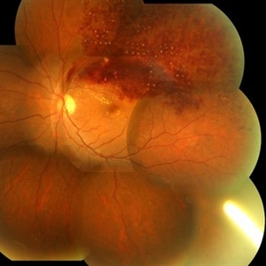
 Coloboma involving the Optic nerve, Retina, and Choroid
Dec 6 2021 by Jesus Lozano, MD
78-year-old woman after prophylactic laser photocoagulation (PLP) for her RE Coloboma involving the optic nerve, retina, and choroid. At 6 month follow up, patient preserved her FC vision as it was before the procedure. Retina attached.
Photographer: Yair Bet Yosef, Hadassah Medical Center. Israel
Imaging device: Optos Silverstone fundus image
Condition/keywords: coloboma, coloboma of choroid, coloboma of macula, coloboma of optic disc, PLP, prophylactic photocoagulation
 Loading…