Search results (832 results)
-
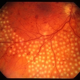
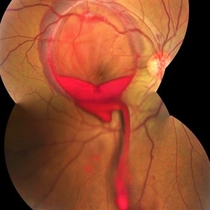
 Bilateral Retinoschisis Retinal Detachment
Sep 15 2012 by Barbara Parolini, MD
Fundus photograph of a case of bilateral retinoschisis and retinal detachment. The border of the external layer breaks and the border of the schisis have been treated with argon laser. An epiretinal membrane formed after the formation of retinal detachment.
Photographer: Dr Rino Frisina, Istituto Clinico S.Anna, Brescia, Italy
Imaging device: optos
Condition/keywords: epiretinal membrane formation, retinoschisis
-
 PRP laser
Mar 29 2013 by Henry J. Kaplan, MD
Right after PRP laser in PDR.
Condition/keywords: laser photocoagulation, pan-retinal photocoagulation (PRP)
-
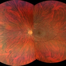 Tigroid Fundus
Aug 31 2021 by Ricardo Leitão Guerra
True color (RGB) confocal scanning laser ophthalmoscopy of a 37-year-old male with myopia highlighting choroidal vessels and vortex veins.
Photographer: Juliana Rio, MD. Leitão Guerra - Oftlamologia, Salvador - Brazil
Imaging device: Zeiss Clarus 7000
Condition/keywords: myopia, retina, tigroid fundus, vortex vein
-
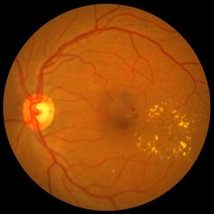 NPDR with CSME
Oct 8 2012 by Jeffrey G. Gross, MD, FASRS
NPDR with CSME with circinate ring of lipid s/p laser.
Condition/keywords: circinate ring, laser, macular edema, nonproliferative diabetic retinopathy
-
 Retinal Detachment Right Eye Optomap
Mar 31 2014 by James B. Soque, CRA, OCT-C, COA, FOPS
36-year-old white male presented with non traumatic retinal detachment OD, with six very distinct demarcation lines and isolated tear, and detachment parameters. Patient underwent PPV OD on 12/3/13 with 20% SF6 gas placement and face down in his first 1 month post op period.
Photographer: James Soque, CRA, COA
Imaging device: Optos Daytona
Condition/keywords: Cryopexy, demarcation line, gas pneumatic displacement, Optomap, Optos, pars plana vitrectomy (PPV), retinal tear, scanning laser ophthalmoscope
-
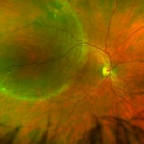
 Stargardts Disease in FAF
Sep 14 2012 by Michael P. Kelly, FOPS
This is a scanning laser ophthalmoscopic FAF image of a patient with Stargardts Disease captured with a Heidelberg Spectralis imaging unit. Note, besides the obvious hyper-autofluorescent areas centrally, the much smaller, and in greater number, pinpoints of hyper-autofluorescence extending from the vascular arcades into the mid-periphery.
Photographer: Michael P. Kelly, FOPS, Director, Duke Eye Center Labs, Duke Universtiy Hospital
Imaging device: Heidelberg Spectralis
Condition/keywords: fundus autofluorescence (FAF), Stargardt disease
-
 Rhegmatogenous Retinal Detachment in Retinopathy of Prematurity
Oct 9 2012 by Audina M. Berrocal, MD FASRS
45-week-old ex-premature 24-week child who had a rhegmatogenous detachment after laser
Photographer: Ditte Hess CRA, BPEI
Imaging device: Ret Cam
Condition/keywords: laser, retinopathy of prematurity (ROP)
-
 Regressed Proliferative Diabetic Retinopathy following PRP
Sep 6 2012 by Sharon Fekrat, MD FACS FASRS
58-year-old man with regressed proliferative diabetic retinopathy in the left eye following panretinal laser photocoagulation. Note attenuated retinal vasculature.
Photographer: Sarah Enfiedjian, Ophthalmic Photographer, Durham VA Medical Center, Durham, NC
Imaging device: Zeiss
Condition/keywords: attenuated vessels, pan-retinal photocoagulation (PRP)
-
 PVR Retinal Detachment following Laser Retinopexy Slide 1
Oct 22 2012 by Ronald C. Gentile, MD
Acute onset total retinal detachment with PVR 10 weeks following laser retinopexy.
Photographer: The New York Eye & Ear Infirmary Department of Medical Imaging
Condition/keywords: laser retinopexy, proliferative vitreoretinopathy (PVR)
-
 Retinal Arterial Macroaneurysm s/p Laser Treatment
Oct 12 2012 by Gregg T. Kokame, MD, MMM, FASRS
Retinal arterial macroaneurysm, s/p laser treatment
Photographer: Jaclyn Pisano, Retina Consultants of Hawaii
Imaging device: Zeiss FF-450 plus
Condition/keywords: laser, retinal arterial macroaneurysm
-
 Diabetic Retinal Hemorrhages in Proliferative Diabetes
Sep 10 2012 by James B. Soque, CRA, OCT-C, COA, FOPS
Fundus Photo of Severe Proliferative Diabetic with Retinal Hemorrhages, Left eye, scattered laser treatment. View: 50 Degrees
Photographer: James Soque, CRA, COA, Island Retina, Shirley, NY
Imaging device: Topcon TRC 50 DX
Condition/keywords: proliferative diabetic retinopathy (PDR)
-
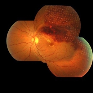
 PDR with Active NVD
Oct 8 2012 by Jeffrey G. Gross, MD, FASRS
PDR with active NVD and preretinal hemorrhage, mild VH and partial PRP.
Condition/keywords: neovascularization of the disc (NVD), preretinal hemorrhage, scatter laser photocoagulation, vitreous hemorrhage
-
---thumb.jpg/image-square;max$300,300.ImageHandler) Proliferative Diabetic Retinopathy (PDR), PRP, Regressed NV
Proliferative Diabetic Retinopathy (PDR), PRP, Regressed NV
Feb 13 2013 by From the Collections of Thomas M. Aaberg, MD and Thomas M. Aaberg Jr., MD
1 year, s/p, PRP.
Condition/keywords: neovascularization (NV), pan-retinal photocoagulation (PRP), post-laser
-
---thumb.JPG/image-square;max$300,300.ImageHandler) Horseshoe Tear With Laser Treatment
Horseshoe Tear With Laser Treatment
Jul 13 2013 by Jason S. Calhoun
Retinal tear which was treated with a laser retinopexy.
Photographer: Jason S. Calhoun, Department of Ophthalmology, Mayo Clinic Jacksonville, Florida
Condition/keywords: laser retinopexy, retinal tear
-
 Valsalva Retinopathy
Jan 26 2017 by JEFFERSON R SOUSA, Tecg.º (Biomedical Systems Technology)
Male patient, 23-years-old, with low visual acuity in the right eye. In the ocular examination of the retinography, intense subhyaloidal hemorrhage. 2 minutes after laser application.
Photographer: JEFFERSON R SOUSA - Suel Abujamra Institute - São Paulo - Brazil
Imaging device: Topcon TRC-50 DX, Imaginet, 35 degree field. Flash 36 / Mosaic with four images.
Condition/keywords: subhyaloid hemorrhage, valsalva retinopathy
-
 Optos Picture With Speculum: Dislocated Natural Lens
Oct 9 2018 by John S. King, MD
55-year-old white female with history of pathologic myopia+, lattice (laser), SB OU (1990s), and dislocated natural lenses OU that had been watched for years. In the fellow eye she developed phacolytic glaucoma and a PPV, PPL was performed. Plan for both eyes are monitoring. I wanted to get a good picture of her lens today with the optos machine, as the other pics had artifact from the lower lid. It worked out well to use a speculum in the left eye. Vision cc is 20/400 J1+ OD and 20/40 J2 OS; aphakic OU; vitreous clear OD; dislocated lens OS (see pic); retinas attached.
Photographer: Maisee Yang
Imaging device: Optos California
Condition/keywords: dislocated crystalline lens, pathologic myopia, scleral buckle, staphyloma
-
---thumb.JPG/image-square;max$300,300.ImageHandler) Horseshoe Tear Before Laser Treatment
Horseshoe Tear Before Laser Treatment
Jul 13 2013 by Jason S. Calhoun
Retinal tear temporally, proceeded with laser retinopexy.
Photographer: Jason S. Calhoun, Department of Ophthalmology, Mayo Clinic Jacksonville, Florida
Condition/keywords: retinal tear
-
 Retinoschisis/Retinal Detachment
Oct 16 2012 by Jeffrey G. Gross, MD, FASRS
Retinoschisis/RD s/p PPV and laser.
Condition/keywords: laser, pars plana vitrectomy (PPV), retinoschisis
-
 Retinal Detachment Repair With Silicone Oil and Scleral Buckle, Fourteen Years Later, With Visual Acuity of 20/25
Sep 12 2016 by Timothy S Fuller, MD
65-year-old woman s/p scleral buckle 14 years ago. Two weeks later, the retina re-detached, and vitrectomy, laser, and silicone oil procedure was performed. Patient remains 20/25 with correction fourteen years later. The cornea is clear, there is no oil emulsification, and there is a stable, moderately inferiorly subluxated PCIOL (as it was prior to RD surgery). IOP is 17 on Cosopt BID.
Photographer: Nicholas Hesse, Texas Retina Associates
Imaging device: Optos
Condition/keywords: laser, scleral buckle, silicone oil
-
 Choroidal Granuloma Secondary to Tuberculosis
Mar 14 2013 by Eduardo Torres-Porras, MD
OCT scan through the granuloma shows attachment of the retinal pigment epithelial-choriocapillaris layer and the neurosensory retina over the granuloma (“contact” sign), inflammatory retinal infiltrate in the deeper retinal layers and subretinal fluid.
Photographer: Eduardo Torres Porras, Laser y ultrasonido ocular de Puebla
Imaging device: Cirrus
Condition/keywords: optical coherence tomography (OCT), tubercular choroidal granuloma
-
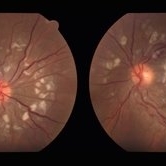 SLE Retinopathy
Nov 14 2016 by Mitzy E Torres Soriano, MD
25-year-old female patient with systemic lupus erythematosus. Photographs show cotton wool spots, intraretinal hemorrhages and vascular tortuosity. FA demonstrated retinal vasculitis and OCT revealed cystoid macular edema. In this case diagnosis of SLE was made after ocular manifestation.
Photographer: Grupo Laser Vision, Rosario, Argentina
Condition/keywords: cotton wool spots, occlusive retinal vasculitis, occlusive vasculitis, systemic lupus erythematosus, vasculopathy
-
 Macular Hole
Sep 27 2012 by Jeffrey G. Gross, MD, FASRS
Macular hole s/p 360 degree laser to fluid cuff.
Condition/keywords: macular hole, subretinal fluid
-
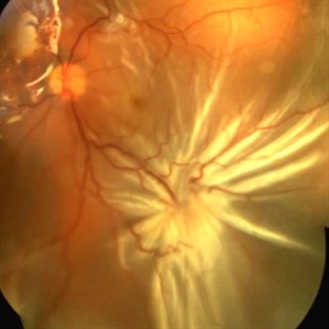 Starfold in Proliferative Vitreoretinopathy
Sep 11 2014 by Thomas A. Ciulla, MD, MBA, FASRS
This patient underwent repair of a rupture globe, extraction of an intraocular foreign body, vitrectomy, laser, and silicone oil placement. When he returned for follow up, he was noted to have a recurrent shallow retinal detachment under the silicone oil, with a prominent star fold at the inferior temporal arcade.
Photographer: Thomas Steele
Condition/keywords: proliferative vitreoretinopathy (PVR)
-
 Branch Retinal Vein Occlusion
Jan 10 2014 by Manish Nagpal, MD, FRCS (UK), FASRS
Fundus photo of a 60-year-old woman who had a ischemic BRVO and underwent sectoral laser along with a Ozurdex implant sequentially in the same sitting. This photo is taken right after the sectoral laser and before the implant is injected
Photographer: Pooja Rathod, Optometrist, Retina Foundation
Imaging device: Topcon
Condition/keywords: branch retinal vein occlusion (BRVO), Ozurdex implant, sectoral laser
-
 Stage IVB
Oct 9 2012 by Audina M. Berrocal, MD FASRS
Progression of ROP to Stage IVB despite laser treatment.
Photographer: Ditte Hess CRA, BPEI
Imaging device: RetCam Digital Imaging
Condition/keywords: retinopathy of prematurity (ROP)
 Loading…