Search results (40 results)
-
 Three Kisses
Mar 18 2025 by Gustavo Uriel Fonseca Aguirre
Cross-section of a B-mode ultrasound showing a kiss-shaped choroidal detachment; three lobes, giving the appearance of three kisses.
Photographer: Gustavo U. Fonseca Aguirre, Hospital Conde de Valenciana, Ciudad de México
Condition/keywords: Kissing Choroidal Detachment
-
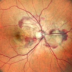 Right Eye Color Photo With Hemorrhages in Case of CNVM With Angioid Streaks
Nov 29 2024 by Anand Temkar
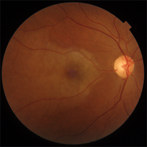
A 45 year old male came with chief complaint of blurring vision in right eyes since past 4 days. His vision is 6/12 in right eye and 6/9 in left eye. His vision was 14 mmHg in right eye and 16 mmHg in left eye. He was diagnosed with Angioid Streaks in both eyes about a year ago, then he developed choroidal neovascularization in his left eye 8 months ago, for which he received AntiVEGF injections x 3. Left eye is a stable eye now. Patient presented with right eye choroidal neovascularization in a case of Angioid Streaks on recent follow up. We have advised him right eye AntiVEGF injections x 3. In this image, the right eye color photo shows bleed from CNVM in case of angioid streaks.
Photographer: Dr.Anand Temkar- Retina Foundation, Ahmedabad
Imaging device: Mirante
Condition/keywords: Angioid Streaks, choroidal neovascular membrane (CNVM)
-
 MIDD (Maternally Inherited Diabetes and Deafness) - Right AF
Nov 30 2024 by John S. King, MD
Both right and left eyes have symmetrical ring of mottled hypo/hyper AF around the fovea and disc. The HyperAF areas correspond to RPE deposits on OCT as well as areas of blockage on FA, and drusenoid deposits seen on fundus photos. Disc drusen in right eye present as HyperAF spot 57 yo WF referred for AMD vs Pattern Dystrophy that was diagnosed 10 years ago. Reported some slow progressive vision loss in both eyes for distance and near. Denies nyctalopia or hemeralopia. Background medical history includes HTN, CVD, and DM. No family history of eye problems. Denied pentosan use. Anterior segment showed moderate cataracts (OD>OS). Posterior segment exam showed macular changes and mild NPDR. The macular appearance showed a symmetrical, paramacular ring of fleck-like drusenoid material with some faint focal areas of RPE hyperplasia. Fundus Photos, AF, OCT were performed as well as a gene test. Further questioning showed revealed that her mother and maternal grandmother had both diabetes mellitus and sensorineural hearing loss. The patient developed diabetes in her teens, and some high frequency hearing loss in her early twenties. She had not had a previous genetic test or diagnosis of MIDD. Gene testing is pending for the mitochondrial component. Invitae's retinal panel, which does not include mitochondrial disorders, only showed a variant of uncertain significance, HMCN1. I discussed this case with Dr. Freund, and it is similar to a the case report : Inoue M, Kiss S, Freund KB. MACULAR PIGMENT RINGS AS THE PRESENTING FINDING OF MITOCHONDRIAL MYOPATHY, ENCEPHALOPATHY, LACTIC ACIDOSIS, AND STROKELIKE EPISODES. Retin Cases Brief Rep. 2015 Fall;9(4):260-4. doi: 10.1097/ICB.0000000000000182. PMID: 26200388.
Photographer: Grace Melton and Carley Gunn
Imaging device: Clarus
Condition/keywords: Macular Dystrophy, Maternally Inherited Diabetes and Deafness, MIDD, Mitochondrial Disorder
-
 Dislocated Crystalline Lens
Mar 19 2024 by Annaka Gooding
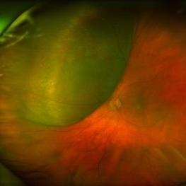
Ultra Wide field fundus photography of a 70 year old male who presented to clinic with a sudden increase of vision due to dropped crystalline lens secondary to severely dense cataract. Patient reported seeing a full black circle in his inferior visual field. Patient's visual acuity at time of visit was 20/100 with a +5.00 diopter lens. The physician recommended surgical intervention, and discussed surgery for PPV/PPL/IOL implantation with an ACIOL.
Photographer: Annaka Gooding, CPO
Imaging device: Optos California RGB
Condition/keywords: dislocated crystalline lens, fundus photography, inferior retina, OPTOS CALIFORNIA RGB, Right Eye, Ultra-wide field retinal imaging
-
 Retinal Fold
Sep 26 2023 by Mauricio Bayram-Suverza, MD
A 38-year-old man underwent vitrectomy in the left eye due to a giant tear in the upper retina. SF6 gas was used as endotamponade. During the post-surgical check-up, it was identified that the patient developed a full-thickness retinal fold due to retinal slippage during fluid-air exchange. As the fold was away from the macular area, it was decided to observe the patient. Three weeks after the surgery, his best-corrected visual acuity was 20/30.
Photographer: Mauricio Bayram-Suverza, Fundación Hospital Nuestra Señora de la Luz
Imaging device: TRC-50DX
Condition/keywords: giant retinal tear, retina surgery complications, Retinal slippage, vitreoretinal surgery
-
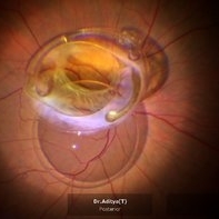 Rescuing IOL CTR Bag Complex
Jun 14 2023 by Aditya S Kelkar, MS, FRCS, FASRS,FRCOphth
INTRAOPERATIVE SNAPSHOPT IN ZEISS ARTEVO 800 OF DROPPED IOL CTR BAG COMPLEX IN A 71 YEAR OLD MALE PATIENT
Photographer: SUBHASREE DUTTA, NATIONAL INSTITUTE OF OPHTHALMOLOGY, PUNE
Imaging device: ZEISS ARTEVO 800
Condition/keywords: dropped capsular IOL bag complex
-
 Candy Stripe Sign
Mar 30 2023 by pedro fernandes souza neto
Candy Stripe Sign, patient with proliferative diabetic retinopathy progressing to vitreous hemorrhage and subsequently to ghost cell glaucoma.
Photographer: Marlos Henrique Oliveira Junior, Federal University of Bahia.
Condition/keywords: dehemoglobinized hemorrhage, diabetes, diabetic glaucoma
-
 Rod Cone dystrophy
Nov 29 2022 by Niloofar Piri, MD
Fundus autofluorescence of the left eye in a 58 yo male with rod cone dystrophy. He presented with night blindness and peripheral vision loss since youth and recent decrease in central vision for the past 10 years. Notice multiple coin shaped hypoautofluorescent pacthes within central 20 degrees which are coalescing centrally. (fundus photo uploaded separately) He has one pathogenic variants of both CEP290 and PRPH2 genes.
Photographer: Sean Kelso, Saint Louis University
Condition/keywords: hereditary retinal degeneration, hereditary retinal dystrophy, rod cone dystrophy
-
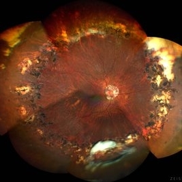
 Choroidal Melanoma
Nov 3 2022 by pedro fernandes souza neto
Transillumination of Enucleation specimen of Choroidal Melanoma: anterior chamber is closed. Total secondary retinal detachment with subretinal serous fluid and some subretinal hemorrhages are present.
Photographer: Eduardo Marback, Federal University of Bahia, Brazil
Condition/keywords: enucleation, melanoma
-
 Posteriorly dislocated IOL
Oct 22 2022 by Vishal Agrawal, MD, FRCS,FACS,FASRS
67 yr old male , post PPV for retinal detachment ( 5 years ) presented with sudden DOV . On examination posteriorly dislocated 4 loop haptic iol - bag complex was noted .
Photographer: Pankaj
Imaging device: CLARUS 700
Condition/keywords: dropped intraocular lens (IOL)
-
 Leukemic optic neuropathy
Oct 28 2022 by pedro fernandes souza neto
Fundus photograph of an 18-year-old woman with Leukemic optic neuropathy.
Photographer: Pedro Fernandes, Universidade Federal da Bahia
Condition/keywords: Leukemic optic neuropathy
-
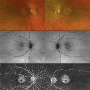 CERKL-related Cone Rod Dystrophy
Jun 27 2022 by Hanna Choi
37-year-old female with cone-rod dystrophy. Developed photophobia and progressive blurry vision in the third decade. VA 20/40 OD, 20/30 OS. The patient is compound heterozygous for pathogenic mutations in the CERKL gene (Arg465Trp and Arg283*).
Photographer: Kaitlynn Silva, New England Retina Consultants
Imaging device: Ultrawide-field Optos Fundus Photography, Autofluorescence, Fluorescein Angiography
Condition/keywords: cone dystrophy, inherited retinal disease, maculopathy
-
 Spontaneously Dropped Lens in a Congenital Rubella Syndrome
Apr 30 2022 by NEIFFER RABELO
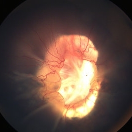
Intraoperative photograph of a 68-year-old patient with congenital rubella syndrome and light perception visual acuity since childhood. The image shows a pigmentary retinopathy and the lens spontaneously displaced into the vitreous cavity. The patient sought care complaining of a total and sporadic loss of vision that was hindering her in daily tasks. Surgery was indicated to remove the lens.
Photographer: Rodrigo Dos Anjos Versiani - Retina Institute - Belo Horizonte - Brazil
Imaging device: ZEISS OPMI LUMERA 700
Condition/keywords: dropped nucleus, retina surgery, rubella retinopathy
-
 Angioid Streaks
Jun 14 2022 by Kingston Rodolfo Ureña-Wong, MD, Opht, MSc
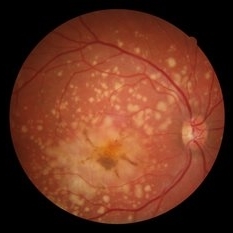
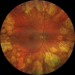
Fundus photograph of an 26-year-old woman with pseudoxanthoma elasticum and angioid streaks. She developed a choroidal neovascular membrane which was treated with anti-VEGF successfully.
Photographer: Kingston Rodolfo Ureña-Wong, Asociación para evitar la ceguera en México, México.
Imaging device: Zeiss Clarus
Condition/keywords: Angioid Streaks
-
 Thioridazine-toxicity
Apr 30 2022 by Niloofar Piri, MD
61 yo male with PMH of longstanding schizophrenia since 20s with secondary intellectual disability presented with decreased vision following a recent stroke. He was found to have bilateral chorio-retinal atrophy involving posterior pole with scalloped edges and coin shaped atrophic area at margins extending into mid-periphery, diagnosis most concerning for intermediate stage thioridazine toxicity given the history. Mother could find handwritten prescriptions from 1990s when he was on Thioridazine 800 mg daily for unknown period of time. Patient had better vision in the left eye which was affected by recent stroke and prompted him to seek medical care. Fundus photograph of the right eye is demonstrated here.
Photographer: Jacob Grodsky, MD
Condition/keywords: drug toxicity, thioridazine toxicity, toxic retinopathy
-
 Cilioretinal Artery Occlusion
Jun 9 2021 by Thirumalesh Mochi Basavaraj, MD
Fundus photograph of 43-year-old women with a Y-shaped clot occluding the trunk and branches of the cilioretinal artery.
Photographer: Puttuswamy , Narayana Nethralaya , Bangalore
Condition/keywords: cilioretinal artery occlusion
-
 Traumatic Lens Drop in Vitreous
Dec 15 2020 by Manish Nagpal, MD, FRCS (UK), FASRS
Patient had come to us status post blunt trauma with the lens dislocated in inferior vitreous.
Photographer: Gayathri Mohan, Retina Fellow, Retina Foundation, Ahmedabad, India
Imaging device: Mirante CSLO
Condition/keywords: dropped nucleus, lens dislocation, traumatic cataract
-
 Morning Glory Disc Anomaly
Nov 11 2020 by Yoshihiro Yonekawa, MD, FASRS
Color fundus photograph of a young boy with morning glory disc anomaly. Notice the concavity surrounding the enlarged disc, radial vasculature, and nasally dragged macula. MRI was negative for moyamoya disease, a known association.
Photographer: Alicia Thresher, Mid Atlantic Retina
Imaging device: Topcon
Condition/keywords: disc coloboma, Morning Glory Syndrome, pediatric retina
-
 Retinal Detachment Associated with Coloboma
Aug 23 2020 by Noy Ashkenazy, MD, MS
Fundus photograph of a 2-year-old boy with a history of CHARGE syndrome. The image nicely illustrates a retinal detachment associated with a congenital coloboma.
Photographer: Giselle DeOliveira
Imaging device: Retcam III
Condition/keywords: CHARGE syndrome, chronic retinal detachment, coloboma, pediatric retina
-
 Torpedo Maculopathy
Jul 29 2020 by Yoshihiro Yonekawa, MD, FASRS
Fundus photograph of a 10-year-old boy with an incidentally identified torpedo maculopathy.
Photographer: Suely Bascope
Imaging device: Topcon
Condition/keywords: macula lesion, pediatric retina, torpedo maculopathy
-
 Proliferative Diabetic Retinopathy with Choroidal Effusion Status Post PRP
Dec 15 2020 by Manish Nagpal, MD, FRCS (UK), FASRS
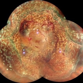
A 17-year-old juvenile diabetic patient came to us with extensive neovascular proliferations and PRP done a week back and had developed 360 degree choroidal effusion as seen in this wide field montage image
Photographer: Sham Talati, Retina Fellow , Retina Foundation, Ahmedabad, India
Imaging device: Mirante CSLO
Condition/keywords: choroidal effusion, diabetic retinopathy, proliferative diabetic retinopathy (PDR)
-
 Butterfly-Shaped Pattern Dystrophy
May 19 2020 by Evgeniia Perevoznikova
Butterfly-shaped pattern dystrophy.
Photographer: ophthalmologist_evgeniya
Condition/keywords: butterfly dystrophy
-
 Large, Dome-Shaped Peripheral Choroidal Melanoma - Widefield Color
Feb 13 2020 by Michael Seider, MD
Large, dome-shaped peripheral choroidal melanoma of the left eye with inferior exudative retinal detachment. Note the lack of obvious orange pigment over the tumor and apparent drusen anteriorly. A lack of ophthalmoscopically obvious lipofuscin is not uncommon among larger choroidal melanomas. B-Scan ultrasonography (transverse, 10 o’clock) confirms a low-moderate internally reflective dome-shaped choroidal lesion with a small adjacent retinal detachment. Ultrasound biomicroscopy (radial, 10 o’clock) confirms no ciliary body involvement of the tumor.
-
 Didanosine Toxicity
Jan 27 2020 by Nimesh A. Patel, MD, FASRS
Patient with history of HIV treated with didanosine. Developed gyrate like peripheral retinal atrophy with central sparing. Vision is 20/25
Imaging device: Clarus
Condition/keywords: AIDS, didanosine, HIV
-
 Congenital Toxoplasmosis
Dec 18 2019 by Yoshihiro Yonekawa, MD, FASRS
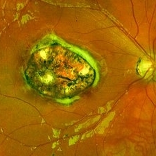
Widefield fundus image of a teenage girl's right eye with an inactive congenital toxoplasmosis macular lesion. Her vision is 20/400 in this eye.
Photographer: Netanya Lerner, COA, Wills Eye Hospital/Mid Atlantic Retina
Imaging device: Optos California
Condition/keywords: congenital toxoplasmosis, pediatric retina
 Loading…