Search results (1464 results)
-
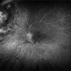
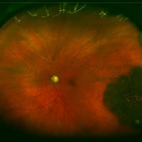
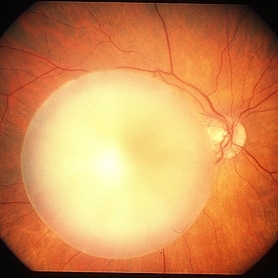
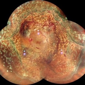 Proliferative Diabetic Retinopathy with Choroidal Effusion Status Post PRP
Dec 15 2020 by Manish Nagpal, MD, FRCS (UK), FASRS
A 17-year-old juvenile diabetic patient came to us with extensive neovascular proliferations and PRP done a week back and had developed 360 degree choroidal effusion as seen in this wide field montage image
Photographer: Sham Talati, Retina Fellow , Retina Foundation, Ahmedabad, India
Imaging device: Mirante CSLO
Condition/keywords: choroidal effusion, diabetic retinopathy, proliferative diabetic retinopathy (PDR)
-
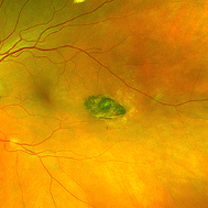
 Cat Scratch
Feb 15 2017 by Hua Gao, MD, PhD, FASRS
A female patient of 57-year-old presented with neuroretinitis due to cat-scratch disease with positive Bartonella henselae antibodies. Two weeks after symptom onset she developed exudates in a "macular star" pattern.
Condition/keywords: cat scratch retinitis
-
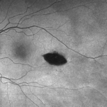 Torpedo Maculopathy
Feb 20 2024 by Soobien Lee
Optos fundus autofluorescence photograph of a 35-year-old asymptomatic female with no ocular or medical history with stable and chronic appearing torpedo-shaped macula lesion in the left eye.
Photographer: Peter Sotirakos, Elman Retina Group
Imaging device: Optos Ultra-Widefield Autoflurescence Imaging
Condition/keywords: autofluorescence imaging, genetics, macula, maculopathy, Optos, torpedo maculopathy
-
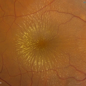
 Torpedo Maculopathy
Feb 20 2024 by Soobien Lee
Optos color fundus photograph of a 35-year-old asymptomatic female with no ocular or medical history with stable and chronic appearing torpedo-shaped macula lesion in the left eye.
Photographer: Peter Sotirakos, Elman Retina Group
Imaging device: Optos Ultra-Widefield Imaging
Condition/keywords: macula, Optos, torpedo maculopathy
-
 Candy Stripe Sign
Mar 30 2023 by pedro fernandes souza neto
Candy Stripe Sign, patient with proliferative diabetic retinopathy progressing to vitreous hemorrhage and subsequently to ghost cell glaucoma.
Photographer: Marlos Henrique Oliveira Junior, Federal University of Bahia.
Condition/keywords: dehemoglobinized hemorrhage, diabetes, diabetic glaucoma
-
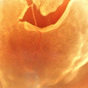
 Choroidal Melanoma
Nov 3 2022 by pedro fernandes souza neto
Transillumination of Enucleation specimen of Choroidal Melanoma: anterior chamber is closed. Total secondary retinal detachment with subretinal serous fluid and some subretinal hemorrhages are present.
Photographer: Eduardo Marback, Federal University of Bahia, Brazil
Condition/keywords: enucleation, melanoma
-
 Central Retinal Artery Occlusion With Cilioretinal Sparing
Apr 4 2018 by Soumya Venkatesh
Fundus photograph of a 23-year-old gentleman presenting with sudden loss of vision 2 days prior to presentation. He underwent all relevant investigations and found to have APLA positive. He also had dengue serology positive. On follow up, his retinal edema reduced unmasking the underlying hemorrhages( flame shaped).
Photographer: Soumya Harapanahalli Venkatesh, JSS university, Karnataka, India
Condition/keywords: central retinal artery occlusion (CRAO), cherry red spot, cilioretinal sparing, retinal ischemia
-
---thumb.jpg/image-square;max$300,300.ImageHandler) Central Retinal Vein Occlusion
Central Retinal Vein Occlusion
Oct 30 2012 by Lihteh Wu, MD
35-year-old hypertensive man with an acute CRVO. Notice the peripapillary cotton wool spots, superficial flame shaped hemorrhages and deeper dot and blot hemorrhages in all 4 quadrants. This is the typical blood and thunder appearance of a CRVO.
Condition/keywords: central retinal vein occlusion (CRVO), cotton wool spots
-
 Dropped IOL
Apr 5 2018 by Mohamed Tawfik, MD
Intra operative photo of a case of dropped IOL after phaco+IOL.
Photographer: Mohamed A,Tawfik
Imaging device: Intra operative Photography Screen shoot
Condition/keywords: dropped intraocular lens (IOL)
-
 Radiation Retinopathy; BRVO with Macular Edema
Apr 26 2023 by Denica Rodriguez
Ultra-wide field fluorescein angiography of a 61 year old male with radiation retinopathy following brachytherapy for choroidal melanoma of his left eye. Following treatment, patient developed a branch retinal vein occlusion both ischemic and non-ischemic. Anti-VEGF injections were recommended. The fine needle biopsy showed a class 2 uveal melanoma. Patient also has diabetic retinopathy affecting both eyes. Patient's vision at the time the image was taken was Dcc 20/80-1.
Photographer: Denica Rodriguez COA, ST
Imaging device: Optos California
Condition/keywords: branch retinal vein occlusion (BRVO), Choroidal melanoma, diabetic retinopathy, FA, fluorescein angiogram (FA), I-125 brachytherapy, macular edema, melanoma, Optos, radiation retinopathy, Retina, ultra-wide field imaging
-
 Retinal Detachment Associated with Coloboma
Aug 23 2020 by Noy Ashkenazy, MD, MS
Fundus photograph of a 2-year-old boy with a history of CHARGE syndrome. The image nicely illustrates a retinal detachment associated with a congenital coloboma.
Photographer: Giselle DeOliveira
Imaging device: Retcam III
Condition/keywords: CHARGE syndrome, chronic retinal detachment, coloboma, pediatric retina
-
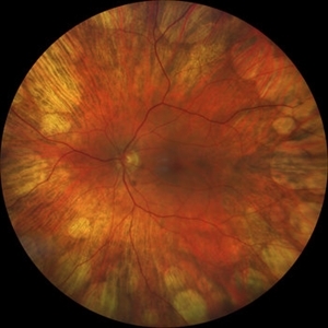
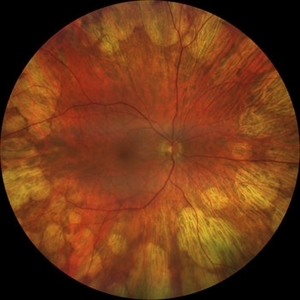
 Rod Cone dystrophy
Nov 29 2022 by Niloofar Piri, MD
Fundus photograph of the left eye in a 58 yo male with rod cone dystrophy. He presented with night blindness and peripheral vision loss since youth and recent decrease in central vision for the past 10 years. Notice waxy pallor of the nerve, severe arterial narrowing and chorioretinal atrophy mainly around the arcades as well as posterior pole along with RPE hyperplastic changes and atrophy. RPE atrophy in midperiphery has coin shaped appearance. FAF has characteristic appearance (uploaded separately) He has one pathogenic variants of both CEP290 and PRPH2 genes.
Photographer: Sean Kelso, Saint Louis University
Condition/keywords: hereditary retinal deg, hereditary retinal dystrophy, Rod cone dystrophy
-
 Rod Cone dystrophy
Nov 29 2022 by Niloofar Piri, MD
Fundus autofluorescence of the left eye in a 58 yo male with rod cone dystrophy. He presented with night blindness and peripheral vision loss since youth and recent decrease in central vision for the past 10 years. Notice multiple coin shaped hypoautofluorescent pacthes within central 20 degrees which are coalescing centrally. (fundus photo uploaded separately) He has one pathogenic variants of both CEP290 and PRPH2 genes.
Photographer: Sean Kelso, Saint Louis University
Condition/keywords: hereditary retinal degeneration, hereditary retinal dystrophy, rod cone dystrophy
-
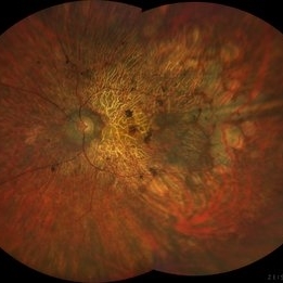
 Spontaneously Dropped Lens in a Congenital Rubella Syndrome
Apr 30 2022 by NEIFFER RABELO
Intraoperative photograph of a 68-year-old patient with congenital rubella syndrome and light perception visual acuity since childhood. The image shows a pigmentary retinopathy and the lens spontaneously displaced into the vitreous cavity. The patient sought care complaining of a total and sporadic loss of vision that was hindering her in daily tasks. Surgery was indicated to remove the lens.
Photographer: Rodrigo Dos Anjos Versiani - Retina Institute - Belo Horizonte - Brazil
Imaging device: ZEISS OPMI LUMERA 700
Condition/keywords: dropped nucleus, retina surgery, rubella retinopathy
-
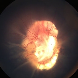
 A Vessel That Would Not Let Go
May 5 2025 by Malvika Singh
Fundus photograph of a retinal detachment showing a horse shoe shaped tear and a bridging vessel.
Photographer: Dr Tejaswita Verma, Retina Foundation, Ahmedabad, India
Imaging device: Mirante SLO/OCT
Condition/keywords: bridging vessel, horseshoe tear
-
 Adenocarcinoma Arising from CHRPE
Sep 17 2015 by Marc C. Peden, MD
49-year-old female referred for presumed ocular melanoma. On examination was noted to have darkly pigmented lesion in the temporal retina of left eye. Lesion had characteristic scalloped edges with central lacunae, however, on ultrasonography was noted to have 1.8mm of elevation with high internal reflectivity. IVFA shows absence of dual circulation with areas of window defect. Findings were consistent with those described by Shields et al., in their April 2001 article in Archives of Ophthalmology.
Photographer: Janet Traynom
Imaging device: Optos P200MA
Condition/keywords: adenocarcinoma arising from CHRPE
-
 Branch Retinal Vein Occlusion with Macular Edema
Aug 23 2012 by Gerardo Garcia-Aguirre, MD
Fundus photograph composition of the left eye, showing flame-shaped and blot hemorrhages in the superotemporal quadrant, with hard exudates surrounding the fovea.
Photographer: Noemí Hernández, Asociación para Evitar la Ceguera en México
Condition/keywords: branch retinal vein occlusion (BRVO), macular edema
-
---thumb.jpg/image-square;max$300,300.ImageHandler) Central Retinal Vein Occlusion
Central Retinal Vein Occlusion
Oct 30 2012 by Lihteh Wu, MD
35-year-old hypertensive man with an acute CRVO. Notice the peripapillary cotton wool spots, superficial flame shaped hemorrhages and deeper dot and blot hemorrhages in all 4 quadrants. This is the typical blood and thunder appearance of a CRVO.
Condition/keywords: central retinal vein occlusion (CRVO), cotton wool spots
-
 Central Retinal Vein Occlusion associated with disc edema
Oct 19 2023 by Gabriel Costa Andrade, PhD
53-year-old woman with an acute CRVO. The patient has a history of breast cancer undergoing treatment with systemic chemotherapy. Notice the peripapillary cotton wool spots, superficial flame shaped hemorrhages and deeper dot and blot hemorrhages in all 4 quadrants.
Photographer: Gabriel Andrade
Condition/keywords: central retinal vein occlusion (CRVO), macular edema, Retina
-
 Coats' Disease - Stage 3A
Aug 21 2019 by Victor M Villegas, MD
Coats' Disease - stage 3A.
Condition/keywords: abnormal retina, Coats' disease, diffuse lipid exudate, edema, foveal hard exudates, pediatic retina, retcam, retinal angioma
-
 Didanosine Toxicity
Jan 27 2020 by Nimesh A. Patel, MD, FASRS
Patient with history of HIV treated with didanosine. Developed gyrate like peripheral retinal atrophy with central sparing. Vision is 20/25
Imaging device: Clarus
Condition/keywords: AIDS, didanosine, HIV
-
 Didanosine Toxicity
Jan 27 2020 by Nimesh A. Patel, MD, FASRS
Patient with history of HIV treated with didanosine. Developed gyrate like peripheral retinal atrophy with central sparing. Vision is 20/25
Imaging device: Clarus
Condition/keywords: AIDS, didanosine, HIV
-
 Dropped Crystalline Lens
Mar 8 2019 by Abdulaziz A. Alshamrani, MD
A 15-year-old female with congenital glaucoma complaining of acute diminution of vision after a blunt trauma.
Condition/keywords: crystalline lens, dropped nucleus, ora serrata
-
 Dropped nucleus
Mar 29 2013 by Henry J. Kaplan, MD
Dropped nucleus on posterior segment.
Condition/keywords: dropped nucleus
-
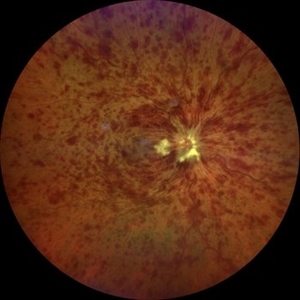
 Leukemic optic neuropathy
Oct 28 2022 by pedro fernandes souza neto
Fundus photograph of an 18-year-old woman with Leukemic optic neuropathy.
Photographer: Pedro Fernandes, Universidade Federal da Bahia
Condition/keywords: Leukemic optic neuropathy
 Loading…