Search results (1464 results)
-
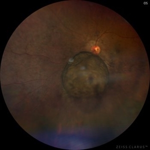
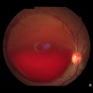
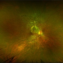
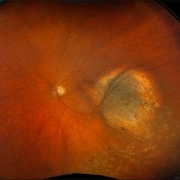 New Choroidal Melanoma
Jul 16 2025 by Virginia Gebhart
78 year old male with a partially amelanotic dome-shaped lesion with RPE changes, hard exudates, overlying intraretinal fluid and minimal SRF temporally. Exam and ultrasound findings consistent with choroidal melanoma. Pt will be scheduled for brachytherapy pending CT scan results.
Photographer: Virginia Gebhart, Retina Consultants of Carolina
Imaging device: Optos California
Condition/keywords: amelanotic melanoma, choroidal melanoma
-
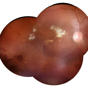
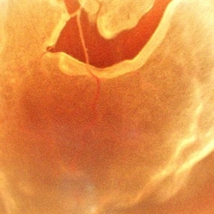
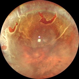 Retinal Detachment with Multiple Horse Shoe Shaped Tears
Jul 14 2025 by Malvika Singh
Fundus photograph of a 46 year old showing a retinal detachment with multiple peripheral horse show shaped tears.
Photographer: Dr Malvika Singh, Retina Foundation, Ahmedabad, India
Imaging device: Mirante SLO/OCT
Condition/keywords: horseshoe tear, retinal detachment
-
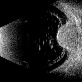
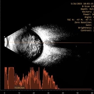
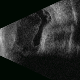
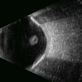
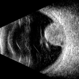 Choroidal Melanoma (USG)
Jul 5 2025 by Gustavo Uriel Fonseca Aguirre
This B-mode transverse ultrasound scan reveals a mushroom-shaped choroidal tumor in the inferior nasal quadrant adjacent to the optic nerve head. The lesion appears solid with homogeneous internal reflectivity and is associated with minimal surrounding subretinal fluid and scleral excavation. It measures 6.54 mm in height × 7.52 mm in base diameter (transverse view) and extends 9.52 mm longitudinally. The vitreous contains abundant punctate opacities consistent with pigment dispersion. The retina and choroid remain attached elsewhere.
Photographer: Gustavo U. Fonseca Aguirre, Hospital Conde de Valenciana, Ciudad de México
Condition/keywords: choroidal melanoma
-
 Choroidal Hemangioma (AF)
Jul 5 2025 by Gustavo Uriel Fonseca Aguirre
This wide-field fundus autofluorescence image demonstrates a mushroom-shaped choroidal melanoma adjacent to the optic nerve head, exhibiting hypo-autofluorescence (melanin). Vitreous pigment dispersion (tobacco dust sign) is evident, indicating tumor activity.
Photographer: Gustavo U. Fonseca Aguirre, Hospital Conde de Valenciana, Ciudad de México
Condition/keywords: choroidal melanoma
-
 Choroidal Melanoma
Jul 5 2025 by Gustavo Uriel Fonseca Aguirre
This 50° central fundus photograph reveals a mushroom-shaped choroidal melanoma adjacent to the optic nerve head. The lesion demonstrates characteristic pigmentation with overlying vitreous pigment dispersion (tobacco dust sign).
Photographer: Gustavo U. Fonseca Aguirre, Hospital Conde de Valenciana, Ciudad de México
Condition/keywords: choroidal melanoma
-
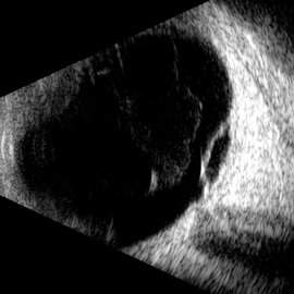
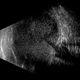
 Choroidal Melanoma
Jul 3 2025 by Gustavo Uriel Fonseca Aguirre
This B-mode transverse ultrasound scan shows asteroid hyalosis with partial posterior vitreous detachment. A dome-shaped choroidal melanoma is observed in the inferior quadrant (preequatorial to equatorial region), appearing as a solid, regularly bordered lesion with heterogeneous internal structure and mild acoustic attenuation. Standardized A-mode reveals medium-to-low internal reflectivity. The tumor measures 11.62 mm in base diameter and 6.60 mm in height. The retina and choroid remain attached, with minimal suprachoroidal fluid in the inferior quadrant.
Photographer: Gustavo U. Fonseca Aguirre, Hospital Conde de Valenciana, Ciudad de México
Condition/keywords: choroidal melanoma
-
 Macular Retinoschisis
Jul 3 2025 by Gustavo Uriel Fonseca Aguirre
This B-mode longitudinal ultrasound scan reveals macular retinoschisis, demonstrating a characteristic splitting of retinal layers with a smooth, dome-shaped elevation. The lesion shows maintained structural integrity of both inner and outer retinal walls without associated subretinal fluid or vitreous traction.
Photographer: Gustavo U. Fonseca Aguirre, Hospital Conde de Valenciana, Ciudad de México
Condition/keywords: macular retinoschisis
-
 Multiple Chorodial Ruptures
Jun 26 2025 by Hector Gabriel Moreno Solano, MD, MHA
Color fundus photograph of the right eye reveals three well-defined, curvilinear choroidal ruptures located temporal to the fovea running parallel. The lesions appear as pale, crescent-shaped bands, with underlying retinal pigment epithelium disruption. One of the ruptures is situated near the foveal center, though without direct involvement.
Photographer: Hector Gabriel Moreno Solano, Instituto Mexicano de Oftalmología “IMO I.A.P”
Imaging device: CLARUS
Condition/keywords: Choroidal Rupture, color fundus photograph, color wide field
-
 Arcus Retinalis
Jun 21 2025 by Moazzam Parvez
Fundus photograph of a 30 year oiled gentleman with multiple dome shaped sub hyaloid haemorrhage with discrete arches retinals around it. Roth spots are also noted on the retina.
Photographer: Moazzam Parvez , Netralayam , Kolkata
Imaging device: Topcon Maestro 2
Condition/keywords: arcus retinalis, Roth spots, Sub hyaloid haemorrhage
-
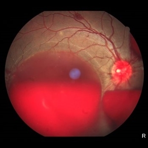
 Sub ILM Hemorrhage
Jun 21 2025 by Moazzam Parvez
Fundus photograph of a 46 year old female presenting with a massive sharply demarcated, dome shaped bleed in her right eye.
Photographer: Moazzam Parvez , Netralayam , Kolkata
Imaging device: Topcon Maestro 2
Condition/keywords: sub ILM hemorrhage
-
 Macular Edema
Jun 4 2025 by Paulina Araujo
The composite fundus photograph of the right eye demonstrates circinate hard exudates in the thickened macular area, along with flame-shaped intraretinal hemorrhages along the inferior temporal arcade.
Photographer: Paulina D.Araujo Martínez, Asociación para Evitar la Ceguera en México I.A.P., Hospital Dr Luis Sánchez Bulnes.
Condition/keywords: macular edema
-
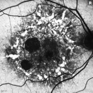
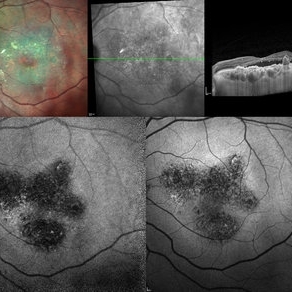 Multi-modal Imaging of Type - 1 CNVM
May 30 2025 by Shivankar Sen, MS, FVRS
Multimodal Imaging of a case of Polypoidal Choroidal Vasculopathy Multicolor Reflectance showing multiple green-hyper-fringent lesions in the macular region (Up Left) Infra-red Autofluorescence and Blue Autofluorescence showing hypo-autofluorescent areas correspondingly revealing the exact extent of the sub-RPE Lesion (Down left and right respectively) Optical Coherence Tomography - Enhanced Depth Imaging showing Thumb-shaped Pigment Epithelial Detachment with presence of Sub-retinal fluid and Hyper-reflective foci (Top Right)
Photographer: Dr. Shivankar Sen
Imaging device: Heidelberg Spectralis HRA+OCT
Condition/keywords: Blue autofluroscence, CNVM, multicolor, near infrared autofluorescence (NIRAF), PCV, reflectance
-
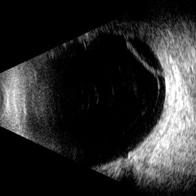
 Asteroid Hyalosis
May 28 2025 by Moazzam Parvez
Ultrasound B-scan image of 63 year-old gentleman with extensive asteroid hyalosis. The bodies stain metachromatically and exhibit birefringence. It appears as discrete, highly echogenic, spherical or star-shaped foci in the vitreous chamber without posterior acoustic shadowing.
Photographer: Dr Moazzam Parvez, Netralayalam, Kolkata
Imaging device: Appaswamy B Scan
Condition/keywords: asteroid hyalosis, birefringence, ul
-
 VKH Pseudotumor – Chronic Subretinal Fibrosis
May 11 2025 by Felipe Murati
Ultra-widefield fundus image from a 36-year-old woman with chronic VKH syndrome showing a pseudotumor-like subretinal fibrotic lesion in the right eye. The lesion developed after multiple relapses and remained stable over a 1-year follow-up with immunosuppressive treatment including prednisone, mycophenolate mofetil, and adalimumab. No active choroiditis or exudative detachment was observed. Multimodal imaging was essential for disease monitoring.
Photographer: Felipe A. Murati, MD, University of Arizona
Imaging device: Optos California ultra-widefield retinal imaging system, single-capture, color fundus modality.
Condition/keywords: adalimumab, chronic inflammation, granulomatous uveitis, OCT, Optos ultra-widefield imaging, pseudotumor, subretinal fibrosis, VKH, Vogt-Koyanagi-Harada
-
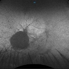
 A Vessel That Would Not Let Go
May 5 2025 by Malvika Singh
Fundus photograph of a retinal detachment showing a horse shoe shaped tear and a bridging vessel.
Photographer: Dr Tejaswita Verma, Retina Foundation, Ahmedabad, India
Imaging device: Mirante SLO/OCT
Condition/keywords: bridging vessel, horseshoe tear
-
 Posterior Nodular Scleritis
Apr 23 2025 by Gustavo Uriel Fonseca Aguirre
This B-mode ultrasound scan demonstrates a posterior scleral nodule accompanied by vitritis, serous retinal detachment, and annular choroidal detachment. The nodule appears as a localized hypoechoic scleral thickening, while the serous retinal detachment shows a smooth convex configuration. The choroidal detachment presents with the characteristic ring-shaped elevation, suggesting significant intraocular inflammation or hypotony.
Photographer: Gustavo U. Fonseca Aguirre, Hospital Conde de Valenciana, Ciudad de México
Condition/keywords: posterior nodular scleritis, posterior scleritis
-
 Retinoschisis
Apr 21 2025 by Gustavo Uriel Fonseca Aguirre
This B-mode longitudinal ultrasound scan reveals a peripheral temporal retinoschisis, demonstrating a characteristic thin, dome-shaped separation of the retinal layers without associated subretinal fluid or vitreous traction. The lesion shows smooth, convex contours with maintained structural integrity of both retinal layers.
Photographer: Gustavo U. Fonseca Aguirre, Hospital Conde de Valenciana, Ciudad de México
Condition/keywords: retinoschisis
-
Hemorrhagic Choroidal Detachment
Apr 14 2025 by Gustavo Uriel Fonseca Aguirre
This B-mode transverse ultrasound scan demonstrates a hemorrhagic choroidal detachment with a characteristic wreath-like configuration, accompanied by concurrent retinal detachment. The choroidal lesion shows dome-shaped elevation with heterogeneous internal reflectivity, while the detached retina appears as a hyperechoic, undulating membrane.
Photographer: Gustavo U. Fonseca Aguirre, Hospital Conde de Valenciana, Ciudad de México
Condition/keywords: hemorrhagic choroidal detachment
-
 Open Funnel (Transversal)
Apr 10 2025 by Gustavo Uriel Fonseca Aguirre
This B-mode transverse ultrasound scan reveals a chronic rhegmatogenous retinal detachment, demonstrating a funnel-shaped configuration with a narrow intraluminal space. Two hyperechoic choroidal calcifications are present, indicative of chronicity.
Photographer: Gustavo U. Fonseca Aguirre, Hospital Conde de Valenciana, Ciudad de México
Condition/keywords: open funnel RD, Retina detachment
-
 Retinopathy of Prematurity
Apr 7 2025 by Gustavo Uriel Fonseca Aguirre
B-mode ultrasound of a 7-month-old premature infant with a history of perinatal supplemental oxygen therapy reveals a total funnel-shaped retinal detachment with significant vasoproliferative tissue causing retinal traction.
Photographer: Gustavo U. Fonseca Aguirre, Hospital Conde de Valenciana, Ciudad de México
Condition/keywords: retinopathy of prematurity
-
 Negative Retinal Detachment
Apr 7 2025 by Gustavo Uriel Fonseca Aguirre
B-mode ultrasound of a 7-month-old premature infant with a history of perinatal supplemental oxygen therapy reveals a total funnel-shaped retinal detachment appearing as a hypoechoic structure, accompanied by significant hyperechoic subretinal hemorrhage. This distinctive echographic pattern creates the characteristic appearance of a "negative retinal detachment."
Photographer: Gustavo U. Fonseca Aguirre, Hospital Conde de Valenciana, Ciudad de México
Condition/keywords: retinopathy of prematurity
-
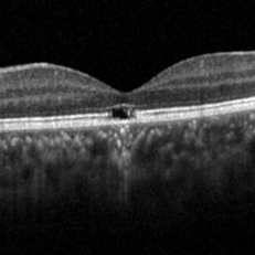 Solar Retinopathy
Apr 1 2025 by Isaac Agranoff
OCT scan of 18-year-old male presenting with 20/40 BCVA OU and bilateral focal outer retinal subfoveal defects. Patient reported long-term history of frequent sungazing, has stopped within past 6-9 months.
Photographer: Isaac Agranoff
Imaging device: Heidelberg Spectralis
Condition/keywords: solar retinopathy
-
 Elmiron Toxicity
Mar 25 2025 by Toolie Winters
Fundus autofluorescence image of a 69-year-old woman with toxic maculopathy OU due to Elmiron usage. Patient stopped using Elmiron in the late 2010s after having been on it for 17 years. The patient has areas of outer retinal and RPE atrophy temporal to fovea that have expanded compared to photos from two years ago. At the time of this appointment, her VA OD was sc20/40-1+2 PH20/30 and VA OS was scCF @ 1 foot.
Photographer: Toolie Winters
Imaging device: Heidelberg Spectralis
Condition/keywords: Elmiron Toxicity, FAF, fundus autofluorescence (FAF), Heidelburg Spectralis, Pentosan Toxicity, Toxic Maculopathy
-
 Hosreshoe Tears on Posterior Pole
Mar 22 2025 by Deepak Bhojwani, MS
A fundus image of an asymptomatic 64 year old male with large horseshoe shaped breaks in inferonasal quadrant on posterior pole, an unusual location for retinal breaks.
Photographer: DR DEEPAK BHOJWANI
Condition/keywords: horseshoe tear, posterior pole break, retinal break
-
 Three Kisses
Mar 18 2025 by Gustavo Uriel Fonseca Aguirre
Cross-section of a B-mode ultrasound showing a kiss-shaped choroidal detachment; three lobes, giving the appearance of three kisses.
Photographer: Gustavo U. Fonseca Aguirre, Hospital Conde de Valenciana, Ciudad de México
Condition/keywords: Kissing Choroidal Detachment
 Loading…