 Initializing download.
-
 By Michael Seider, MD
The Southern California Permanente Medical Group - Uploaded on Feb 13, 2020.
- Last modified by Caroline Bozell on Jul 17, 2020.
- Image of the week
-
Jul 19, 2020
View all images of the week - Rating
- Appears in
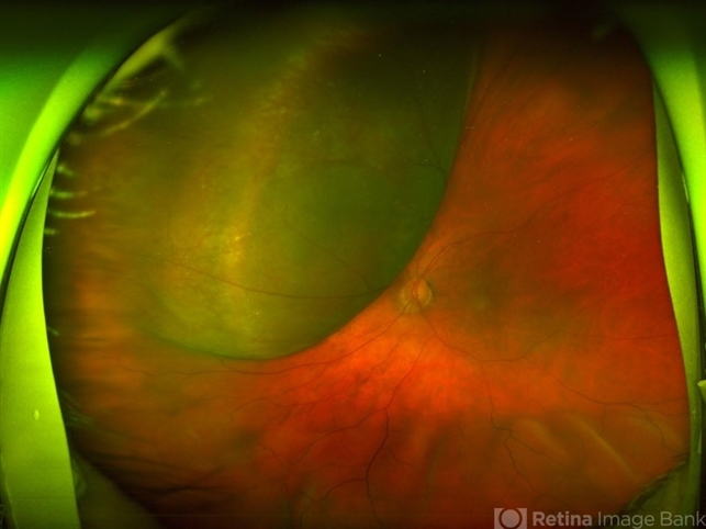
- Large Dome Shaped Peripheral Choroidal Melanoma
- Description
- Large, dome-shaped peripheral choroidal melanoma of the left eye with inferior exudative retinal detachment. Note the lack of obvious orange pigment over the tumor and apparent drusen anteriorly. A lack of ophthalmoscopically obvious lipofuscin is not uncommon among larger choroidal melanomas. B-Scan ultrasonography (transverse, 10 o’clock) confirms a low-moderate internally reflective dome-shaped choroidal lesion with a small adjacent retinal detachment. Ultrasound biomicroscopy (radial, 10 o’clock) confirms no ciliary body involvement of the tumor.