Search results (1465 results)
-
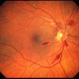
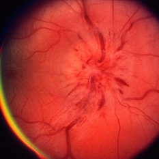 Papillitis
May 2 2013 by Henry J. Kaplan, MD
Anterior optic neuropathy or papillitis in the right eye; notice the blurred optic disc margin, engorged capillaries and flame shaped hemorrhages at the margin.
Condition/keywords: optic disc edema, optic disc swelling, papillitis
-
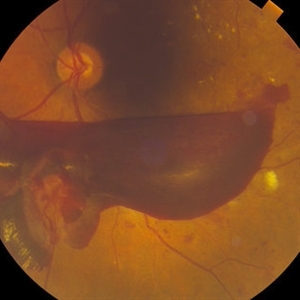
 "Boat-Shaped" Preretinal Hemorrhage
Feb 21 2019 by Mitzy E Torres Soriano, MD
Color fundus photograph showing preretinal (subhyaloid) hemorrhage in a diabetic patient with proliferative diabetic retinopathy.
Photographer: Andrea Vitale, MD
Condition/keywords: preretinal hemorrhage, proliferative diabetic retinopathy (PDR), subhyaloid hemorrhage
-
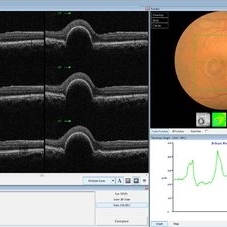 PED due to CSCR
Sep 2 2012 by Hamid Ahmadieh, MD
OCT image of a 37-year-old man with a serous PED secondary to CSCR.
Photographer: Hamid Ahmadieh, Ophthalmic Research Center, Labbafinejad Medical Center
Imaging device: Heidelberg Spectralis
Condition/keywords: central serous chorioretinopathy (CSCR), optical coherence tomography (OCT), pigment epithelial detachment (PED)
-
 Spontaneous Flattening of Drusenoid PED
Jul 1 2014 by John S. King, MD
Consult to r/o ExAMD; observed; scans about a year apart.
Photographer: Wayne A Ladlee Jr
Imaging device: Cirrus
Condition/keywords: drusenoid PED, macular drusenoid lesion, pigment epithelial detachment (PED)
-
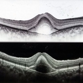
 Fibrovascular PED
Feb 21 2014 by Roy Schwartz, MD
72-year-old female with fibrovascular PED. Upper picture - PED with sub RPE hyper-reflective substance, in a multi-layered pattern, corresponding to fibrovascular PED. CME. Lower picture - PED flattened, a denser sub RPE hyperreflective substance is seen. CME resolved.
Condition/keywords: fibrovascular pigment epithelial detachment (PED), neovascular age-related macular degeneration (AMD), optical coherence tomography (OCT), ranibizumab
-
 "Flower Cataract"
Jul 11 2013 by Jason S. Calhoun
Patient presents with a cataract shaped like a flower. Patient had surgery to remove cataract.
Photographer: Jason S. Calhoun, Department of Ophthalmology, Mayo Clinic Jacksonville, Florida
Condition/keywords: cataract
-
 Boat-Shaped Hemorrhage
Mar 1 2014 by Homayoun Tabandeh, MD, FASRS
Boat-shaped hemorrhage in a patient with retro-hyaloid hemorrhage associated with proliferative diabetic retinopathy.
Condition/keywords: diabetic retinopathy
-
 Epiretinal Membrane
Oct 11 2012 by Michael P. Kelly, FOPS
This is a patient with idiopathic panuveitis who developed a visually significant epiretinal membrane. Pars plana vitrectomy with membrane peeling was performed to remove the epiretinal proliferation. I recommend magnifying the image to see the exquisite detail centrally.
Photographer: Michael P. Kelly, FOPS Director, Duke Eye Center Labs, Duke Universtiy Hospital
Imaging device: Zeiss 450Plus
Condition/keywords: epiretinal membrane (ERM), panuveitis
-
---thumb.jpg/image-square;max$300,300.ImageHandler) Pattern Dystrophy
Pattern Dystrophy
Aug 7 2013 by From the Collections of Thomas M. Aaberg, MD and Thomas M. Aaberg Jr., MD
Typical butterfly shaped pattern dystrophy, right eye #1.
Condition/keywords: butterfly dystrophy, pattern macular dystrophy
-
---thumb.jpg/image-square;max$300,300.ImageHandler) Pediculosis Pubis (Crab Lice)
Pediculosis Pubis (Crab Lice)
Oct 11 2012 by Jeffrey G. Gross, MD, FASRS
Pediculosis pubis (Crab lice) with nits infestation of lashes.
Condition/keywords: nits infestation of lashes, pediculosis pubis (crab lice)
-
 Pigment Epithelial Detachment late FA with small occult CNV
Jul 6 2012 by Tarek S. Hassan, MD, FASRS
72-year-old man with VA loss and metamorphopsia of 2 months duration. PED found, testing done to rule out CNV. Very suspicious for CNV in superonasal fovea/parafovea.
Condition/keywords: choroidal neovascularization (CNV), pigment epithelial detachment (PED)
-
 "Flower Cataract"
Jul 11 2013 by Jason S. Calhoun
Patient presents with a cataract shaped like a flower. Patient had surgery to remove cataract.
Photographer: Jason S. Calhoun, Department of Ophthalmology, Mayo Clinic Jacksonville, Florida
Condition/keywords: cataract
-
 Massive Submacular Hemorrhage
Sep 28 2012 by Joseph M. Civantos, MD
82-year-old gentleman who developed this massive submacular hemorrhage 3 days after his 16th Lucentis injection. Visual acuity dropped from 20/80 to LP.
Condition/keywords: subretinal hemorrhage
-
 CHRPE - grouped pigmentation
Jan 11 2013 by Alex P. Hunyor, MD
Congenital grouped pigmentation of the RPE ("bear tracks").
Condition/keywords: bear tracks, congenital hypertrophy of the retinal pigment epithelium (CHRPE)
-
 Subfoveal Choroidal Neovascularization, In Stereo
Sep 28 2012 by Michael P. Kelly, FOPS
Subfoveal Choroidal Neovascularization.
Photographer: Michael P. Kelly, FOPS Director, Duke Eye Labs, Duke University Hospital, Duke Eye Center, Durham, NC
Imaging device: Zeiss FF4
Condition/keywords: pigment epithelial detachment (PED), stereo pair, subfoveal choroidal neovascularization
-
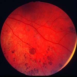
 Pancytopenia
Mar 29 2013 by Henry J. Kaplan, MD
Multiple blot and flame shaped hemorrhages in a patient with pancytopenia #1.
Condition/keywords: pancytopenia, retinal hemorrhage
-
 Hemorrhagic Pigment Epithelial Detachment
Dec 14 2016 by Hashim Ali Khan, OD, FAAO
OCT of a 20-year-old female after trauma with tennis-ball, showing a hemorrhagic PED. RPE is elevated. The second Hyper-reflective band corresponding to Bruchs membrane (BM complex) is visible.
Condition/keywords: pigment epithelial detachment (PED), subretinal hemorrhage
-
 Berlin's Edema With Hemmorrhagic PED
Dec 12 2018 by Surendra Prakash, MBBS, MS, FELLOWSHIP IN VITREO RETINA
Fundus photograph of 24-year-old male having Berlin's edema with multiple sub RPE hemorrhage due to blunt trauma by football.
Photographer: DR SURENDRA PRAKASH
Condition/keywords: Berlin's edema, hemorrhagic detachment of retinal pigment epithelium
-
 Fibrovascular Retinal Pigment Epithelial Detachment - Color Fundus
Jul 16 2014 by James B. Soque, CRA, OCT-C, COA, FOPS
69-year-old white female with Hx of 10 anti-VEFG treatment injections of right eye, VA 20/200, now stable, off drug for 10 months.
Photographer: James B Soque, CRA COA
Imaging device: Topcon TRC 50 DX with MERGE software, 5 MP dig camera
Condition/keywords: color fundus photograph, fibrovascular pigment epithelial detachment (PED), pigment epithelial atrophy, retina
-
 Lattice Degeneration
Nov 9 2012 by Norman Byer
In this 54-year-old woman, lattice degeneration has led to a large horseshoe tractional tear around the posterior side on one end of the lesion resulting in a clinical retinal detachment. Note the very attenuated blood column passing through the white sheath vessel that crosses the tear. This demonstrates that the white blood vessels and a fragment of attached tissue are the only structures which have escaped the tearing effect of the strong vitreoretinal traction which occurred. This usually is true, although, in some cases this bridging vessel may bleed.
Condition/keywords: bridging vessel, lattice degeneration, tractional retinal tear, white sheath vessel
-
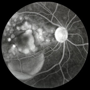 Vogt-Koyanagi-Harada with Multiple PEDs
Oct 10 2012 by Jeffrey G. Gross, MD, FASRS
VKH with multiple PEDs, FA mid phase.
Condition/keywords: FA mid phase, pigment epithelial detachment (PED)
-
 Central Serous Chorioretinopathy (CSC)
Oct 16 2012 by S. Natarajan, MD, FASRS, FRCS (GLASGOW) , FICO, D.Sc, FELA
Middle-aged male came with small PED 4 months back; now this has progressed to a larger PED with SRF underneath the fovea.
Photographer: Prof. Dr. S. Natarajan
Condition/keywords: central serous chorioretinopathy (CSCR), central serous retinopathy (CSR), pigment epithelial detachment (PED), subretinal fibrosis
-
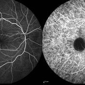 PED due to CSCR 4
Sep 2 2012 by Hamid Ahmadieh, MD
Early phase FA & ICG images of a 37-year-old man with a serous PED secondary to CSCR
Photographer: Hamid Ahmadieh, Ophthalmic Research Center, Labbafinejad Medical Center
Imaging device: Heidelberg Spectralis
Condition/keywords: central serous chorioretinopathy (CSCR), indocyanine green (ICG) angiography, pigment epithelial detachment (PED)
-
 Pigment Epithelial Detachment
Jul 6 2012 by Tarek S. Hassan, MD, FASRS
72-year-old man with VA loss and metamorphopsia of 2 months duration. PED found, testing done to rule out CNV.
Condition/keywords: metamorphopsia, pigment epithelial detachment (PED)
-
 Branch Retinal Vein Occlusion with Macular Edema
Aug 23 2012 by Gerardo Garcia-Aguirre, MD
Fundus photograph composition of the left eye, showing flame-shaped and blot hemorrhages in the superotemporal quadrant, with hard exudates surrounding the fovea.
Photographer: Noemí Hernández, Asociación para Evitar la Ceguera en México
Condition/keywords: branch retinal vein occlusion (BRVO), macular edema
 Loading…