Search results (1463 results)
-
 PED
PED
Oct 17 2014 by Avris Romario Diparaja Siahaan
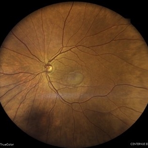
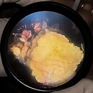
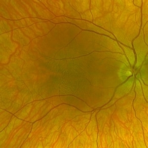
A red free fundus photograph of a 74-year-old man with pigment epithelial detachment in his left eye.
Photographer: Avris Romario Diparaja Siahaan, Klinik Mata Nusantara
Imaging device: Heidelberg Spectralis
Condition/keywords: pigment epithelial detachment (PED), red-free
-
 Polyploidal Choroidal Vasculopathy
Dec 27 2024 by Tejaswita Verma
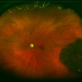
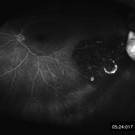
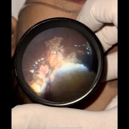
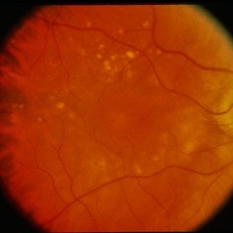
Fundus image of a 74 year old woman with CF1mt vision in right eye showing large PED in a case of PCV. There was associated full thickness macular hole in the same eye.
Photographer: DR. TEJASWITA VERMA
Imaging device: MIRANTE
Condition/keywords: PED, polypoidal choroidal vasculopathy (PCV)
-
 Serous Pigment Epithelial Detachments
Mar 20 2024 by Jeffrey Barker
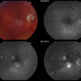
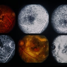
A four up of a color and fluorescein angiogram of a 48 year old male with serous pigment epithelial detachments.
Photographer: Jeffrey P. Barker, B.S.
Condition/keywords: PED
-
 Subretinal Neovascular Membrane with PED
Apr 17 2024 by Akansha Sharma
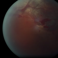
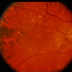
Color fundus photograph of a 72 year old male with ped along with subretinal bleed around it.
Photographer: Dr. Akansha Sharma, Bharati Eye Hospital
Condition/keywords: CNVM, PED, SRNVM, subretinal neovascularization (SRNV), wet age-related macular degeneration (wet AMD)
-
 Acute Posterior Multifocal Placoid Pigment Epitheliopathy
Jan 4 2019 by Cláudia Farinha
Composite image of both eyes of a 27-year-old male with APMPPE. In the fundus photograph, multiple yellowish placoid lesions are observed in the posterior pole in both eyes. The ICGA revealed more lesions than those observed in fundoscopy, and these were hypofluorescent through the angiogram as expected. The en face OCTA segmented at the level of the choriocapillaris revealed areas of ischemia in close correspondence with the hypofluorescent lesions (image superimposed in ICGA ). The OCT b-scan with superimposed flow shows disruption and hyperreflectivity of the external retinal layers in the affected areas and again the absence of flow in the choriocapillaris underneath. A systemic study was carried out to exclude other inflammatory and infectious causes of placoid retinochoroidopathy. The clinical picture resolved after approximately one month from the onset, without recurrence.
Photographer: Pedro Melo, Ophthalmology Department, Centro Hospitalar e Universitário de Coimbra, Coimbra Portugal
Condition/keywords: acute posterior multifocal placoid pigment epitheliopathy (APMPPE), white dot syndrome
-
 Adenocarcinoma Arising from CHRPE
Sep 17 2015 by Marc C. Peden, MD
49-year-old female referred for presumed ocular melanoma. On examination was noted to have darkly pigmented lesion in the temporal retina of left eye. Lesion had characteristic scalloped edges with central lacunae, however, on ultrasonography was noted to have 1.8mm of elevation with high internal reflectivity. IVFA shows absence of dual circulation with areas of window defect. Findings were consistent with those described by Shields et al., in their April 2001 article in Archives of Ophthalmology.
Photographer: Janet Traynom
Imaging device: Optos P200MA
Condition/keywords: adenocarcinoma arising from CHRPE
-
 Adenocarcinoma Arising from CHRPE
Sep 17 2015 by Marc C. Peden, MD
49-year-old female referred for presumed ocular melanoma. On examination was noted to have darkly pigmented lesion in the temporal retina of left eye. Lesion had characteristic scalloped edges with central lacunae, however, on ultrasonography was noted to have 1.8mm of elevation with high internal reflectivity. IVFA shows absence of dual circulation with areas of window defect. Findings were consistent with those described by Shields et al., in their April 2001 article in Archives of Ophthalmology.
Photographer: Janet Traynom COT
Imaging device: Optos P200MA
Condition/keywords: adenocarcinoma arising from CHRPE
-
 Bergmeister's papilla
Jan 19 2023 by pedro fernandes souza neto
Fundus photography of an 35-year-old woman, asymptomatic and found incidentally the Bergmeister's papilla.
Photographer: Pedro Fernandes, Universidade Federal da Bahia
Condition/keywords: Bergmeister's Papillae, congenital prepapillary vascular anomaly
-
 Calcified Retinoblastoma after intra-arterial chemotherapy
Jan 19 2024 by Hector Gabriel Moreno Solano, MD, MHA
Fundus photography of a 5- Year-old Mexican child with bilateral retinoblastoma following unilateral enucleation and 4 cycles of intra-arterial chemotherapy in her only remaining eye. The image shows a succesfully treated tumor with a completely calcificied regression.
Photographer: Hector Solano, Hospital General de Zona #20 IMSS, Puebla
Imaging device: SmartPhone (IPhone 11 ProMax)
Condition/keywords: pediatic retina, pediatric tumor, retinoblastoma
-
 Calcified Retinoblastoma After Intra-arterial Chemotherapy
Apr 6 2024 by Hector Gabriel Moreno Solano, MD, MHA
Fundus photography of a 5 yea-old Mexican child with bilateral retinoblastoma following unilateral enucleation and 4 cycles of intra-arterial chemotherapy in her only remaining eye. The image shows a successfully treated tumor with a completely calcificied regression.
Photographer: Héctor Gabriel Moreno-Solano, MD, MHA
Imaging device: SmartPhone (IPhone 11 pro Max)
Condition/keywords: pediatric retina, pediatric tumor, retinoblastoma
-
 Candy Stripe Sign
Mar 30 2023 by pedro fernandes souza neto
Candy Stripe Sign, patient with proliferative diabetic retinopathy progressing to vitreous hemorrhage and subsequently to ghost cell glaucoma.
Photographer: Marlos Henrique Oliveira Junior, Federal University of Bahia.
Condition/keywords: dehemoglobinized hemorrhage, diabetes, diabetic glaucoma
-
 Choroidal Melanoma
May 24 2023 by pedro fernandes souza neto
Transillumination of Enucleation specimen of Choroidal Melanoma
Photographer: Isabela Valladares Cesar Evangelista, Centro Oftalmológico de Minas Gerais
Condition/keywords: Choroidal melanoma
-
 Choroidal Melanoma
Nov 3 2022 by pedro fernandes souza neto
Transillumination of Enucleation specimen of Choroidal Melanoma: anterior chamber is closed. Total secondary retinal detachment with subretinal serous fluid and some subretinal hemorrhages are present.
Photographer: Eduardo Marback, Federal University of Bahia, Brazil
Condition/keywords: enucleation, melanoma
-
 Choroidal Melanoma
Nov 3 2022 by pedro fernandes souza neto
Enucleation specimen of Choroidal Melanoma: anterior chamber is closed. Total secondary retinal detachment with subretinal serous fluid and some subretinal hemorrhages are present.
Photographer: Eduardo Marback, Federal University of Bahia, Brazil
Condition/keywords: enucleation
-
 Choroidal rupture Subretinal and Vitreous Hemorrhage Secondary to Blunt Trauma
Dec 30 2012 by Humberto Ruiz-Garcia, MD
Fundus photograph of a 23-year-old male, who suffered blunt trauma while working out with resistance rubber bands. The patient presented with "3-ball" hyphema which solved 48 hours with head up positioning and topical steroid and cyclopegic.
Photographer: Pedro Ruiz-Orozco, MD, Clinica Santa Lucia, Guadalajara, Mexico
Condition/keywords: choroidal rupture, vitreous hemorrhage
-
 Coats Disease
Sep 13 2013 by Maria Ana Martinez-Castellanos, MD
Peripheral fundus angiogram in a 2-years-old boy with Coat's disease.
Photographer: Maria A. Martinez-Castellanos. Asociacion para Evitar la Ceguera en Mexico
Imaging device: RetCAm II
Condition/keywords: pediatic retina, serous retinal detachment, vascular anomaly, vascular occlusions
-
 Diabetic Retinopahty
Nov 2 2022 by pedro fernandes souza neto
Fundus photograph of a 40-year-old man with diabetes and hypertension shows hard exudates, difuse intraretinal hemorrhages and splinter hemorrhages.
Photographer: Pedro Fernandes, Universidade Federal da Bahia, Brazil.
Condition/keywords: diabetic mellitus, hypertensive retinopathy, retinopathy
-
 Drusen
Apr 9 2014 by Howard Schatz, MD
Pediatric drusen. RE 20/40 LE 20/100. Drusen.
-
 Foveal hypoplasia - "thumb print" sign
Jul 14 2023 by Pedro S Tetelbom, MD
Right eye fundus photograph of a 26-year-old female with ocular albinism and foveal hypoplasia. "Thumb print" sign can be seen due the reflectance pattern of the Henle Fiber Layer.
Photographer: Johnny Ferrell, Jones Eye Institute, University of Arkansas for Medical Sciences
Imaging device: Optos Silverstone
Condition/keywords: foveal hypoplasia
-
 GHPC
GHPC
May 13 2013 by Howard Schatz, MD
PED in GHPC, 20/200.
Condition/keywords: geographic helicoid peripapillary choroidopathy (GHPC)
-
 Intraocular Foreign Body
Nov 8 2022 by pedro fernandes souza neto
Intraocular foreign body after stone trauma. CT scan image showing intraocular foreign body location.
Photographer: Pedro Fernandes, Federal University of Bahia
Condition/keywords: intraocular foreign body, trauma
-
 Leukemic optic neuropathy
Oct 28 2022 by pedro fernandes souza neto
Fundus photograph of an 18-year-old woman with Leukemic optic neuropathy.
Photographer: Pedro Fernandes, Universidade Federal da Bahia
Condition/keywords: Leukemic optic neuropathy
-
 Myopic Choroidal Neovascularization
Apr 7 2023 by pedro fernandes souza neto
Fundus photography of an 40-year-old man with myopic choroidal neovascularization.
Photographer: Pedro Fernandes, Federal University of Bahia
Condition/keywords: myopia, myopic choroidal neovascularization (CNV)
-
 PED - ARMD Treated with Surround Laser
Dec 17 2014 by David Callanan, MD
Female patient, PED - ARMD treated with surround laser.
Condition/keywords: pigment epithelial detachment (PED)
-
 PED - ARMD Treated with Surround Laser
Dec 17 2014 by David Callanan, MD
Female patient, PED - ARMD treated with surround laser.
Condition/keywords: pigment epithelial detachment (PED)
 Loading…