Search results (210 results)
-
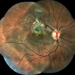
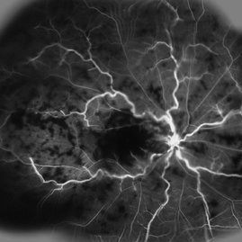
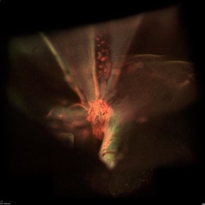
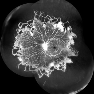 Vascular Non Perfusion in Takayasu Arteritis
Feb 6 2024 by SHILPI H NARNAWARE, ICO ( Retina) , FAICO ( Vitreo-Retina)
A case of 16 year-old female with combined RD in RE. Fundus examination & FFA revealed 360 degrees non-perfusion in periphery in non-symptomatic eye.
Photographer: Shilpi Narnaware, Sarakshi Netralaya , Nagpur, Maharashtra , India
Imaging device: Mirante ( by Nidek)
Condition/keywords: CNP areas, takayasu arteritis
-
 Sickle Cell Retinopathy
Nov 5 2022 by Mateus Queiroz Corrêa, MD
19 -year-old young man with combined rhegmatogenous and tractional retinal detachment secondary to a proliferative sickle retinopathy ( stage V)
Photographer: Mateus Corrêa, Sorocaba Eye Bank Hospital
Imaging device: Optos California
Condition/keywords: Retinal detachment, sickle cell retinopathy
-
 Epiretinal Membrane/Macular Pucker With Combined Hamartoma of Retina and RPE
Jul 8 2015 by Emmanuel Chang, MD PhD FACS FASRS
10-year-old with history of progressive severe distortion in the left eye over the past year.
Photographer: Retina and Vitreous of Texas
Imaging device: Heidelberg Autofluorescence
Condition/keywords: combined hamartoma, epiretinal membrane (ERM), retinal pigment epithelium (RPE) hamartoma
-
 Combined Harmartoma of the Retina and RPE
Apr 4 2023 by Jeffrey Barker
32 Year old Male with a Combined Hamartoma of the Retina and RPE.
Photographer: Jeffrey P. Barker, B.S.
Condition/keywords: hamartoma
-
 Fraternal Twins
May 22 2023 by Gustavo M. Hüning, MD, MBA, FASRS
Intrasurgical photograph using a non-contact system and 3D visualization system of a 65-year-old woman who suffered an ocular trauma.
Photographer: Gustavo M. Hüning, Hüning Clínica do Olhar, Santa Maria - Brazil
Imaging device: Alcon Luxor combined with Alcon nGenuity
Condition/keywords: dislocated intraocular lens (IOL), implant, pars plana vitrectomy (PPV)
-
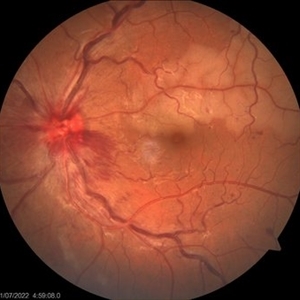 Combined central retinal vein occlusion and branch retinal arteriolar occlusion
Sep 13 2022 by Ruchir Mehta, DO, DNB, FRCS
Fundus photograph of left eye of a 63 years old female with known type 2 DM and HTN showing combined central retinal venous occlusion and superior branch retinal arteriolar occlusion
Photographer: Ruchir Mehta, Mehta Superspeciality Eye Hospital, Jamnagar, Gujarat, India
Imaging device: Zeiss Visucam 500
Condition/keywords: branch retinal artery occlusion (BRAO), central retinal vein occlusion (CRVO), COMBINED
-
 Combined Hamartoma
Oct 5 2016 by Guruprasad S. Ayachit, MBBS,MS
Fundus photograph of a 9-year-old boy with an ill-defined lesion extending from nasal to the disc going on to include the papillomacular bundle; 14X10 mm in greatest dimensions. There is a thick epiretinal membrane causing distortion and straightening of temporal vascular arcade.
Photographer: Shravan Masurkar, M M Joshi Eye Institute, Hubli
Imaging device: Topcon TRC50DX
Condition/keywords: combined hamartoma
-
 Combined Hamartoma of Retina and Retinal Pigment Epithelium
Mar 26 2018 by Hashim Ali Khan, OD, FAAO
Fundus photograph of a 12-year-old boy with combined hamartoma of retina and retinal pigment epithelium.
Condition/keywords: combined hamartoma, retinal pigment epithelium
-
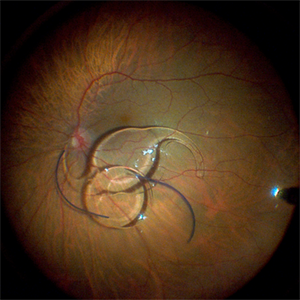
 Dislocation of the Crystalline Lens with a Retinal Detachment
Apr 21 2025 by Hrishikesh Naik, MS
An intraoperative screen grab shows a dislocation of the crystalline lens along with an associated rhegmatogenous retinal detachment in a case of Marfan’s syndrome. The case was managed by a combined PPV-SB procedure. A vitrectomy cutter is seen at the left.
Photographer: Hrishikesh Naik
Condition/keywords: intraoperative, lens dislocation, Marfan's syndrome, Retinal Detachment, vitrectomy
-
 Early Phase of FA in a Case of Combined CRAO and CRVO
May 15 2014 by Manish Nagpal, MD, FRCS (UK), FASRS
30-year-old anemic lady presented with a acute loss of vision. Her vision was just hand movements in the affected eye and the other eye was normal.
Photographer: pooja barot, Optometrist, Retina Foundation, Ahmedabad
Condition/keywords: central retinal artery occlusion (CRAO), central retinal vein occlusion (CRVO), macular edema
-
 Exudative Macular Detachment After Intensive Laser Photocoagulation
Mar 12 2016 by Sjakon G Tahija, MD
Fundus photograph of 44-year-old man with exudative detachment of the macula after vitrectomy and ILM peeling for proliferative diabetic retinopathy combined with intensive endolaser photocagulation.
Photographer: Avris Siahaan, Klinik Mata Nusantara
Condition/keywords: exudative detachment, pan-retinal photocoagulation (PRP)
-
 Fluorescein Angiography of a Combined CRAO AND CRVO
Mar 24 2017 by Manish Nagpal, MD, FRCS (UK), FASRS
Young girl 22-year-old came with sudden decreased vision and had a combined CRAO and CRVO whose FA revealed extensive ischemia and delayed filling.
Photographer: pranita chaudhary
Condition/keywords: central retinal artery occlusion (CRAO), central retinal vein occlusion (CRVO)
-
 Montage OF a Combined Case of CRVO and CRAO
May 15 2014 by Manish Nagpal, MD, FRCS (UK), FASRS
30-year-old anemic lady presented with a acute loss of vision. Her vision was just hand movements in the affected eye and the other eye was normal.
Photographer: pooja barot, Optometrist, Retina Foundation, Ahmedabad
Condition/keywords: central retinal artery occlusion (CRAO), central retinal vein occlusion (CRVO), macular edema
-
 Outer Retinal Tear in Schisis-Detachment
Mar 25 2016 by Gregory R. Blaha, MD, PhD
Large outer retinal tear in combined retinoschisis-detachment. The retinal vessels are visible going over the retinal break.
Photographer: Janice Neal, Gurley Eye Care Associates
Imaging device: Topcon Mark II
Condition/keywords: retinal tear, retinoschisis
-
 Pseudophakic RRD, S/P Buckle/Vit. w/ Residual Gas Fish Eggs OD
May 23 2018 by Hosam Attia, MD
71-year-old male, s/p combined buckle vitrectomy for recurrent, macula-off, rhegmatogenous retinal detachment, with residual gas fish eggs OD.
Imaging device: Optos California Ultra-Wide Field Fundus Camera
Condition/keywords: encircling scleral buckle, gas bubble, intraocular gas, intravitreal gas bubble
-
 Total Rhegmatogenous and Tractional Retinal Detachment Following Choroidal Melanoma Laser Ablation Treatment
Sep 22 2020 by Sophia El Hamichi, MD
A 69-year-old female, with a history of choroidal melanoma in her left eye with exudative detachment, underwent tumor laser ablation. She then developed a complex combined tractional and rhegmatogenous retinal detachment with a giant retinal tear.
Photographer: Belinda Rodriguez, Murray Ocular Oncology and Retina, Miami
Condition/keywords: tractional retinal detachment
-
 Tractional and Rhegmatogenous Retinal Detachment
Dec 30 2016 by Manish Nagpal, MD, FRCS (UK), FASRS
Combined tractional and rhegmatogenous retinal detachment seen in a case of vasculitis. Two small breaks are noted in the extreme nasal area next to an area of proliferation.
Photographer: Avijit Vishnoi
Condition/keywords: tractional retinal detachment, vasculitis
-
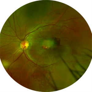
 Combined Hamartoma of the Retina and Retinal Pigment Epithelium (CHRRPE)
Jan 21 2020 by Pierre-Henry Gabrielle, MD
Fundus photograph of a 17-year-old man with combined hamartomas of the retina and retinal pigment epithelium (CHRRPE) at the posterior pole of the left eye.
Photographer: Pierre-Henry Gabrielle, Ophthalmology department, Dijon University Hospital, France
Imaging device: Zeiss Visucam
Condition/keywords: combined hamartoma, fundus photograph
-
 Hamartoma of the Retina
May 29 2018 by JEFFERSON R SOUSA, Tecg.º (Biomedical Systems Technology)
A 4-year-old male patient attended the clinic for evaluation. In the mapping examination and retina and retinography, important alterations were observed in the posterior pole of the left eye. This in turn was sent to perform the ocular ultrasonography examination, which together with the previous examinations, confirmed changes that suggested diagnosis of: COMBINED HAMARTOMA OF RETINA AND PIGMENTARY EPITHELIUM.
Photographer: JEFFERSON R SOUSA - Study Center and Ophthalmological Research Dr. Andre M V Gomes, Institute Dr. Suel Abujamra São Paulo-Brazil
Imaging device: Topcon TRC-50 DX, Imaginet 5.0, angle de 35º . Flash 36 / Mosaic with 9 images.
Condition/keywords: combined hamartoma, retinal pigment epithelium (RPE) hamartoma, tumor
-
 Pseudophakic RRD, S/P Buckle/Vit. w/ Residual Gas Fish Eggs OD
May 23 2018 by Hosam Attia, MD
71-year-old male, s/p combined buckle vitrectomy for recurrent, macula-off, rhegmatogenous retinal detachment, with residual gas fish eggs OD.
Imaging device: Optos California Ultra-Wide Field Fundus Camera
Condition/keywords: encircling scleral buckle, gas bubble, intraocular gas, intravitreal gas bubble
-
 Combined Hamartoma
Feb 29 2016 by Andrea Arriola-Lopez, MD MSc
40 year-old man with diminished VA since 6 month ago. Fundus examination revealed macular folds, yellow-whitish elevated lesion at the fovea and a subretinal hemorrhage.
Photographer: Andrea Elizabeth Arriola-Lopez MD, MSc
Imaging device: OPTOS Dakota
Condition/keywords: combined hamartoma, macula, subretinal hemorrhage
-
 Combined Hamartoma of Retina and RPE
May 10 2019 by Deepak Bhojwani, MS
A 31-year-old male came with incidental finding of poor vision in left eye on his screening eye examination (done for job purpose). His left eye depicts classic combined hamartoma of retina and RPE in left eye. Right examination was unremarkable.
Photographer: Deepak Bhojwani, Raghudeep Eye Hospital, Ahmedabad
Imaging device: Zeiss Viscam 500
Condition/keywords: combined hamartoma, retina, retinal pigment epithelium, tumor
-
 Combined Hamartoma of the Retinal Pigment Epithelium Case 1
Oct 5 2012 by Ronald C. Gentile, MD
A peripapilary combined hamartoma of the retinal pigment epithelium involving the nasal disc margin. This tumor is slightly elevated, charcoal grey in color with grey-white tissue on it surface. The underlying retinal vessels are obscured.
Photographer: The New York Eye & Ear Infirmary Department of Medical Imaging
Condition/keywords: hamartoma, retinal pigment epithelium
-
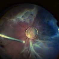
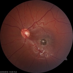
 360 retinotomy for combined closed funnel tractional and rhematogenous retinal detachment
Jan 1 2023 by Malek Yassine, MD
This is Fundus Autofluorecence, showing the residual hypoautofluorescent spots on the exposed choroid, relating to the previous panretinal photocoagulation, as well as the limits of the retinotomy with continuous laser which appeasr hypoautofluorecent with hyperautofluorecent margins.
Photographer: Malek Yassine, HMOED, Agadir, Morocco.
Imaging device: Zeiss Clarus
Condition/keywords: combined retinal detachment, rhegmatogenous retinal detachment, tractional retinal detachment
-
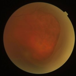
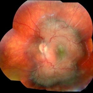
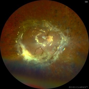 360 Retinotomy in a closed Funnel combined Tractional and rhegmatogenous retinal detachment
Jan 1 2023 by Malek Yassine, MD
This is the results at 6 months of a Bimanual 23 G-PPV with a very extensive and posterior 360 retinotomy for the management of a combined longstanding closed funnel RD, with submacular membranes, intraretinal PVR. Preop VA was a doubtful light perception. Borders of the retinotomy are stable at 6 months under 1300 Cs Silicon oil with some pigmented PVR developping the edges. Macula appears spared. Silicon oil emulsification droplets are well visualized beneath the superior temporal arcade.
Imaging device: Zeiss Clarus 700
Condition/keywords: combined retinal detachment, retinotomy, silicone oil
 Loading…