Search results (210 results)
-
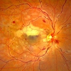
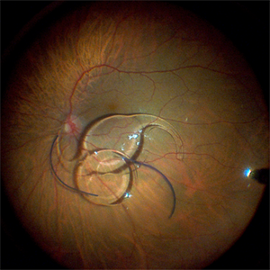
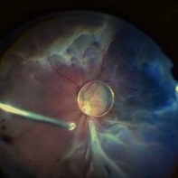 Dislocation of the Crystalline Lens with a Retinal Detachment
Apr 21 2025 by Hrishikesh Naik, MS
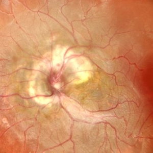
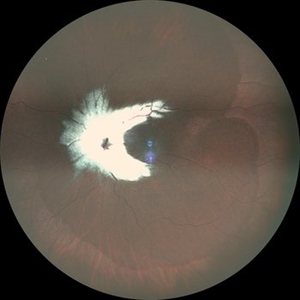
An intraoperative screen grab shows a dislocation of the crystalline lens along with an associated rhegmatogenous retinal detachment in a case of Marfan’s syndrome. The case was managed by a combined PPV-SB procedure. A vitrectomy cutter is seen at the left.
Photographer: Hrishikesh Naik
Condition/keywords: intraoperative, lens dislocation, Marfan's syndrome, Retinal Detachment, vitrectomy
-
 Uveal Effusion Syndrome in a Nanophthalmic Eye
Apr 3 2025 by Gustavo Uriel Fonseca Aguirre
Combined B-mode ultrasound with A-mode vector analysis in a nanophthalmic eye (axial length: 14.49 mm).
Photographer: Gustavo U. Fonseca Aguirre, Hospital Conde de Valenciana, Ciudad de México
Condition/keywords: nanophthalmos, uveal effusion syndrome
-
 Combined Traction and Rhegmatogenous Retinal Detachment From Proliferative Diabetic Retinopathy
Mar 27 2025 by Nikhil K Bommakanti, MD
A middle-aged patient presented with a combined traction and rhegmatogenous retinal detachment.
Condition/keywords: Active PDR Tractional retinal Detachment, PDR, Retinal Detachment, rrd, TRD
-
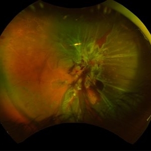
 Combined Retinal Detachment With a Butterfly Shaped Configuration
Mar 13 2025 by S. Natarajan, MD, FASRS, FRCS (GLASGOW) , FICO, D.Sc, FELA
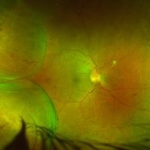
A 46 year old female presented to us with diminished vision in both the eyes. Her blood glucose levels were deranged. She had bilateral proliferative diabetic retinopathy and pan retinal photocoagulation was done elsewhere. Left eye showed a combined retinal detachment with fibrovascular proliferation on the disc and along inferior arcade with a convex configuration of retinal detachment. Patient was planned for surgical intervention. The image shows a butterfly like configuration of combined retinal detachment with the subretinal fluid pocket appearing like the wings of the butterfly.
Photographer: ASHWINI SUTAR ADITYA JYOT EYE HOSPITAL
Imaging device: Mirante ( PLEASE SELECT COVER PAGE )
Condition/keywords: retinal detachment with a butterfly shaped
-
 Combined Hamartoma of the Retina and RPE
Jan 23 2025 by Tejaswita Verma
A 10 year old boy presented with 6/60 vision and LE exotropia with the fundus lesion suggesting a chronic etiology and ILM folds.
Photographer: DR. TEJASWITA VERMA
Imaging device: MIRANTE
Condition/keywords: combined hamartoma of retina and RPE
-
 Combined Pathology
Oct 26 2024 by rahul saradge
53 year old male patient was presented with a complaints of diminished vision in LE since 1 month. The BCVA in RE was 6/36p and LE was CF 1/2m. Ocular dilated examination showed RE temporal CD with ?CRVO,OIS and OS showed TRD and old Hemi CRVO. Patient was injected with PST tricot followed by PRP laser at an interval of 1 week. Patient improved to BCVA 6/9.
Photographer: Aishwarya Bangar Isha Netralaya Thane
Imaging device: optos
Condition/keywords: choroidal detachment, crvo, ois, optos, pan retinal photocoagulation, tractional retinal detachment
-
 Combined Traction Rhegmatogenous Detachment
Oct 17 2024 by Hemanth Murthy, MBBS, MD, FASRS
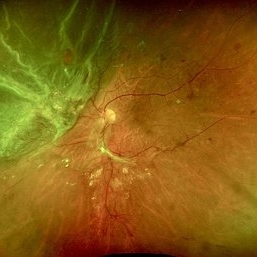
A 68 year old male presented with a shadow in the left eye since 3 days. He was a known diabetic and hypertensive for 20 years. Vision was 20/40 in right eye and 20/60 in left eye. Fundus examination showed Proliferative diabetic retinopathy in right eye and Proliferative diabetic retinopathy with combined traction rhegmatogenous detachment in left eye.
Photographer: Mr Veda Vyas
Condition/keywords: combined retinal detachment, proliferative diabetic retinopathy (PDR)
-
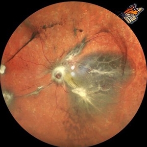
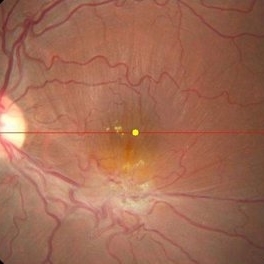
 Combined Retinal Detachment With Macular Hole
Sep 28 2024 by Tejaswita Verma
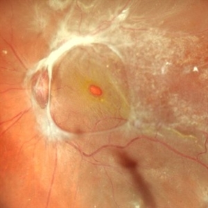
Fundus image of the LE of a 67 year old diabetic, hypertensive female with CF 3metres vision showing combined RD with FTMH, in a pseudophakic eye. She was lost to follow up status post 2 anti VEGF injections received 8 months back due to typhoid fever.
Photographer: DR. TEJASWITA VERMA
Imaging device: MIRANTE
Condition/keywords: full thickness macular hole, proliferative diabetic retinopathy (PDR), tractional retinal detachment
-
 Myelinated Nerve Fibres With Combined Hamartoma of Retina and RPE
Jul 31 2024 by Tejaswita Verma
Fundus image of a 20 year old female who presented with metamorphopsia ,slightly blurred vision. BCVA was 6/9, epiretinal membrane present on central fundus examination with myelinated nerve fibres.
Photographer: DR. TEJASWITA VERMA
Imaging device: MIRANTE
Condition/keywords: combined hamartoma of retina and RPE, myelinated nerve fibers
-
 Post Combined Surgery of Cataract, TRD & Vitreous Hemorrhage
Jun 27 2024 by Sanauddin Samejo , Diploma (Ophthalmic Technician Training Course)
A 27 year-old diabetic female visited the clinic one week after combined surgery of cataract, tractional retinal detachment and vitreous hemorrhage.
Photographer: Sanauddin Samejo, Burjeel Hospital, Abu Dhabi, UAE
Imaging device: Silver Stone Optos
Condition/keywords: Combined Surgery Cataract Tractional Retinal Detachment Vitreous Hemorrhage, POST SURGERY, Retinal Detachment, TRD
-
 Hollenhorst Plaque
Jun 25 2024 by Virginia Gebhart
75 year female with complaint of shadow in the bottom of her vision for many years. Hollenhorst plaque on superior pole of the disc and sclerotic superotemporal arteriole. Also DBHs superiorly most likely due to combined BRAO/BRVO.
Photographer: Virginia Gebhart
Imaging device: Topcon 50DX
Condition/keywords: branch retinal artery occlusion (BRAO), branch retinal vein occlusion (BRVO), hollenhorst plaque, sclerotic arteriole
-
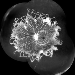
 Combined Retinal Artery Macro Aneurysm with Retinal Vein Occlusion
Jun 25 2024 by Aniruddh Soni, DO DNB FLVPEI
Color Fundus and Fluorescein Angiography Montage of a 55 year Lady with Combined Retinal Artery Macro Aneurysm with Retinal Vein Occlusion.
Photographer: Dr Aniruddh Soni, Anupam Eye Hospital, Jaipur, INDIA
Condition/keywords: macroaneurysm, Vein Occlusion
-
 Combined Retinal Artery Macro Aneurysm with Retinal Vein Occlusion
Jun 25 2024 by Aniruddh Soni, DO DNB FLVPEI
Left Eye Color Fundus and Fluorescein angiography Monatge of a 55 Year Female showing Retinal Artery Macro Aneurysm with Retinal Vein Occlusion.
Photographer: Dr Aniruddh Soni, Anupam Eye Hospital, Jaipur, INDIA
Condition/keywords: macroaneurysm, Vein Occlusion
-
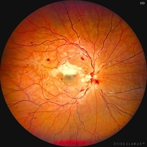 Combined Central Retinal Vein Occlusion with Branch Retinal Artery Occlusion
Apr 29 2024 by KANWALJEET HARJOT MADAN, M.S. (Ophthalmology), FAICO (Vitreous - Retina)
This is fundus photograph of a 46-year male patient who presented with sudden diminution of vision in his right eye (RE) for 3 days. He was hypertensive but non diabetic. On examination, his best corrected vision in RE was 6/12. His left eye (LE) was normal. His fundus examination in RE revealed multiple intra retinal hemorrhages in all quadrants with tortuosity of veins suggestive of central retinal vein occlusion (CRVO) with mild disc edema. An ischemic area was seen superior to fovea suggestive of branch retinal artery occlusion. OCT depicted thickening of inner retinal layers with little evidence of macular edema. Hematological and cardio vascular investigations were done. He had bilateral thickening of intimal and medial walls of carotid arteries. He was under cardiology treatment. His vision improved to 6/6.
Photographer: Dr. Kanwaljeet Harjot Madan, M.S. (Ophthalmologist) Fellow in Vitrous & Retina. Thind Eye Hospital, Jalandhar City. Punjab. India
Condition/keywords: branch retinal artery occlusion (BRAO), central retinal vein occlusion (CRVO)
-
 Combined Central Retinal Vein Occlusion with Branch Retinal Artery Occlusion
Apr 28 2024 by KANWALJEET HARJOT MADAN, M.S. (Ophthalmology) Fellow in Vitreous & Retina
This is fundus photograph of a 46-year male patient who presented with sudden diminution of vision in his right eye (RE) for 3 days. He was hypertensive but non diabetic. On examination, his best corrected vision in RE was 6/12. His left eye (LE) was normal. His fundus examination in RE revealed multiple intra retinal hemorrhages in all quadrants with tortuosity of veins suggestive of central retinal vein occlusion (CRVO) with mild disc edema. An ischemic area was seen superior to fovea suggestive of branch retinal artery occlusion. OCT depicted thickening of inner retinal layers with little evidence of macular edema. Hematological and cardio vascular investigations were done. He had bilateral thickening of intimal and medial walls of carotid arteries. He was under cardiology treatment. His vision improved to 6/6.
Photographer: Dr Kanwaljeet Harjot Madan
Condition/keywords: branch retinal artery occlusion (BRAO), central retinal vein occlusion
-
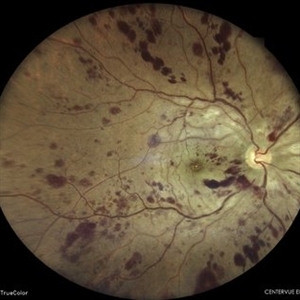 Combined Central Retinal Artery and Vein Occlusion
Apr 8 2024 by Akansha Sharma
Color fundus photograph of a 63 year old male with combined central retinal artery and vein occlusion with carotid artery stenosis and infarct in the brain.
Photographer: Dr. Akansha Sharma, Bharati Eye Hospital
Condition/keywords: central retinal artery occlusion (CRAO), central retinal vein occlusion (CRVO), CRAO
-
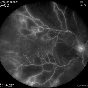 Combined Central Retinal Artery and Vein Occlusion
Apr 8 2024 by Akansha Sharma
Fundus fluorescein angiography of a 63 year old male with combined central retinal artery and vein occlusion with carotid artery stenosis and infarct in the brain demonstrating late filling.
Photographer: Dr. Akansha Sharma, Bharati Eye Hospital
Condition/keywords: central retinal artery occlusion (CRAO), central retinal vein occlusion (CRVO), CRAO
-
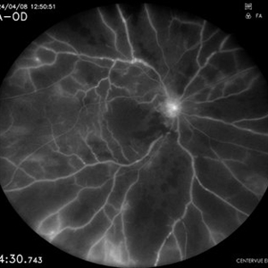 Combined Central Retinal Artery and Vein Occlusion
Apr 8 2024 by Akansha Sharma
Fundus fluorescein angiography of a 63 year old male with combined central retinal artery and vein occlusion with carotid artery stenosis and infarct in the brain demonstrating late filling.
Photographer: Dr. Akansha Sharma, Bharati Eye Hospital
Condition/keywords: central retinal artery occlusion (CRAO), central retinal vein occlusion (CRVO), CRAO
-
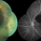
 Vascular Non Perfusion in Takayasu Arteritis
Feb 6 2024 by SHILPI H NARNAWARE, ICO ( Retina) , FAICO ( Vitreo-Retina)
A case of 16 year-old female with combined RD in RE. Fundus examination & FFA revealed 360 degrees non-perfusion in periphery in non-symptomatic eye.
Photographer: Shilpi Narnaware, Sarakshi Netralaya , Nagpur, Maharashtra , India
Imaging device: Mirante ( by Nidek)
Condition/keywords: CNP areas, takayasu arteritis
-
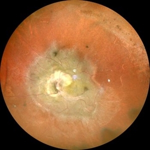
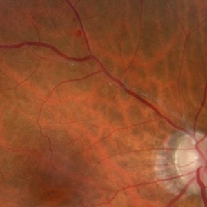
 Combined hamartoma of retina and retinal pigment epithelium
Aug 8 2023 by Navneet Mehrotra, DNB
A 20 year old female presented with decreased vision and metamorphopsia noticed in her left eye for one year. Other eye was normal. BCVA was 6/12 in her left eye.
Photographer: Dharti, Retina Care , Ahmedabad
Condition/keywords: Combined pigment epithelial and retinal hamartoma
-
 Fraternal Twins
May 22 2023 by Gustavo M. Hüning, MD, MBA, FASRS
Intrasurgical photograph using a non-contact system and 3D visualization system of a 65-year-old woman who suffered an ocular trauma.
Photographer: Gustavo M. Hüning, Hüning Clínica do Olhar, Santa Maria - Brazil
Imaging device: Alcon Luxor combined with Alcon nGenuity
Condition/keywords: dislocated intraocular lens (IOL), implant, pars plana vitrectomy (PPV)
-
 Cutter Segmentation in a case of Diabetic Combined Retinal Detachment | Intra-Operative Still
Apr 25 2023 by Veer Singh, MS, FVRS, FMRF, FICO (Retina)
Cutter Segmentation in a case of Diabetic Combined Retinal Detachment | Intra-Operative Still Patient underwent Vitrectomy with Silicone Oil
Photographer: Dr. Veer Singh
Condition/keywords: combined retinal detachment, cutter, diabetic retinopathy, intraoperative, pars plana vitrectomy (PPV)
-
 Combined Harmartoma of the Retina and RPE
Apr 4 2023 by Jeffrey Barker
32 Year old Male with a Combined Hamartoma of the Retina and RPE.
Photographer: Jeffrey P. Barker, B.S.
Condition/keywords: hamartoma
 Loading…