 Initializing download.
-
 By JEFFERSON R SOUSA, Tecg.º (Biomedical Systems Technology)
Lens Oftalmologia - Hospital Beneficiência Portuguesa
Co-author(s): Dra. Luíza Salles de Moura Mendonça - Uploaded on May 29, 2018.
- Last modified by Caroline Bozell on May 29, 2018.
- Rating
- Appears in
- Miscellaneous
- Condition/keywords
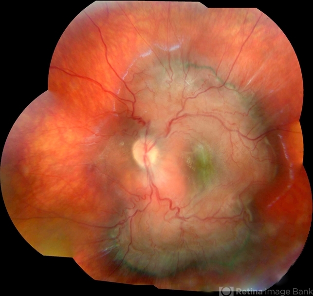
- combined hamartoma, retinal pigment epithelium (RPE) hamartoma, tumor
- Photographer
- JEFFERSON R SOUSA - Study Center and Ophthalmological Research Dr. Andre M V Gomes, Institute Dr. Suel Abujamra São Paulo-Brazil
- Imaging device
-
Fundus camera
Topcon TRC-50 DX, Imaginet 5.0, angle de 35º . Flash 36 / Mosaic with 9 images. - Description
- A 4-year-old male patient attended the clinic for evaluation. In the mapping examination and retina and retinography, important alterations were observed in the posterior pole of the left eye. This in turn was sent to perform the ocular ultrasonography examination, which together with the previous examinations, confirmed changes that suggested diagnosis of: COMBINED HAMARTOMA OF RETINA AND PIGMENTARY EPITHELIUM.

---thumb.jpg/image-square;max$79,0.ImageHandler "Vitrectomy Choroidal Mass")
---thumb.jpg/image-square;max$79,0.ImageHandler "Combined Hamartoma")


