Search results (10 results)
-
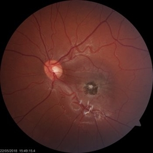
 Post Combined Surgery of Cataract, TRD & Vitreous Hemorrhage
Jun 27 2024 by Sanauddin Samejo , Diploma (Ophthalmic Technician Training Course)
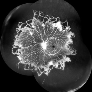
A 27 year-old diabetic female visited the clinic one week after combined surgery of cataract, tractional retinal detachment and vitreous hemorrhage.
Photographer: Sanauddin Samejo, Burjeel Hospital, Abu Dhabi, UAE
Imaging device: Silver Stone Optos
Condition/keywords: Combined Surgery Cataract Tractional Retinal Detachment Vitreous Hemorrhage, POST SURGERY, Retinal Detachment, TRD
-
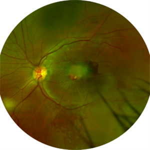
 Vascular Non Perfusion in Takayasu Arteritis
Feb 6 2024 by SHILPI H NARNAWARE, ICO ( Retina) , FAICO ( Vitreo-Retina)
A case of 16 year-old female with combined RD in RE. Fundus examination & FFA revealed 360 degrees non-perfusion in periphery in non-symptomatic eye.
Photographer: Shilpi Narnaware, Sarakshi Netralaya , Nagpur, Maharashtra , India
Imaging device: Mirante ( by Nidek)
Condition/keywords: CNP areas, takayasu arteritis
-
 Fraternal Twins
May 22 2023 by Gustavo M. Hüning, MD, MBA, FASRS
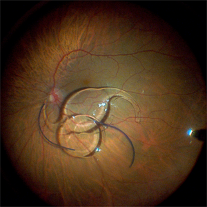
Intrasurgical photograph using a non-contact system and 3D visualization system of a 65-year-old woman who suffered an ocular trauma.
Photographer: Gustavo M. Hüning, Hüning Clínica do Olhar, Santa Maria - Brazil
Imaging device: Alcon Luxor combined with Alcon nGenuity
Condition/keywords: dislocated intraocular lens (IOL), implant, pars plana vitrectomy (PPV)
-
 Combined central retinal vein occlusion and branch retinal arteriolar occlusion
Sep 13 2022 by Ruchir Mehta, DO, DNB, FRCS
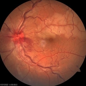
Fundus photograph of left eye of a 63 years old female with known type 2 DM and HTN showing combined central retinal venous occlusion and superior branch retinal arteriolar occlusion
Photographer: Ruchir Mehta, Mehta Superspeciality Eye Hospital, Jamnagar, Gujarat, India
Imaging device: Zeiss Visucam 500
Condition/keywords: branch retinal artery occlusion (BRAO), central retinal vein occlusion (CRVO), COMBINED
-
 Sickle Cell Retinopathy
Nov 5 2022 by Mateus Queiroz Corrêa, MD
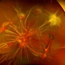
19 -year-old young man with combined rhegmatogenous and tractional retinal detachment secondary to a proliferative sickle retinopathy ( stage V)
Photographer: Mateus Corrêa, Sorocaba Eye Bank Hospital
Imaging device: Optos California
Condition/keywords: Retinal detachment, sickle cell retinopathy
-
 Combined Hamartoma of the Retina and Retinal Pigment Epithelium (CHRRPE)
Jan 21 2020 by Pierre-Henry Gabrielle, MD
Fundus photograph of a 17-year-old man with combined hamartomas of the retina and retinal pigment epithelium (CHRRPE) at the posterior pole of the left eye.
Photographer: Pierre-Henry Gabrielle, Ophthalmology department, Dijon University Hospital, France
Imaging device: Zeiss Visucam
Condition/keywords: combined hamartoma, fundus photograph
-
 Combined Hamartoma
Oct 5 2016 by Guruprasad S. Ayachit, MBBS,MS
Fundus photograph of a 9-year-old boy with an ill-defined lesion extending from nasal to the disc going on to include the papillomacular bundle; 14X10 mm in greatest dimensions. There is a thick epiretinal membrane causing distortion and straightening of temporal vascular arcade.
Photographer: Shravan Masurkar, M M Joshi Eye Institute, Hubli
Imaging device: Topcon TRC50DX
Condition/keywords: combined hamartoma
-
 Combined Hamartoma
Feb 29 2016 by Andrea Arriola-Lopez, MD MSc
40 year-old man with diminished VA since 6 month ago. Fundus examination revealed macular folds, yellow-whitish elevated lesion at the fovea and a subretinal hemorrhage.
Photographer: Andrea Elizabeth Arriola-Lopez MD, MSc
Imaging device: OPTOS Dakota
Condition/keywords: combined hamartoma, macula, subretinal hemorrhage
-
 Epiretinal Membrane/Macular Pucker With Combined Hamartoma of Retina and RPE
Jul 8 2015 by Emmanuel Chang, MD PhD FACS FASRS
10-year-old with history of progressive severe distortion in the left eye over the past year.
Photographer: Retina and Vitreous of Texas
Imaging device: Heidelberg Autofluorescence
Condition/keywords: combined hamartoma, epiretinal membrane (ERM), retinal pigment epithelium (RPE) hamartoma
-
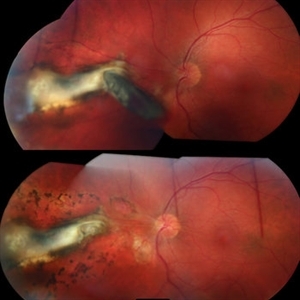 IOFB Combined
Mar 12 2015 by Ahmad B. Tarabishy, MD
A 26-year-old gentleman presented with a metallic intraocular foreign body embedded in the nasal retina (above). Post-operative appearance two weeks after vitrectomy, foreign body removal, endolaser, and gas (below).
Photographer: Jessica Armbruster
Imaging device: Topcon TRC-50EX
Condition/keywords: encapsulated intraocular foreign body, non metallic retained intraocular foreign body (RIOFB), penetrating trauma
 Loading…