Search results (210 results)
-
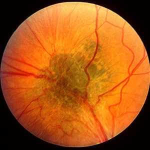
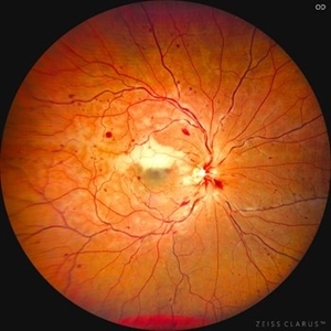
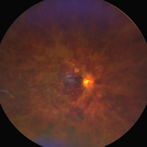 Combined central retinal vein and artery occlusion
Jun 19 2022 by T. P . VIGNESH, MBBS,MS
Fundus photo of a 67 year old female patient, revealing multiple superficial retinal haemorrhages and occlusion of both central retinal vein and central retinal artery.
Photographer: Bharathi Singaravel
Imaging device: Zeiss Clarus
Condition/keywords: central retinal artery occlusion (CRAO), COMBINED, ischemic CRVO
-
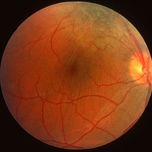
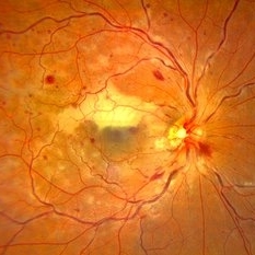
 Combined central retinal vein occlusion and branch retinal arteriolar occlusion
Sep 13 2022 by Ruchir Mehta, DO, DNB, FRCS
Fundus photograph of left eye of a 63 years old female with known type 2 DM and HTN showing combined central retinal venous occlusion and superior branch retinal arteriolar occlusion
Photographer: Ruchir Mehta, Mehta Superspeciality Eye Hospital, Jamnagar, Gujarat, India
Imaging device: Zeiss Visucam 500
Condition/keywords: branch retinal artery occlusion (BRAO), central retinal vein occlusion (CRVO), COMBINED
-
 360 retinotomy for combined closed funnel tractional and rhematogenous retinal detachment
Jan 1 2023 by Malek Yassine, MD
This is Fundus Autofluorecence, showing the residual hypoautofluorescent spots on the exposed choroid, relating to the previous panretinal photocoagulation, as well as the limits of the retinotomy with continuous laser which appeasr hypoautofluorecent with hyperautofluorecent margins.
Photographer: Malek Yassine, HMOED, Agadir, Morocco.
Imaging device: Zeiss Clarus
Condition/keywords: combined retinal detachment, rhegmatogenous retinal detachment, tractional retinal detachment
-
 360 Retinotomy in a closed Funnel combined Tractional and rhegmatogenous retinal detachment
Jan 1 2023 by Malek Yassine, MD
This is the results at 6 months of a Bimanual 23 G-PPV with a very extensive and posterior 360 retinotomy for the management of a combined longstanding closed funnel RD, with submacular membranes, intraretinal PVR. Preop VA was a doubtful light perception. Borders of the retinotomy are stable at 6 months under 1300 Cs Silicon oil with some pigmented PVR developping the edges. Macula appears spared. Silicon oil emulsification droplets are well visualized beneath the superior temporal arcade.
Imaging device: Zeiss Clarus 700
Condition/keywords: combined retinal detachment, retinotomy, silicone oil
-
 CHRRPE
Apr 26 2020 by Dipak Nag, MBBS, FCPS, MSc, FRF
CHRRPE
Condition/keywords: combined hamartoma, retinal pigment epithelium
-
 Combined Hamartoma
Feb 20 2013 by From the Collections of Thomas M. Aaberg, MD and Thomas M. Aaberg Jr., MD
No history.
Condition/keywords: combined hamartoma
-
 Combined Hamartoma
Feb 20 2013 by From the Collections of Thomas M. Aaberg, MD and Thomas M. Aaberg Jr., MD
No history.
Condition/keywords: combined hamartoma
-
 Combined Hamartoma of Retina and RPE
Mar 29 2013 by Henry J. Kaplan, MD
Greenish lesion on the arcade with epiretinal membrane formation, vessels inside the lesion are contracted and those outside are distracted.
Condition/keywords: combined hamartoma
-
 Combined Hamartoma of Retina and RPE
Mar 29 2013 by Henry J. Kaplan, MD
Hamartoma visible as a grreen lesion on superior arcade with ERM formation and dragging of the macula.
Condition/keywords: combined hamartoma
-
 Combined Artery and Vein Occlusion
Jun 27 2013 by Jason S. Calhoun
Patient comes in with decreased vision in both eyes. VA 20/200-OD, 20/60-OS. Fundus exam shows great amount of macular edema due to artery and vein occlusions. There is some neovascularization on the optic nerve in the right eye. Patient was treated with Eylea injection in the left eye today and will return for Eylea injection in the right eye.
Photographer: Jason S. Calhoun, Mayo Clinic Jacksonville, Florida
Imaging device: TOPCON TRC 50-EX
Condition/keywords: branch retinal artery occlusion (BRAO), branch retinal vein occlusion (BRVO)
-
 Combined Artery and Vein Occlusion
Jun 27 2013 by Jason S. Calhoun
Patient came in with decreased vision in both eyes. VA 20/200-OD, 20/60-OS. Fundus exam shows great amount of macular edema due to artery and vein occlusions. There is some neovascularization on the optic nerve in the right eye. Patient was treated with Eylea injection in the left eye today and will return for Eylea injection in the right eye.
Photographer: Jason S. Calhoun, Mayo Clinic Jacksonville, Florida
Imaging device: TOPCON TRC 50-EX
Condition/keywords: branch retinal vein occlusion (BRVO), central retinal vein occlusion (CRVO)
-
 Combined BRAO and BRVO
Aug 26 2019 by Narciso F. Atienza, MD, MBA, FASRS, FPCS, FPAO.
OCT of macula shows disorganization in the outer retinal layers.
Photographer: Narciso F Atienza, Jr. MD, MBA
Imaging device: Topcon 3D OCT
Condition/keywords: branch retinal artery occlusion (BRAO), branch retinal vein occlusion (BRVO)
-
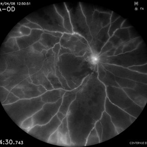 Combined Central Retinal Artery and Vein Occlusion
Apr 8 2024 by Akansha Sharma
Fundus fluorescein angiography of a 63 year old male with combined central retinal artery and vein occlusion with carotid artery stenosis and infarct in the brain demonstrating late filling.
Photographer: Dr. Akansha Sharma, Bharati Eye Hospital
Condition/keywords: central retinal artery occlusion (CRAO), central retinal vein occlusion (CRVO), CRAO
-
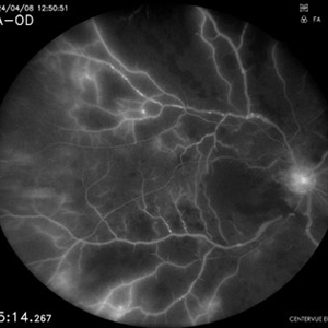 Combined Central Retinal Artery and Vein Occlusion
Apr 8 2024 by Akansha Sharma
Fundus fluorescein angiography of a 63 year old male with combined central retinal artery and vein occlusion with carotid artery stenosis and infarct in the brain demonstrating late filling.
Photographer: Dr. Akansha Sharma, Bharati Eye Hospital
Condition/keywords: central retinal artery occlusion (CRAO), central retinal vein occlusion (CRVO), CRAO
-
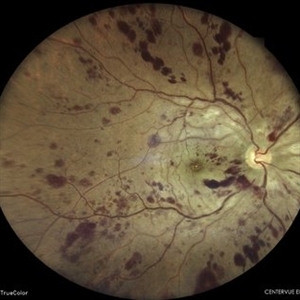 Combined Central Retinal Artery and Vein Occlusion
Apr 8 2024 by Akansha Sharma
Color fundus photograph of a 63 year old male with combined central retinal artery and vein occlusion with carotid artery stenosis and infarct in the brain.
Photographer: Dr. Akansha Sharma, Bharati Eye Hospital
Condition/keywords: central retinal artery occlusion (CRAO), central retinal vein occlusion (CRVO), CRAO
-
 Combined Central Retinal Artery Occlusion with Central Retinal Venous Occlusion
Mar 22 2023 by VIRAL SHAH
26 YEARS OLD MALE PATIENTS HAS COMPLAIN OF DIMNESS OF VISION SINCE 3 DAYS IN RIGHT EYE. HE IS SUFFERING FROM ANEMIA
Photographer: VIRAL SHAH
Condition/keywords: VASCULAR SHEATHING WITH HOLLENHORST PLAQUE
-
 Combined Central Retinal Vein Occlusion with Branch Retinal Artery Occlusion
Apr 29 2024 by KANWALJEET HARJOT MADAN, M.S. (Ophthalmology), FAICO (Vitreous - Retina)
This is fundus photograph of a 46-year male patient who presented with sudden diminution of vision in his right eye (RE) for 3 days. He was hypertensive but non diabetic. On examination, his best corrected vision in RE was 6/12. His left eye (LE) was normal. His fundus examination in RE revealed multiple intra retinal hemorrhages in all quadrants with tortuosity of veins suggestive of central retinal vein occlusion (CRVO) with mild disc edema. An ischemic area was seen superior to fovea suggestive of branch retinal artery occlusion. OCT depicted thickening of inner retinal layers with little evidence of macular edema. Hematological and cardio vascular investigations were done. He had bilateral thickening of intimal and medial walls of carotid arteries. He was under cardiology treatment. His vision improved to 6/6.
Photographer: Dr. Kanwaljeet Harjot Madan, M.S. (Ophthalmologist) Fellow in Vitrous & Retina. Thind Eye Hospital, Jalandhar City. Punjab. India
Condition/keywords: branch retinal artery occlusion (BRAO), central retinal vein occlusion (CRVO)
-
 Combined Central Retinal Vein Occlusion with Branch Retinal Artery Occlusion
Apr 28 2024 by KANWALJEET HARJOT MADAN, M.S. (Ophthalmology) Fellow in Vitreous & Retina
This is fundus photograph of a 46-year male patient who presented with sudden diminution of vision in his right eye (RE) for 3 days. He was hypertensive but non diabetic. On examination, his best corrected vision in RE was 6/12. His left eye (LE) was normal. His fundus examination in RE revealed multiple intra retinal hemorrhages in all quadrants with tortuosity of veins suggestive of central retinal vein occlusion (CRVO) with mild disc edema. An ischemic area was seen superior to fovea suggestive of branch retinal artery occlusion. OCT depicted thickening of inner retinal layers with little evidence of macular edema. Hematological and cardio vascular investigations were done. He had bilateral thickening of intimal and medial walls of carotid arteries. He was under cardiology treatment. His vision improved to 6/6.
Photographer: Dr Kanwaljeet Harjot Madan
Condition/keywords: branch retinal artery occlusion (BRAO), central retinal vein occlusion
-
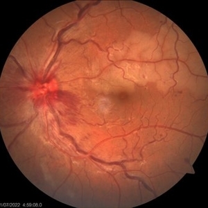
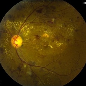
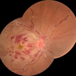
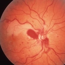
 Combined Cilioretinal Artery and Central Retinal Vein Occlusion
May 14 2016 by Ines Leal
Combined cilioretinal artery and central retinal vein occlusion in an otherwise 49-year-old healthy female patient. Color fundus photography shows whitening of the retina in the distribution of the cilioretinal artery and intraretinal hemorrhages with tortuous and engorged veins.
Photographer: Inês Leal, MD, Department of Ophthalmology, Faculty of Medicine, Universidade de Lisboa,
Condition/keywords: cilioretinal artery occlusion, venous occlusion
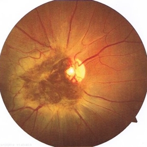
-
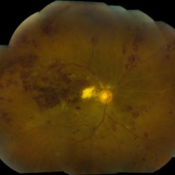
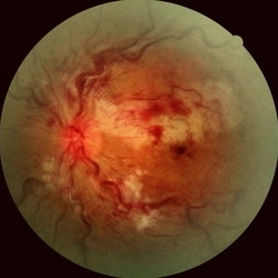
 Combined CRAO and CRVO
May 15 2014 by Manish Nagpal, MD, FRCS (UK), FASRS
30-year-old anemic lady presented with a acute loss of vision. Her vision was just hand movements in the affected eye and the other eye was normal.
Photographer: pooja barot, Optometrist, Retina Foundation, Ahmedabad
Condition/keywords: central retinal artery occlusion (CRAO), central retinal vein occlusion (CRVO), macular edema
-
 Combined CRVO and BRAO
Mar 27 2019 by Gary R. Cook, MD, FACS
Right eye of a 56-year-old white male with a combined perfused CRVO (venous dilation and dot & blot hemorrhages in all 4 quadrants) and a superotemporal BRAO with peripapillary hemorrhages and cotton wool spots, and an area of retinal whitening inside of the ST arcade. V.A.= 20/70.
Imaging device: Topcon VT-50
Condition/keywords: branch retinal artery occlusion (BRAO), central retinal vein occlusion (CRVO)
-
---thumb.JPG/image-square;max$300,300.ImageHandler) Combined Dry and Wet AMD
Combined Dry and Wet AMD
Nov 18 2013 by Mallika Goyal, MD
Right eye fundus of 60-year-old male with extensive RPE atrophy and macular scarring from AMD. No fluid at macula following anti-VEGF therapy and PDT.
Photographer: Mallika Goyal, MD, Apollo Health City, Hyderabad
-
---thumb.JPG/image-square;max$300,300.ImageHandler) Combined Dry and Wet AMD
Combined Dry and Wet AMD
Nov 18 2013 by Mallika Goyal, MD
Left eye fundus of 60-year-old male with extensive RPE atrophy and macular scarring from AMD. No fluid at macula following anti-VEGF therapy and PDT.
Photographer: Mallika Goyal, MD, Apollo Health City, Hyderabad
 Loading…