Search results (210 results)
-
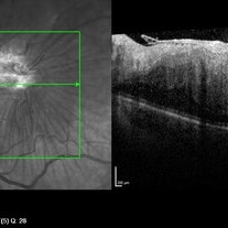
 Epiretinal Membrane/Macular Pucker With Combined Hamartoma of Retina and RPE
Jul 8 2015 by Emmanuel Chang, MD PhD FACS FASRS
10-year-old with history of progressive severe distortion in the left eye over the past year.
Photographer: Retina and Vitreous of Texas
Imaging device: Heidelberg Autofluorescence
Condition/keywords: combined hamartoma, epiretinal membrane (ERM), retinal pigment epithelium (RPE) hamartoma
-
 Horseshoe Tear
Nov 9 2012 by Norman Byer
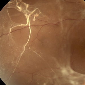
This horseshoe tear was the cause of the detachment in this 54-year-old man. The orange area on the right half of the slide represents the area of scleral indentation. Please note that most of the tear lies over the indented area and appears orange. However, the extreme left side of the tear is brownish black in color because it is exactly superimposed over the dark shadow that always lies just beyond the indented area. The ability of scleral indentation to produce this color change combined with a sharp demarcation between the blackish area and the yellowish edge of intact retina is a pathognomonic sign of a full thickness retinal break.
Condition/keywords: scleral indentation
-
 Subretinal Bands in CTRRD
Mar 1 2014 by Homayoun Tabandeh, MD, FASRS
Fundus photograph of a diabetic man with subretinal and pre-retinal bands and membranes associated with combined tractional rhegmatogenous retinal detachment due to proliferative diabetic retinopathy.
Photographer: Daniel Rivas
Condition/keywords: proliferative diabetic retinopathy (PDR), subretinal bands, tractional retinal detachment
-
 Retinoschisis
Nov 9 2012 by Norman Byer
This 51-year-old woman has retinoschisis with a large outer layer hole which has a white posterior rolled border. The left side of the posterior border of this hole can be seen to lie quite close to the inner layer showing that the outer layer is detached. This, therefore, is actually a combined schisis detachment which may safely be observed without treatment. This is an asymptomatic process, and the detachment of the outer layer is almost always localized and self limited.
Condition/keywords: intact inner layer, localized detachment of outer layer, outer layer hole, retinoschisis, rolled edges of retina, schisis detachment, white posterior
-
 Lattice Lesion
Nov 9 2012 by Norman Byer
This lattice lesion in a 30- year-old woman also shows combined features with a reddish crater above and a parallel snailtrack appearance just below it. Please note especially another interesting feature. From the left end of the lesion, there is a faint thin yellow line slanting down toward the right just below the shadow of the scleral indentation. This line identifies the dome of the pocket of liquified vitreous which is present over every lesion of lattice degeneration.
Condition/keywords: lattice degeneration, lattice lesion, liquefied vitreous, reddish crater, scleral indentation, snail track
-
 Combined Hamartoma of Retina and RPE
Mar 29 2013 by Henry J. Kaplan, MD
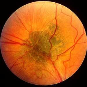
Greenish lesion on the arcade with epiretinal membrane formation, vessels inside the lesion are contracted and those outside are distracted.
Condition/keywords: combined hamartoma
-
 Retinal Schisis Detachment
Nov 9 2012 by Norman Byer
This 57-year-old man has a combined retinal schisis detachment caused by an outer layer hole in the upper right. On the right half of this photograph, the outer layer is detached and represented by the prominent yellow line which is lying against the inner layer. On the left half the inner layer appears very thin and the schisis cavity lies just behind it as it was originally. This, therefore, represents a localized detachment of the outer layer and thus a true secondary retinal detachment. The reason these cases remain localized and nonprogressive is that the only fluid available to the subretinal space is that which is contained within the schisis cavity. Furthermore, this fluid tends to be quite viscous and is not readily passed through the retinal breaks. A clinical symptomatic progressive retinal detachment cannot occur unless the retinal schisis cavity is very large or a break occurs in the inner layer also.
Condition/keywords: intact inner layer, localized detachment of outer layer, outer layer hole, retinal schisis detachment, retinoschisis, secondary retinal detachment
-
 Combined Hamartoma of the Retinal Pigment Epithelium Case 1
Oct 5 2012 by Ronald C. Gentile, MD
A peripapilary combined hamartoma of the retinal pigment epithelium involving the nasal disc margin. This tumor is slightly elevated, charcoal grey in color with grey-white tissue on it surface. The underlying retinal vessels are obscured.
Photographer: The New York Eye & Ear Infirmary Department of Medical Imaging
Condition/keywords: hamartoma, retinal pigment epithelium
-
 Combined CRAO and CRVO
May 15 2014 by Manish Nagpal, MD, FRCS (UK), FASRS
30-year-old anemic lady presented with a acute loss of vision. Her vision was just hand movements in the affected eye and the other eye was normal.
Photographer: pooja barot, Optometrist, Retina Foundation, Ahmedabad
Condition/keywords: central retinal artery occlusion (CRAO), central retinal vein occlusion (CRVO), macular edema
-
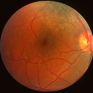
 Combined Hamartoma of the Retina and Retinal Pigment Epithelium
Dec 22 2015 by P. Mahesh Shanmugam, MBBS, DO, FRCSEd, PhD, FAICO
A fundus photo of 11-year-old boy with CHRRPE, gliotic epiretinal membrane overlying deep greyish lesion. Retinal wrinkling and dragging of macula is seen.
Condition/keywords: combined hamartoma, retinal pigment epithelium (RPE) hamartoma
-
 Optic Nerve Malformation/Possible Combined Hamartoma of Retinal Pigment Epithelium
Feb 19 2013 by From the Collections of Thomas M. Aaberg, MD and Thomas M. Aaberg Jr., MD
No history; Possible Bergmeister's papilla.
Condition/keywords: optic nerve malformation, retinal pigment epithelium
-
 Retinoschisis Detachment
Nov 9 2012 by Norman Byer
Combined retinoschisis detachment, so-called schisis detachment, in a 47-year-old woman. The large outer layer hole in the center has a posterior yellow border which represents the position of the outer layer. Please observe superior to the hole the dark convexity of the scleral indentation. Just below the hole at the middle of the slide and going to the left the yellow zone comes to lie right against the inner layer and a fluid filled cavity lies deep to the outer layer. At this point, therefore, there is a true neurosensory detachment of the retina. On the right side of the hole, the yellow line slants up and to the right and lies close to the pigment epithelium. On the right side of the photograph, the original schisis cavity can be seen separating the yellow line of the outer layer above from the inner retinal layer below. The mechanism of this detachment is that some of the fluid from the schisis cavity passes through the outer layer hole and detaches the outer layer. This lesion has not been treated and has remained exactly the same for 13 years. A similar symmetrical "schisis-detachment" is present in the fellow eye.
Condition/keywords: neurosensory detachment of retina, outer layer hole, pigment epithelium, retinoschisis, schisis detachment, scleral indentation
-
 Lattice Combined with Tiny Round Hole
Nov 9 2012 by Norman Byer
This 45 year-old man shows the snail track form of lattice combined with a tiny round hole. There is a tiny subclinical retinal detachment confined to the lesion itself.
Condition/keywords: glial vitreous tuft, lattice degeneration, round hole, snail track
-
 Lattice Lesion
Nov 9 2012 by Norman Byer
This lattice lesion in a 36-year-old woman shows a snail track feature on the left combined with a reddish crater and retinal hole to the right. The hole has caused a small subclinical detachment. The next slide pair will show more of this lesion.
Condition/keywords: lattice lesion, reddish crater, retinal hole, snail track, subclinical detachment
-
 Combined Hamartoma of the Retinal Pigment Epithelium Case 2
Oct 5 2012 by Ronald C. Gentile, MD
Magnified view of the peripapilary combined hamartoma of the retinal pigment epithelium involving the inferior disc margin. This tumor and slightly elevated, charcoal grey to light grey in color with grey-white tissue on it surface. The underlying retinal vessels are obscured with some epiretinal vitreous membranes.
Photographer: The New York Eye & Ear Infirmary Department of Medical Imaging
Condition/keywords: hamartoma, retinal pigment epithelium
-
 Fluorescein Angiography of a Combined CRAO AND CRVO
Mar 24 2017 by Manish Nagpal, MD, FRCS (UK), FASRS
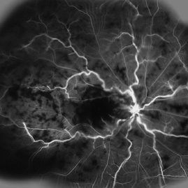
Young girl 22-year-old came with sudden decreased vision and had a combined CRAO and CRVO whose FA revealed extensive ischemia and delayed filling.
Photographer: pranita chaudhary
Condition/keywords: central retinal artery occlusion (CRAO), central retinal vein occlusion (CRVO)
-
 Montage OF a Combined Case of CRVO and CRAO
May 15 2014 by Manish Nagpal, MD, FRCS (UK), FASRS
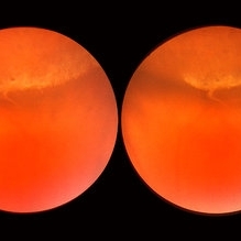
30-year-old anemic lady presented with a acute loss of vision. Her vision was just hand movements in the affected eye and the other eye was normal.
Photographer: pooja barot, Optometrist, Retina Foundation, Ahmedabad
Condition/keywords: central retinal artery occlusion (CRAO), central retinal vein occlusion (CRVO), macular edema
-
 Geographic Atrophy
Mar 27 2013 by Michael P. Kelly, FOPS
This is a combined FAF/SD-OCT in EDI mode of a patient with geographic atrophy and foveal sparing.
Photographer: Michael P. Kelly, FOPS. Director, Duke Eye Labs, Duke University Eye Center
Imaging device: Heidelberg Spectralis
Condition/keywords: enhanced depth imaging, foveal sparing, fundus autofluorescence (FAF), geographic atrophy, optical coherence tomography (OCT)
-
 Combined Hamartoma of the Retina and RPE
Jul 8 2015 by Emmanuel Chang, MD PhD FACS FASRS
10-year-old with history of progressive severe distortion in the left eye over the past year.
Photographer: Retina and Vitreous of Texas
Imaging device: Heidelberg Spectralis
Condition/keywords: combined hamartoma, epiretinal membrane (ERM), retinal pigment epithelium (RPE) hamartoma
-
 Combined Hamartoma of Retina and RPE
Mar 29 2013 by Henry J. Kaplan, MD
Hamartoma visible as a grreen lesion on superior arcade with ERM formation and dragging of the macula.
Condition/keywords: combined hamartoma
-
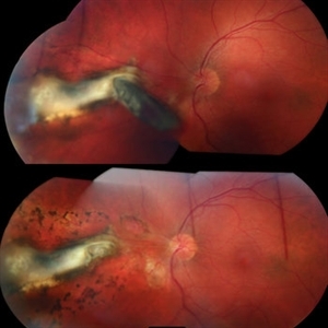 IOFB Combined
Mar 12 2015 by Ahmad B. Tarabishy, MD
A 26-year-old gentleman presented with a metallic intraocular foreign body embedded in the nasal retina (above). Post-operative appearance two weeks after vitrectomy, foreign body removal, endolaser, and gas (below).
Photographer: Jessica Armbruster
Imaging device: Topcon TRC-50EX
Condition/keywords: encapsulated intraocular foreign body, non metallic retained intraocular foreign body (RIOFB), penetrating trauma
-
 Combined Harmartoma of the Retinal Pigment Epithelium Case 1
Oct 5 2012 by Ronald C. Gentile, MD
Magnified view of the nasal peripapilary combined hamartoma of the retinal pigment epithelium. The tumor is slightly elevated, charcoal grey in color with grey-white tissue on its surface.
Photographer: The New York Eye & Ear Infirmary Department of Medical Imaging
Condition/keywords: hamartoma, retinal pigment epithelium
-
 Combined Hamartoma of the Retina and RPE
Apr 16 2015 by Rita Couceiro, MD, MS
Fundus photograph, red-free picture (top images) and fluorescein angiography pictures (bottom images) of a 6-year-old girl with a combined hamartoma of the retina and RPE in the right eye.
Condition/keywords: hamartoma
-
 Combined Hamartoma of the Retinal Pigment Epithelium Case 2
Oct 5 2012 by Ronald C. Gentile, MD
A peripapilary combined hamartoma of the retinal pigment epithelium involving the inferior disc margin. This tumor and slightly elevated, charcoal grey to light grey in color with grey-white tissue on it surface. The underlying retinal vessels are obscured with some epiretinal membrane and some striae extending to the inferior nasal macula.
Photographer: The New York Eye & Ear Infirmary Department of Medical Imaging
Condition/keywords: hamartoma, retinal pigment epithelium
-
 Exudative Macular Detachment After Intensive Laser Photocoagulation
Mar 12 2016 by Sjakon G Tahija, MD
Fundus photograph of 44-year-old man with exudative detachment of the macula after vitrectomy and ILM peeling for proliferative diabetic retinopathy combined with intensive endolaser photocagulation.
Photographer: Avris Siahaan, Klinik Mata Nusantara
Condition/keywords: exudative detachment, pan-retinal photocoagulation (PRP)
 Loading…