Search results (38 results)
-
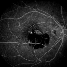
 CHRPE and Bear Tracks
Jan 7 2025 by Drew Mitchell
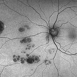
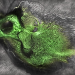
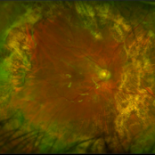
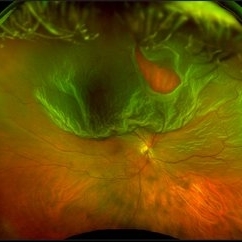
Fundus Autofluorescence of a CHRPE and Bear Tracks.
Photographer: Drew Mitchel, OCT-C
Imaging device: Optos Silverstone
Condition/keywords: bear tracks, CHRPE, congenital hypertrophy of the retinal pigment epithelium (CHRPE)
-
 Choroidal Melanoma with Serous Retinal Detachment
Dec 20 2024 by Daniel Davis, OCT-C
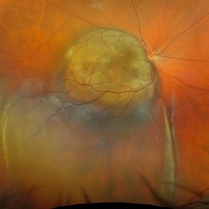
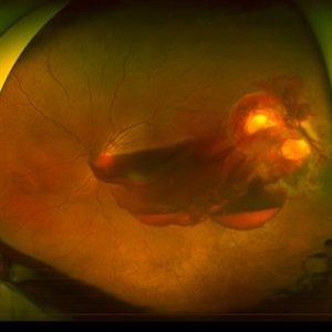
67 year old male presenting with large pigmented choroidal mass with serous retinal detachment.
Photographer: Daniel Davis, OCT-C, The Retina Institute
Imaging device: Optos California
Condition/keywords: Retina detachment
-
 Who Stole My Blood Supply?
Jan 25 2025 by Muna Bhende, MD
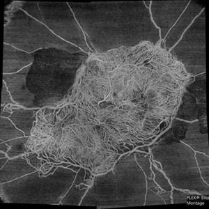
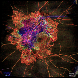
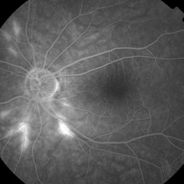
Superficial capillary plexus slab montage image of a young female diabetic with florid proliferation . There is no flow in the capillaries anterior to the tangle of new vessels indicating severe retinal ischemia.
Photographer: Mohanapriya L , Medical Research Foundation, Sankara Nethralaya, Chennai, India
Imaging device: PLEX elite 9000
Condition/keywords: florid type PDR, OCTA
-
 MIDD (Maternally Inherited Diabetes and Deafness) - Right AF
Nov 30 2024 by John S. King, MD
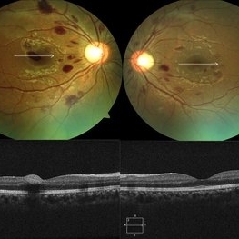
Both right and left eyes have symmetrical ring of mottled hypo/hyper AF around the fovea and disc. The HyperAF areas correspond to RPE deposits on OCT as well as areas of blockage on FA, and drusenoid deposits seen on fundus photos. Disc drusen in right eye present as HyperAF spot 57 yo WF referred for AMD vs Pattern Dystrophy that was diagnosed 10 years ago. Reported some slow progressive vision loss in both eyes for distance and near. Denies nyctalopia or hemeralopia. Background medical history includes HTN, CVD, and DM. No family history of eye problems. Denied pentosan use. Anterior segment showed moderate cataracts (OD>OS). Posterior segment exam showed macular changes and mild NPDR. The macular appearance showed a symmetrical, paramacular ring of fleck-like drusenoid material with some faint focal areas of RPE hyperplasia. Fundus Photos, AF, OCT were performed as well as a gene test. Further questioning showed revealed that her mother and maternal grandmother had both diabetes mellitus and sensorineural hearing loss. The patient developed diabetes in her teens, and some high frequency hearing loss in her early twenties. She had not had a previous genetic test or diagnosis of MIDD. Gene testing is pending for the mitochondrial component. Invitae's retinal panel, which does not include mitochondrial disorders, only showed a variant of uncertain significance, HMCN1. I discussed this case with Dr. Freund, and it is similar to a the case report : Inoue M, Kiss S, Freund KB. MACULAR PIGMENT RINGS AS THE PRESENTING FINDING OF MITOCHONDRIAL MYOPATHY, ENCEPHALOPATHY, LACTIC ACIDOSIS, AND STROKELIKE EPISODES. Retin Cases Brief Rep. 2015 Fall;9(4):260-4. doi: 10.1097/ICB.0000000000000182. PMID: 26200388.
Photographer: Grace Melton and Carley Gunn
Imaging device: Clarus
Condition/keywords: Macular Dystrophy, Maternally Inherited Diabetes and Deafness, MIDD, Mitochondrial Disorder
-
 From Ora to Ora
Aug 26 2024 by Nassim Alejandro Abreu Arbaje, MD
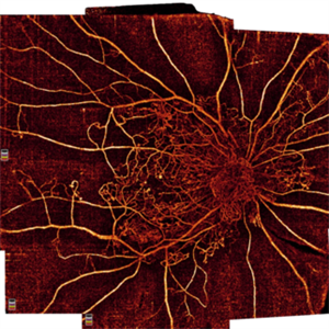
Ultra-wide field OCT angiography of a 39 year-old healthy male. The photo attempts to explore retinal vasculature up to the ora serrata.
Photographer: Johel Arrieta, TowardPi
Imaging device: TowardPi BMizar 400khz
Condition/keywords: OCT Angiography, OCTA, ultra-wide field imaging
-
 Von Hippel Lindau With Retinal Capillary Hemangioma
Nov 2 2023 by Marcelo Zas, MD PhD
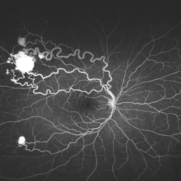
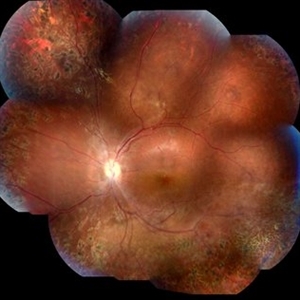
This wide-field AGF image shows the vascular tumors with their corresponding afferent and efferent vessels.
Photographer: Mariano Cotic MD
Imaging device: Silverstone SS OCT Optos
Condition/keywords: tumor
-
 Benign Familial Fleck Retina
Feb 2 2023 by Hemanth Murthy, MBBS, MD, FASRS
12 year boy first born of consanguineous marriage, came for routine eye check up with BCVA 20/40 OU. He has no night blindness. His OCT showed thickening of the RPE with dome like elevations involving the ellipsoid layer. Dark adapted ERG showed normal 'b' wavesPhotopic ERG showed reduced 'a' and b waves.
Photographer: Veda Vyas
Imaging device: Optos Daytona
Condition/keywords: Benign familial fleck retina
-
 Proliferative Diabetic Retinopathy with Macular Isquemia by OCT Angiography
Oct 14 2022 by JORGE SOBERANES
Depth-encoded OCT angiography of a patient with proliferative diabetic retinopathy showing vascular changes and extensive ischemia including macular area.
Photographer: Jorge I. Soberanes, Asociación para Evitar la Ceguera en México.
Imaging device: PLEX Elite 9000, Zeiss
Condition/keywords: diabetic retinopathy, OCT Angiography
-
 submacular perfluorocarbon liquid
Sep 7 2022 by JEFFERSON R SOUSA, Tecg.º (Biomedical Systems Technology)
A 63-year-old male patient underwent vitreoretinal surgery with the use of perfluorocarbon. From a technological point of view, extended-field retinography presents many points of focus variation due to the difficulty of establishing a diffuse focus, as it is a recent post-operative case. In OCT Fundus Enface, although it has a low resolution, it is extremely important for documenting the presence of perfluor. Best seen in structural OCT.
Photographer: JEFFERSON ROCHA DE SOUSA - Retinal Department at Instituto Dr. Suel Abujamra Sao Paulo-Brazil
Imaging device: Optical Coherence Tomography system OCT CIRRUS 5000, Protocol, HD 5 Line
Condition/keywords: perfluorocarbon fluid, post-vitrectomy, submacular perfluorocarbon liquid (PFO), vitrectomy
-
 Central Retinal Vein Occlusion by OCT Angiography
Jun 13 2022 by JORGE SOBERANES
A 63 year old man with a central retinal vein oclussion. In the OCT angiogram we could observe retinal isquemia, neovascularization and arteriovenous shunts.
Photographer: Jorge I. Soberanes MD
Imaging device: PLEX Elite 9000, Zeiss
Condition/keywords: Central vein oclussion, neovascularization, OCT angiography, retina, Shunts
-
 Extra-scleral Extension of Choroidal Melanoma
Dec 23 2021 by Jessica Norkus
89-year-old female with extra-scleral extension of choroidal metastatic melanoma. Patient hadn't been seen by any eye doctor in 3 years prior to this visit. Noticed scleral darkening about 6 months ago, with vision loss noted for about 4-5 months. Presented with LP vision. Emergent MRI of brain/orbit showed no extension beyond what is seen at slit lamp. CT C/A/P w/ contrast ordered and found 2 hepatic lesions, concerning for potential mets. Patient referred to medical oncology.
Photographer: Jessica Norkus, COA, OSC
Imaging device: Topcon TRC 50DX
Condition/keywords: external photography, extrascleral extension, metastatic cancer, metastatic lesion
-
 Green Goblin Detachment
Jan 13 2022 by Netan Choudhry, MD, FRCS(C) FASRS
Tractional retinal detachment with macular hole in a 76-year-old female.
Photographer: John Golding BA, Vitreous Retina Macula Specialists of Toronto, OCTane Imaging Lab
Imaging device: Multicolor fundus photo taken on the Spectralis OCT2 (Heidelberg Engineering GmbH).
Condition/keywords: macular hole, Multispectral imaging, tractional retinal detachment
-
 Proliferative Diabetic Retinopathy
Oct 16 2021 by Timur Shaimov
32 y.o. female with Type 1 Diabetes with no glucose compensation for several years. A manual montage of several 8x8 mm OCT angiograms were obtained for this Widefield OCTA image.
Photographer: Timur Shaimov
Imaging device: RTVue xR Avanti
Condition/keywords: OCT Angiography, proliferative diabetic retinopathy (PDR)
-
 Retinoschisis
Mar 28 2021 by JEFFERSON R SOUSA, Tecg.º (Biomedical Systems Technology)
A 14-year-old male patient was admitted for visual evaluation. Visual acuity s/c in the right eye and 20/80 in the left eye. According to family members, he reported low vision since childhood. He had already undergone treatment with photocoagulation in another service to which he had a diagnostic hypothesis of Coats' disease. Laboratory tests were requested (HIV, TOXO, TOXOCARIASIS, ECA, VDRL, PPD). In the evaluation it was observed important exudation in the posterior pole, some vascular irregularities in the right eye. In the left eye, there is retinoschisis affecting the entire posterior pole and the region nasal to the optic disc, macula with a characteristic aspect of a cartwheel. Well exemplified by OCT-A (Structrure Deep: IPL - 25, OPL - 25).
Photographer: JEFFERSON R SOUSA - Study Center and Ophthalmological Research Dr. Andre M V Gomes, Institute Dr. Suel Abujamra São Paulo-Brazil
Imaging device: Topcon TRC-50 DX, Imaginet 4.0, angle de 50 graus. Flash 50w-s
Condition/keywords: Coats' disease, retinoschisis
-
 A Motor Vehicle Accident Causing Valsalva Retinopathy OD, While Racing A Side By Side 4 Wheel Off-Road Vehicle
Apr 29 2020 by John S. King, MD
43-year-old white male who was injured while racing a side by side 4-wheel off-road vehicle (see Video: https://imagebank.asrs.org/file/53854/sxs-crash-during-a-race-causing-valsalva-retinopathy-od). He presented about three weeks after the injury. He was being seen by his local eye doctor who wanted an evaluation for the retinal heme and scotoma. His main complaint was a central/parcentral scotoma described as a greyish area in vision. Va 20/50 OD, nomotensive, no APD (by technician), anterior segment u/r; see picture for the fundus exam - of note there are superficial/preretinal heme, with layering of the heme superiorly, and small superficial heme at nasal edge of the optic disc; in the parafoveal region nasally there is some mottling of the RPE that may indicate an area of prior commotio retinae (also possible to have TON), which may account for his scotoma. Really bad accident (video), and amazingly, he had no LOC or injuries other than the right retina. Helmet and racing harness seat belt were used.
Photographer: Asli Ahmed
Imaging device: Topcon 50
Condition/keywords: valsalva retinopathy
-
 Submacular PFO
Feb 20 2020 by Kevin J. Blinder, MD, FASRS
This is a 53-year-old gentleman that was referred to us for a second opinion with an inoperable RD with PVR after 3 failed attempts. We performed a PPV, membranectomy, scleral buckling procedure, with silicone oil injection. This case did not require PFO. You can imagine our surprise when we discovered submacular PFO postoperatively. It is very difficult to see the PFO on the Optos. The infrared shows it clearly, with confirmation of the submacular space on the SD-OCT.
Photographer: Jarrod Wehmeier, The Retina Institute; St. Louis, MO
Imaging device: optos
Condition/keywords: submacular perfluorocarbon liquid (PFO)
-
 Flame of the Forest
Apr 9 2020 by Daraius N Shroff, MS FMRF FRCS
A 54-year-old man with DM for 15 years. The left eye had a visual acuity of 20/40. Wide field swept source OCTA revealed branching out central neovascular trunk vessels from the disc with terminal loops, along with exuberant proliferation of irregular small-calibre fine new vessels. The patient underwent OCTA guided pan retinal photocoagulation.
Photographer: Anuj Choudhary, Shroff Eye Centre, New Delhi
Imaging device: Zeiss Plex Elite 9000
Condition/keywords: proliferative diabetic retinopathy (PDR)
-
 Acute Macular Neuroretinopathy
Dec 11 2019 by Lauren Whaley
34-year-old female patient presented with changes in vision after recent upper respiratory infection. Referring doctor originally thought it was a blood pressure issue. She noticed a "C" shape in her vision. Infrared image was captured showing exactly what patient was describing! Doctor confirmed with this image that it was AMN.
Photographer: Lauren R. Whaley, COA
Imaging device: Heidelberg Spectralis
Condition/keywords: 30 degrees, acute macular neuroretinopathy, Heidelburg Spectralis, left eye, macula, near infrared autofluorescence (NIRAF)
-
 Metastatic NSCLCA to the Choroid: Initial Appearance
May 27 2019 by John S. King, MD
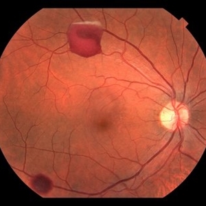
60-year-old white male non-smoker presented to Dr. Zocchi with acute transient decreased vision in the right eye. Background history includes metastatic NSCLC (adenocarcinoma). Acuity OD 20/60, and posterior segment had two small, yellow, choroidal lesions, above the nerve and IT arcade (these had a fairly smooth and dome shaped appearance on the OCT, and top lesion had mild SRF) (see photo)
Photographer: Shelly Blair
Imaging device: Optos CA
Condition/keywords: choroidal metastasis, lung cancer metastasis
-
 Volume Rendering Structural and Angiographic Optical Coherence Tomography Angiography Image of a Retinal Capillary Microaneurysm, A Newly Described Entity.
May 21 2019 by Richard F. Spaide, MD
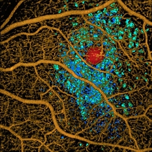
This is a newly described entity in which patients develop solitary aneurysms that are much larger than typical microaneurysms and they are supplied by capillaries. The aneurysm is shown in red. The associated macular edema produced cystoid spaces in Henle’s fiber layer, rendered as teal and in the inner nuclear layer as blue.
Photographer: Richard F. Spaide, MD
Condition/keywords: aneurysm, optical coherence tomography (OCT), volume rendering
-
 Optos Giant Tear within Retinal Detachment
Apr 30 2019 by Lauren Whaley
Noticed an inferior visual field defect on a patient with history of vitreous hemorrhage. Decided to take an Optos image and this is what we found. Doctor performed pneumatic retinopexy in office and patient recovering well.
Photographer: Lauren R. Whaley
Imaging device: Optos
Condition/keywords: Optos, retinal tear, subretinal fluid
-
 Penetrating Trauma with Retinal Detachment
Apr 30 2019 by Olivia Rainey
Ultra-wide field pseudocolor image of a 39-year-old female with penetrating trauma resulting in a retinal detachment with an intraretinal hemorrhage affecting the left eye. Patient was struck with a champagne glass in October of 2018, which lacerated the eyelid and globe. Patient was "seeing red" when she first came to the office and after multiple surgeries she was seeing 20/20 at her last check in April 2019.
Photographer: Olivia Rainey
Imaging device: Optos
Condition/keywords: hemorrhage, left eye, Optos, penetrating trauma, ruptured globe, ultra-wide field imaging
-
 Susac's Syndrome
Feb 13 2018 by John S. King, MD
Background: 46-year-old WF with CML (stable on Sprycel) saw her PCP for headaches without known cause; Headaches worsened and became confused, disoriented, off balance, and impaired short term memory. Heme-oncology ordered MRI that showed abnormal signal in the cerebellum and other parts of the brain, and LP has elevated protein. LP did show positive tau test, but fortunately, was a false positive for CJD. IV and PO steroids started and symptoms improved. MRI showed much improvement one month since starting steroids. 3 weeks later had a scotoma in right eye and eye doctor did not find anything at that time to cause it. Tinnitus developed (and some intermittent vertigo before that) and ENT referred back to eye doctor, who then referred the patient to Dr. Zocchi. He found a CWS and BRAO OD, and bilateral arteritis. She had some additional work-up for vasculitis. Given the retinal arteritis, cochlear issues, and MRI findings, Dr.Zocchi suspected Susac's Syndrome. She was started on multiple regimens including prednisone, IVIG, azathiprine, and MTX, and has had the best reponse to IVIG (FA shows a recurrence/worsening while adjusting IMT). She is stable and doing well with 20/20 vision in both eyes.
Photographer: Kay Dalby
Imaging device: Topcon
Condition/keywords: retinal vasculitis, Susac's syndrome
-
 Dengue-Associated-Retinopathy (Anaemic Retinopathy)
Jan 16 2018 by Deepak Bhojwani, MS
22-year-old male with systemic dengue fever and anaemia presenting with roth spots in both eyes (OD>OS). Horizontal raster OCT scans showing intraretinal foveal hameorrhage in right eye.
Photographer: Dr Deepak Bhojwani, Raghudeep Eye Hospital , Ahmedabad
Imaging device: Zeiss- HD- OCT
Condition/keywords: anaemic retinopathy, Roth spots
-
 Subretinal Hemorrhage Due to SRNVM, Fluorescein Angiogram Photograph
Dec 1 2016 by James B. Soque, CRA, OCT-C, COA, FOPS
89-year-old white male with NVAMD and new subretinal hemorrhage, fluorescein angiogram, early phase, of the right eye. Currently receiving anti VEGF treatment.
Photographer: James Soque, CRA, OCT-C, COA, Island Retina, Shirley, NY
Imaging device: Topcon TRC 50 DX, with MERGE software
Condition/keywords: hemorrhage, Hot spot, neovascular age-related macular degeneration (AMD), subretinal hemorrhage, subretinal blood, wet age-related macular degeneration (wet AMD)
 Loading…