Search results (1541 results)
-
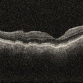
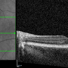
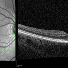
 Acute and Chronic OCT of BRAO
Sep 9 2021 by Aleksandra V. Rachitskaya, MD, FASRS
Acute and chronic OCT findings in BRAO. Acutely, inner retinal hyper-reflectivity is seen. Chronically, retina atrophy ensues.
Condition/keywords: BRAO, OCT
-
 Acute Posterior Multifocal Placoid Pigment Epitheliopathy
Feb 20 2024 by Soobien Lee
A 20-year-old caucasian female with viral prodrome and vision loss OS>OD secondary to Acute Posterior Multifocal Placoid Pigment Epitheliopathy (APPME). OCT of the left macula shows bacillary layer detachment.
Photographer: Kim Seay, Elman Retina Group
Condition/keywords: acute posterior multifocal placoid pigment epitheliopathy (APMPPE), bacilliary layer detachment, OCT, Uveitis, white dot syndrome
-
 Acute Posterior Multifocal Placoid Pigment Epitheliopathy
Feb 20 2024 by Soobien Lee
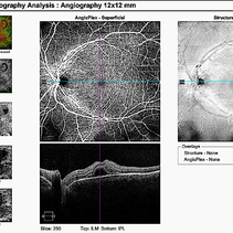
12x12mm OCT Angiography of a 20-year-old caucasian female with viral prodrome and vision loss OS>OD secondary to Acute Posterior Multifocal Placoid Pigment Epitheliopathy (APPME). Imaging shows multifocal flow voids.
Photographer: Kim Seay, Elman Retina Group
Imaging device: 12x12mm OCT-Angiography
Condition/keywords: acute posterior multifocal placoid pigment epitheliopathy (APMPPE), bacillary layer detachment, OCT, OCT Angiography, Uveitis, white dot syndrome
-
 Both Eyes OCT in Case of Right Eye Choroidal Hemangioma
Nov 29 2024 by Anand Temkar
BE OCT of a 42 year old male, showing the elevation of the right eye retina along with the cystic spaces and subretinal fluid.
Photographer: Dr.Anand Temkar- Retina Foundation, Ahmedabad
Imaging device: Mirante
Condition/keywords: OCT
-
 Branch Retinal Artery Occlusion
Dec 15 2022 by Christopher R. Adam, M.D.
OCT
Condition/keywords: branch retinal artery occlusion (BRAO), BRAO
-
 Branch Retinal Vein Occlusion
Dec 15 2022 by Christopher R. Adam, M.D.
OCT
Condition/keywords: branch retinal vein occlusion (BRVO), BRVO, cystoid macular edema (CME)
-
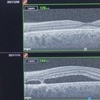
 Bullseye Maculopathy
Jan 22 2024 by Kali Jend
Optical coherence tomography of a 73-year-old female with Bullseye Macular Changes affecting her left eye. Patient reports having a family history of this condition and denies prior Plaquenil or Elmiron use. Compared to previous imaging, the patient's condition progressed in the left eye from 2020 to 2023. Patient has a history of fluctuating Diabetic Macular Edema and a current Epiretinal Membrane as well. Patient's vision was Ncc20/60 at the time the image was taken.
Photographer: Kali Jend
Imaging device: Heidelberg Spectralis
Condition/keywords: bullseye maculopathy, epiretinal membrane (ERM), heidelberg spectralis, left eye, macular pucker, OCT, optical coherence tomography (OCT)
-
 CCH
CCH
Feb 6 2025 by Jack B Margines, MD, MHCI
SD-OCT of an extramacular Circumscribed Choroidal Hemangioma in an asymptomatic 53 year-old female
Photographer: Ryan Milam, University of California, Irvine Gavin Herbert Eye Institute
Imaging device: Zeiss Cirrus
Condition/keywords: Circumscribed Choroidal Hemangioma, OCT
-
 Choroidal folds i/c/o hypotony
Nov 23 2023 by Anand Temkar
OCT showing choroidal folds in a follow up case of filtration surgery with mitomycin c and anterior vitrectomy elsewhere.
Photographer: Dr.Anand Temkar- Retina Foundation, Ahmedabad
Imaging device: Mirante
Condition/keywords: choroidal folds, hypotony, OCT
-
 Color Fundus Photograph of Macular Infarction Secondary to Subonjunctival Gentamicin Injection
May 16 2014 by Arwa Azmeh, MD, PhD
A 20-year-old male suffered from diplopia since age one. He was diagnosed to have acquired fourth nerve palsy in his left eye. VA at time of diagnosis was 20/20 in OU and Fundus exam was WNL in OU. His history revealed no other complaints. 3 days ago he underwent left superior oblique tucking for relief of his diplopia.The surgery was uneventful and at the end of surgery subconjunctival gentamicin was injected. Immediately following surgery his VA in OS decreased from 20/20 to complete loss of central vision and sensation of HM from the periphery. He was referred to us 3 days after surgery. At time of referral fundus exam of his left eye revealed macular infarction with cherry red spot appearance with few retinal hemorrhages, mild optic disc edema and CWS surrounding optic disc. Peripheral retina had normal color and appearance. The vitreous was clear. Anterior segment was quiet. IOP was WNL. Macular OCT was consistent with macular infarction. FA revealed delay in central retinal artery filling as fluorescein started to appear in the arteries at the level of the optic disc at 28 sec, and in the retinal veins at 38 sec. Macular area remained to be non-perfused throughout the whole FA. In late phases staining of blood vessels walls was noticed. The "wipe out" of large vessels and capillaries persisted in the central area. OCT through foveal area showed diffuse thickening of the retina with severe elevation in the fovea, reduced backscattering from the outer layers of the retina and enhanced reflectivity from the inner retina, due to ischemia. Complete blood count and cardiovascular study were WNL. The final diagnosis was macular infarction secondary to subconjunctival gentamicin injection.
Imaging device: OCT
Condition/keywords: macular infarction, subconjunctival gentamicin
-
 CSR Treated with Focal Laser: FFA
Dec 6 2021 by Nizamuddin HM Shaik, MD, FRCS
FFA of 35-year-old lady with CSR treated with focal laser.
Photographer: Mahmoud , Ophthalmology Technecian, International Medical Center
Imaging device: OCT
Condition/keywords: central serous chorioretinopathy (CSCR), FFA, focal laser, laser photocoagulation
-
 CSR Treated with Focal Laser: Fundus Photo
Dec 6 2021 by Nizamuddin HM Shaik, MD, FRCS
Fundus photograph of 35-year old lady with CSR treated with focal laser.
Photographer: Mahmoud , Ophthalmology Technician, International Medical Center
Imaging device: OCT
Condition/keywords: central serous chorioretinopathy (CSCR), focal laser, laser photocoagulation
-
 CSR Treated with Focal Laser: Fundus, FFA, OCT Images
Dec 6 2021 by Nizamuddin HM Shaik, MD, FRCS
Fundus photograph , FFA and OCT ( Pre and Post ) of a 35-year-old lady with CSR treated with focal laser.
Photographer: Mahmoud , Ophthalmology Technician, International Medical Center
Imaging device: OCT
Condition/keywords: central serous chorioretinopathy (CSCR), laser photocoagulation
-
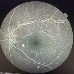
 Early-FA-phase-of-macular-infarction-secondary-to-subconjunctival-gentamycin-injection
May 16 2014 by Arwa Azmeh, MD, PhD
A 20-year-old male suffered from diplopia since age one. He was diagnosed to have acquired fourth nerve palsy in his left eye. VA at time of diagnosis was 20/20 in OU and Fundus exam was WNL in OU. His history revealed no other complaints. 3 days ago he underwent left superior oblique tucking for relief of his diplopia.The surgery was uneventful and at the end of surgery subconjunctival gentamicin was injected. Immediately following surgery his VA in OS decreased from 20/20 to complete loss of central vision and sensation of HM from the periphery. He was referred to us 3 days after surgery. At time of referral fundus exam of his left eye revealed macular infarction with cherry red spot appearance with few retinal hemorrhages, mild optic disc edema and CWS surrounding optic disc. Peripheral retina had normal color and appearance. The vitreous was clear. Anterior segment was quiet. IOP was WNL. Macular OCT was consistent with macular infarction. FA revealed delay in central retinal artery filling as fluorescein started to appear in the arteries at the level of the optic disc at 28 sec, and in the retinal veins at 38 sec. Macular area remained to be non-perfused throughout the whole FA. In late phases staining of blood vessels walls was noticed. The "wipe out" of large vessels and capillaries persisted in the central area. OCT through foveal area showed diffuse thickening of the retina with severe elevation in the fovea, reduced backscattering from the outer layers of the retina and enhanced reflectivity from the inner retina, due to ischemia. Complete blood count and cardiovascular study were WNL. The final diagnosis was macular infarction secondary to subconjunctival gentamicin injection.
Imaging device: OCT
Condition/keywords: macular infarction, subconjunctival gentamicin
-
 Endophthalmitis
Jul 5 2024 by Zach Seim
OCT of a 59 year old male with Fungal Endophthalmitis. The fungus can be seen breaking through in the OCT.
Photographer: Zach Seim
Imaging device: Heidelberg OCT
Condition/keywords: endophthalmitis, fungal, OCT
-
 Focal Laser Treatment for Central Serous Retinopathy: FFA
Dec 6 2021 by Nizamuddin HM Shaik, MD, FRCS
FFA of a 35-year-old lady with CSR treated with focal laser.
Photographer: Mahmoud , Ophthalmology Technecian, International Medical Center
Imaging device: OCT
Condition/keywords: central serous chorioretinopathy (CSCR), laser photocoagulation
-
 Giant Retinal Tear
Oct 14 2022 by Angela Rico
40 year old female who presented with Giant Retinal tear
Photographer: Angela Rico M.D.
Imaging device: OCT
Condition/keywords: retinal tear
-
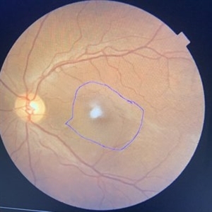
 Idiopathic Choroidal Neovascularization
Mar 2 2023 by Corey Grant
Optical coherence tomography and ultra-wide field fundus photograph of a 51 year old male with idiopathic choroidal neovascularization affecting his right eye. The patient had no symptoms at the time of the appointment and his vision was Dcc20/20-2 OU. The physcian stated that there wasn't active exudation on the exam or ocular imaging and based on the clinical findings, he has recommended we defer any treatments.
Photographer: Corey Grant
Imaging device: Heidelberg Spectralis, OPTOS California
Condition/keywords: choroidal neovascularization (CNV), CNVM, fundus photograph, OCT, optical coherence tomography (OCT), Optos, Right Eye, ultra-wide field imaging
-
 Late FA Phase of Macular Infarction Secondary to Subconjunctival Gentamicin Injection
May 16 2014 by Arwa Azmeh, MD, PhD
A 20-year-old male suffered from diplopia since age one. He was diagnosed to have acquired fourth nerve palsy in his left eye. VA at time of diagnosis was 20/20 in OU and fundus exam was WNL in OU. His history revealed no other complaints. 3 days ago he underwent left superior oblique tucking for relief of his diplopia.The surgery was uneventful and at the end of surgery subconjunctival gentamicin was injected. Immediately following surgery his VA in OS decreased from 20/20 to complete loss of central vision and sensation of HM from the periphery. He was referred to us 3 days after surgery. At time of referral fundus exam of his left eye revealed macular infarction with cherry red spot appearance with few retinal hemorrhages, mild optic disc edema and CWS surrounding optic disc. Peripheral retina had normal color and appearance. The vitreous was clear. Anterior segment was quiet. IOP was WNL. Macular OCT was consistent with macular infarction. FA revealed delay in central retinal artery filling as fluorescein started to appear in the arteries at the level of the optic disc at 28 sec, and in the retinal veins at 38 sec. Macular area remained to be non-perfused throughout the whole FA. In late phases staining of blood vessels walls was noticed. The "wipe out" of large vessels and capillaries persisted in the central area. OCT through foveal area showed diffuse thickening of the retina with severe elevation in the fovea, reduced backscattering from the outer layers of the retina and enhanced reflectivity from the inner retina, due to ischemia. Complete blood count and cardiovascular study were WNL. The final diagnosis was macular infarction secondary to subconjunctival gentamycin injection.
Imaging device: OCT
Condition/keywords: macular infarction, subconjunctival gentamicin
-
 Macular OCT Image of a Patient With Central Retinal Artery Occlusion
Jul 7 2024 by Thiago Mazzeo
This is a macular OCT image of a patient that presented sudden visual loss in the right eye (Light perception) after leaving the hospital due to uncontrolled systemic arterial hypertension.
Photographer: Thiago Mazzeo
Imaging device: Zeiss Cirrus 5000
Condition/keywords: Central Retinal Artery Occlusion, macular changes, OCT
-
 Neovascular AMD with Ring Shaped lesions
Jul 12 2023 by Gregg T. Kokame, MD, MMM, FASRS
Vertical OCT Scan - Neovascular AMD with Active CNV Ring shaped lesions underneath the RPE inverted U-shaped elevation
Photographer: Jaclyn Pisano
Imaging device: Zeiss Cirrus 6000
Condition/keywords: edema, lesion, OCT, Sub-retinal fluid, wet age-related macular degeneration (wet AMD)
-
 Neovascular AMD with Ring Shaped lesions
Jul 12 2023 by Gregg T. Kokame, MD, MMM, FASRS
Horizontal OCT Scan - Neovascular AMD with Active CNV Ring shaped lesions underneath the RPE inverted U-shaped elevation
Photographer: Jaclyn Pisano
Imaging device: Zeiss Cirrus 6000
Condition/keywords: inverted u-shaped elevation, lesion, macular edema, OCT, Sub-retinal fluid, wet age-related macular degeneration (wet AMD)
-
 New Subretinal Hemorrhage in AMD
Jan 8 2025 by Drew Mitchell
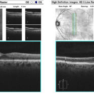
HD 1 line 100x OCT scan of a New Subretinal Hemorrhage in a established patient with AMD.
Photographer: Drew Mitchell, OCT-C
Imaging device: Zeiss Cirrus 6000
Condition/keywords: age-related macular degeneration (AMD), OCT, subretinal hemorrhage
-
 NIR and OCT Left Eye Early Parafoveal Changes Plaquenil
Jan 14 2025 by Kyle D Kovacs, MD
58 year old woman with 11 year history of plaquenil use with early parafoveal outer retinal attenuation and bull's eye on near-infrared imaging. Left eye
Condition/keywords: OCT, plaquenil toxicity
-
 NIR and OCT Right Eye Early Parafoveal Changes Plaquenil
Jan 14 2025 by Kyle D Kovacs, MD
58 year old woman with 11 year history of plaquenil use with early parafoveal outer retinal attenuation and bull's eye on near-infrared imaging. Right eye.
Condition/keywords: OCT, plaquenil toxicity
 Loading…