Search results (1541 results)
-
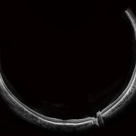 Wide-Field-OCT-montage
Jan 8 2018 by Netan Choudhry, MD, FRCS(C) FASRS
This is an SD-OCT montage image of a 55 year old male with optic neuropathy representing a wide-field OCT spanning 130 degrees.
Photographer: John Golding, Vitreous Retina Macula Specialists of Toronto
Imaging device: Heidelberg Spectralis OCT system
Condition/keywords: wide angle imaging
-
 Flame of the Forest
Apr 9 2020 by Daraius N Shroff, MS FMRF FRCS
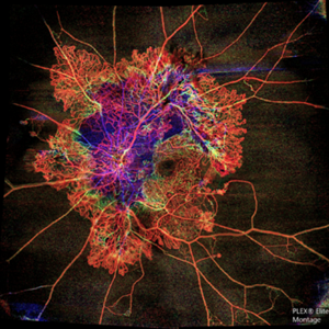
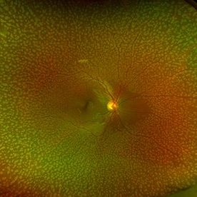
A 54-year-old man with DM for 15 years. The left eye had a visual acuity of 20/40. Wide field swept source OCTA revealed branching out central neovascular trunk vessels from the disc with terminal loops, along with exuberant proliferation of irregular small-calibre fine new vessels. The patient underwent OCTA guided pan retinal photocoagulation.
Photographer: Anuj Choudhary, Shroff Eye Centre, New Delhi
Imaging device: Zeiss Plex Elite 9000
Condition/keywords: proliferative diabetic retinopathy (PDR)
-
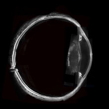 Whole Eye OCT
Jan 4 2019 by Netan Choudhry, MD, FRCS(C) FASRS
Swept-Source OCT montage of a 45-year-old male with Alports disease and posterior subcapsular cataract.
Photographer: John Golding BA, Vitreous Retina Macula Specialists of Toronto
Imaging device: Topcon DRI Triton
Condition/keywords: Alports disease, optical coherence tomography (OCT), swept source
-
 Acute Posterior Multifocal Placoid Pigment Epitheliopathy
Feb 20 2024 by Soobien Lee
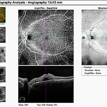
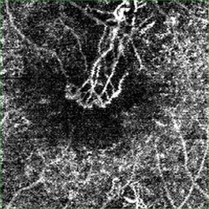
12x12mm OCT Angiography of a 20-year-old caucasian female with viral prodrome and vision loss OS>OD secondary to Acute Posterior Multifocal Placoid Pigment Epitheliopathy (APPME). Imaging shows multifocal flow voids.
Photographer: Kim Seay, Elman Retina Group
Imaging device: 12x12mm OCT-Angiography
Condition/keywords: acute posterior multifocal placoid pigment epitheliopathy (APMPPE), bacillary layer detachment, OCT, OCT Angiography, Uveitis, white dot syndrome
-
 Acute Posterior Multifocal Placoid Pigment Epitheliopathy
Feb 20 2024 by Soobien Lee
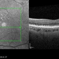
A 20-year-old caucasian female with viral prodrome and vision loss OS>OD secondary to Acute Posterior Multifocal Placoid Pigment Epitheliopathy (APPME). OCT of the left macula shows bacillary layer detachment.
Photographer: Kim Seay, Elman Retina Group
Condition/keywords: acute posterior multifocal placoid pigment epitheliopathy (APMPPE), bacilliary layer detachment, OCT, Uveitis, white dot syndrome
-
 Choroidal Excavation
Jun 2 2019 by Nelson Chamma Capelanes, MD
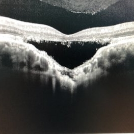
SD-OCT of a 32-year-old woman showing a subfoveal choroidal excavation associated with chronic central serous chorioretinopathy.
Photographer: Nelson Chamma Capelanes, Promacula, Brazil
Imaging device: Heidelberg Spectralis SD-OCT
Condition/keywords: choroidal excavation, chronic central serous chorioretinopathy (CSCR), pachychoroid
-
 Retinal Arterio-Venous Malformations
Apr 7 2017 by Deepak Bhojwani, MS
Multimodal imaging of a 16-year-old boy with retinal arterio-venous malformations(AVM). He also had cerebral AVM's on MRI-contrast studies suggesting Wyburn-Mason syndrome.
Photographer: DEEPAK BHOJWANI, RAGHUDEEP EYE HOSPITAL, AHMEDABAD.
Imaging device: Zeiss VISUCAM
Condition/keywords: color fundus photograph, FA early phase, optical coherence tomography (OCT), Wyburn-Mason
-
 Tractional Retinal Detachment
Sep 27 2012 by Virgilio Morales-Canton, MD
OCT image of a 42-year-old male patient with a localized traction of the superior macula secondary to proliferative diabetic retinopathy.
Imaging device: Cirrus
Condition/keywords: tractional retinal detachment
-
 Acute Macular Neuroretinopathy
Dec 11 2019 by Lauren Whaley
34-year-old female patient presented with changes in vision after recent upper respiratory infection. Referring doctor originally thought it was a blood pressure issue. She noticed a "C" shape in her vision. Infrared image was captured showing exactly what patient was describing! Doctor confirmed with this image that it was AMN.
Photographer: Lauren R. Whaley, COA
Imaging device: Heidelberg Spectralis
Condition/keywords: 30 degrees, acute macular neuroretinopathy, Heidelburg Spectralis, left eye, macula, near infrared autofluorescence (NIRAF)
-
 Central Retinal Vein Occlusion by OCT Angiography
Jun 13 2022 by JORGE SOBERANES
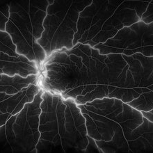
A 63 year old man with a central retinal vein oclussion. In the OCT angiogram we could observe retinal isquemia, neovascularization and arteriovenous shunts.
Photographer: Jorge I. Soberanes MD
Imaging device: PLEX Elite 9000, Zeiss
Condition/keywords: Central vein oclussion, neovascularization, OCT angiography, retina, Shunts
-
 Plaquenil Toxicity
Apr 30 2013 by Theodore Leng, MD, MS, FASRS
SD-OCT scan from a 44-year-old woman with bilateral plaquenil toxicity. There is damage visible in the outer retina in a perifoveal distribution.
Condition/keywords: hydroxychloroquine toxicity, plaquenil toxicity
-
 Submacular PFO
Feb 20 2020 by Kevin J. Blinder, MD, FASRS
This is a 53-year-old gentleman that was referred to us for a second opinion with an inoperable RD with PVR after 3 failed attempts. We performed a PPV, membranectomy, scleral buckling procedure, with silicone oil injection. This case did not require PFO. You can imagine our surprise when we discovered submacular PFO postoperatively. It is very difficult to see the PFO on the Optos. The infrared shows it clearly, with confirmation of the submacular space on the SD-OCT.
Photographer: Jarrod Wehmeier, The Retina Institute; St. Louis, MO
Imaging device: optos
Condition/keywords: submacular perfluorocarbon liquid (PFO)
-
 Active CNVM
Jul 11 2016 by Manish Nagpal, MD, FRCS (UK), FASRS
Colour photo showing an active CNVM.
Photographer: pooja barot
Condition/keywords: choroidal neovascular membrane (CNVM), optical coherence tomography (OCT)
-
 Active CNVM on Angio OCT
Jul 11 2016 by Manish Nagpal, MD, FRCS (UK), FASRS
Angio OCT picture showing neovascularization corresponding to the area of CNVM.
Photographer: pooja barot
Condition/keywords: choroidal neovascular membrane (CNVM), optical coherence tomography (OCT)
-
 Benign Familial Fleck Retina
Feb 2 2023 by Hemanth Murthy, MBBS, MD, FASRS
12 year boy first born of consanguineous marriage, came for routine eye check up with BCVA 20/40 OU. He has no night blindness. His OCT showed thickening of the RPE with dome like elevations involving the ellipsoid layer. Dark adapted ERG showed normal 'b' wavesPhotopic ERG showed reduced 'a' and b waves.
Photographer: Veda Vyas
Imaging device: Optos Daytona
Condition/keywords: Benign familial fleck retina
-
 Central Retinal Vein Occlusion
Jan 21 2022 by Olivia Rainey
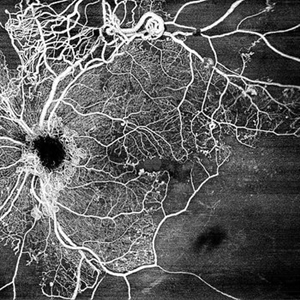
Ultra-widefield fluorescein angiogram of a 23-year-old female with a Central Retinal Vein Occlusion affecting her left eye. The patient presented on 12/22/2021 cc20/40-2 vision in the left eye. The patient reported recent trauma of being hit with a fist on both sides of face followed by vision loss. The patient has history of Hashimoto's thyroid disease. The following labs have been ordered, PT, PTT, CBC, antithrombin III activity, protein C, protein S, Factor V Leiden mutation, Prothrombin (G20210A), lipid panel, HbA1c, quantiferon gold, RPR, and CXR.
Photographer: Olivia Rainey, OCT-C, COA
Imaging device: Optos California
Condition/keywords: central retinal vein occlusion (CRVO), disc leakage, fluorescein angiogram (FA), fluorescein leakage, left eye, non-ischemic central retinal vein occlusion (CRVO), Optos, trauma, ultra-wide field imaging
-
 Chorioretinitis with Overlying Vitreous Stranding/Vitritis
Mar 23 2023 by Isaac Agranoff
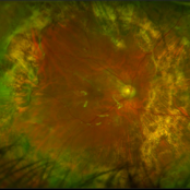
Fundus photograph of a 37-year-old woman presenting with chorioretinitis with overlying vitreous stranding/vitritis that has remained unchanged for multiple years. Patient presented with irritation and blurred vision and her vision was 20/40 OD. The OCT revealed evidence of low-grade inflammation and the recommend treatment was anti-inflammatory eye drops at this time and to obtain second opinion with another physician in the office.
Photographer: Isaac Agranoff, Technician
Imaging device: Optos California
Condition/keywords: chorioretinal scar, chorioretinitis, inflammation, Optos, ultra-wide field imaging, vitritis
-
 Cuticular Drusen
Jan 17 2024 by John Lee
Heidelberg SD-OCT of a 65-year-old woman with age-related macular degeneration demonstrating classic sawtooth appearance of cuticular drusen.
Photographer: Natasha Vinson
Imaging device: Heidelberg Spectralis
Condition/keywords: age-related macular degeneration (AMD), cuticular drusen
-
 Dry AMD, Advanced Atrophic without Subfoveal Involvement
Oct 12 2021 by Kelli Nyenhuis
OCT Heidelberg photograph of a 79-year-old woman with AMD advancing that has been observed over the last 10 years.
Photographer: Kelli Nyenhuis, OMA
Imaging device: Heidelberg
Condition/keywords: dry age-related macular degeneration (dry AMD)
-
 Foveoschisis secondary to high myopia
Mar 13 2015 by Niloofar Piri, MD
Infrared and HD-OCT of the right eye in a 55-year-old African American female with high myopia (more than -6.00 D), BCVA: 20/25 OU Cartwheel appearance of the fovea in the infrared imaging is visible. HD- OCT demonstartes schisis in different layers of the retina (both NFL and OPL; notice stretching of the Muller cells); VMT is also present . Outer retinal layers are preserved which explains the good vision . She had the same findings in OS.
Photographer: Niloofar Piri, MD
Imaging device: Heidelberg Spectralis
Condition/keywords: high myopia, retinoschisis
-
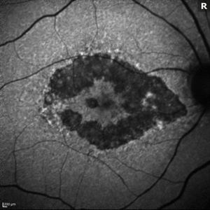
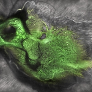 Green Goblin Detachment
Jan 13 2022 by Netan Choudhry, MD, FRCS(C) FASRS
Tractional retinal detachment with macular hole in a 76-year-old female.
Photographer: John Golding BA, Vitreous Retina Macula Specialists of Toronto, OCTane Imaging Lab
Imaging device: Multicolor fundus photo taken on the Spectralis OCT2 (Heidelberg Engineering GmbH).
Condition/keywords: macular hole, Multispectral imaging, tractional retinal detachment
-
 Macula off Rhegmatogenous Retinal Detachment
Aug 28 2012 by Sharon Fekrat, MD FACS FASRS
62 year old man with a rhegmatogenous retinal detachment involving the foveal center in his left eye as depicted on this Zeiss Stratus OCT image.
Photographer: Michael P. Kelly, FOPS Director, Duke Eye Labs, Duke University Eye Center, Durham, NC
Imaging device: Zeiss Stratus
-
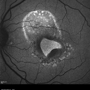 Macular Tear
May 14 2014 by Avris Romario Diparaja Siahaan
Blue autofluorescence (BAF) a 40-year-old man with macular tear (had a photocoagulation laser).
Photographer: Avris Romario Diparaja Siahaan
Imaging device: Heidelberg HRA + OCT Spectralis
Condition/keywords: autofluorescence imaging, macular hole
-
 Myopic Traction Maculopathy
Mar 17 2025 by Drew Mitchell
HD 1 line 100x 9 mm scan of a right eye with MTM at stage 3c. Macular Schisis Detachment.
Photographer: Drew Mitchell OCT-C
Imaging device: Zeiss Cirrus 5000
Condition/keywords: full thickness macular hole, Macular hole, myopic foveoschisis, myopic macular schisis, myopic traction maculopathy, PVD
-
 OCT Image of Epiretinal Membrane
Aug 29 2017 by Carolyn Daley
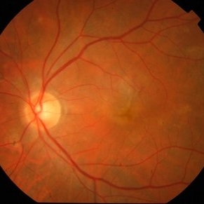
OCT photograph of a 64-year-old women with an epiretinal membrane in the right eye. Patient has not noticed any decline in vision so surgery was not recommended at this time.
Photographer: Carolyn Daley
Imaging device: Heidelberg Spectralis
Condition/keywords: epiretinal membrane (ERM), optical coherence tomography (OCT)
 Loading…