Search results (1541 results)
-
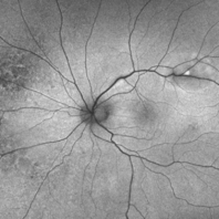
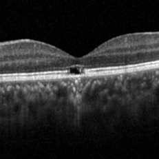
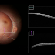
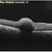 Sub-Internal Limiting Membrane Hemorrhage
Apr 17 2025 by Malvika Singh
OCT of a 41 year-old, male, with a central retinal vein occlusion and a foveal sub-internal limiting membrane hemorrhage.
Photographer: Dr Malvika Singh, Retina Foundation, Ahmedabad, India
Imaging device: Mirante SLO/OCT
Condition/keywords: optical coherence tomography (OCT), SUB ILM hemorrhage
-
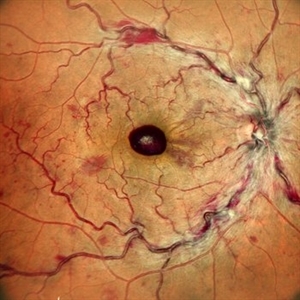
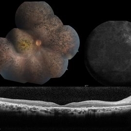
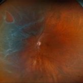
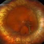
 Ultra-Wide Field Image of Central Retinal Vein Occlusion with Foveal Hemorrhage
Apr 17 2025 by Malvika Singh
Ultra- wide field fundus photograph of a 41 year-old male, with a central retinal vein occlusion and a foveal sub-internal limiting membrane hemorrhage.
Photographer: Dr Malvika Singh, Retina Foundation, Ahmedabad, India
Imaging device: Mirante SLO/OCT
Condition/keywords: central retinal vein occlusion (CRVO), macular hemorrhage, Ultra-wide field retinal imaging
-
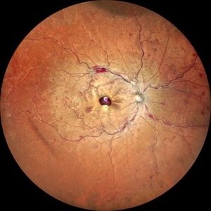
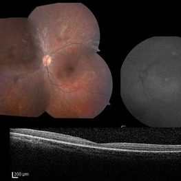
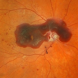
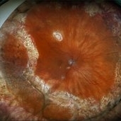
 Central Retinal Vein Occlusion with Foveal Hemorrhage
Apr 17 2025 by Malvika Singh
Fundus photograph of a 41 year-old, male, with a central retinal vein occlusion and a foveal sub-internal limiting membrane hemorrhage.
Photographer: Dr Malvika Singh, Retina Foundation, Ahmedabad, India
Imaging device: Mirante SLO/OCT
Condition/keywords: central retinal vein occlusion (CRVO), macular hemorrhage
-
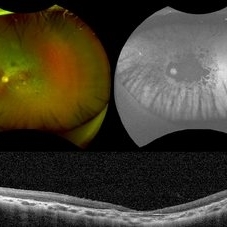
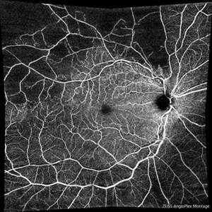
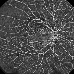 Branch Retinal Vein Occlusion
Apr 10 2025 by Rinat Sutiushev
Ultra-Widefield OCT Angiography of a 77-year-old woman with ischemic occlusion of the superior temporal branch of the central retinal vein with non-proliferative diabetic retinopathy.
Photographer: Rinat Sutiushev, Ophthalmological center “Vision”, Saint Petersburg
Imaging device: TOWARDPI BMIZAR – 400KHZ FULL RANGE SS-OCTA
Condition/keywords: branch retinal vein occlusion (BRVO), nonproliferative diabetic retinopathy, retina
-
 Blister Retinal Detachment Superotemporal with a Flap Tear
Apr 10 2025 by Daniela Bogenschutz
Autofluorescence of a 70-year-old male with a superotemporal retinal detachment prior to having an OCT with unusual findings. Patient states symptoms were "starburst" in his vision in the location of the retinal detachment with the retinal tear. Surgery was scheduled immediately to avoid further progression.
Photographer: Daniela Bogenschutz, OSC; Retina Consultants of the Carolinas, PA
Condition/keywords: Retinal Detachment, retinal detachment with single break
-
 LCA type 12
Apr 10 2025 by Joshua Friedman
LCA type 12 due to pathogenic mutations in RDH12. 13-year-old male with a visual acuity of 20/80 and 20/300 in the right and left eye, respectively. There is extensive pigment migration in the peripheral retina and macula. Like RPE65, there is widespread hypoautofluorescent signal, however, the peripapillary retina is uniquely spared in this form of LCA. On OCT, there is almost complete loss of the retina centrally.
Photographer: Stephen Tsang, MD, PhD
Condition/keywords: Leber Congenital Amaurosis
-
 LCA Type 2
Apr 10 2025 by Joshua Friedman
LCA Type 2 (RPE65) showing characteristic hypoautofluorescence and retinal thinning. 8F with best corrected visual acuity of 20/400 (OD) and 20/150 (OS). Small white intraretinal spots and RPE mottling are visible on color fundus photography. Blue light autofluorescence reveals near-complete loss of signal, while OCT demonstrates widespread outer retinal thinning.
Photographer: Stephen Tsang, MD, PhD
Condition/keywords: Leber Congenital Amaurosis
-
 LCA type 10
Apr 10 2025 by Joshua Friedman
LCA type 10 due to mutations in CEP290. 36-year-old male with best corrected visual acuity of light perception in both eyes since childhood. On color fundus imaging, there is a mix of polymorphous white flecks and pigmentary changes. On autofluorescence imaging, there is almost complete loss of macular RPE. On OCT, there is complete loss of inner and outer retinal layers, the greatest losses occurring centrally.
Photographer: Stephen Tsang, MD, PhD
Condition/keywords: Leber Congenital Amaurosis
-
 Severe NPDR with Subhyaloid Hemorrhage
Apr 9 2025 by Kimberly Wakester
Optomap RGB of an 47 year-old man with severe NPDR with subhyaloid hemorrhage in the right eye.
Photographer: Kimberly Wakester, COA, OCT-C
Imaging device: Optos California
Condition/keywords: severe NPDR, subhyaloid hemorrhage
-
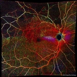
 Branches Starved of Flow, Yet Nature Strives to Grow
Apr 1 2025 by rohan jain
Tufts of NVE's in a case of Branch Retinal Vein Occlusion
Photographer: Dr. ROHAN JAIN
Condition/keywords: branch retinal vein occlusion (BRVO), capillary nonperfusion, non-perfused branch retinal vein occlusion (BRVO), non-perfusion, NVE, OCT Angiography, ST BRVO
-
 Solar Retinopathy
Apr 1 2025 by Isaac Agranoff
OCT scan of 18-year-old male presenting with 20/40 BCVA OU and bilateral focal outer retinal subfoveal defects. Patient reported long-term history of frequent sungazing, has stopped within past 6-9 months.
Photographer: Isaac Agranoff
Imaging device: Heidelberg Spectralis
Condition/keywords: solar retinopathy
-
 Retinal Detachment with Retinal Tear
Mar 31 2025 by Kimberly Wakester
Optomap RGB of an 48-year-old woman with a retinal detachment with retinal tear in the left eye. Surgery was recommended. Patient is to continue follow up care post operatively.
Photographer: Kimberly Wakester, COA, OCT-C
Imaging device: Optos California
Condition/keywords: Retinal Detachment, retinal tear
-
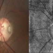 The Halloween Smile
Mar 27 2025 by Shrishti mishra
A 73 year old male with Le optic disc pit . On color fundus photo a single pit can be noted whereas on oct enface is- os interface 2 optic disc pits are noted which resembles a halloween smile .
Photographer: Mr Sudhakar
Imaging device: Zeiss cirrus6000
Condition/keywords: OCT, oct en face, optic disc pit
-
 Completed Bleb with OCT Through Fovea
Mar 25 2025 by Robert Andrew Sisk, MD, FACS, FASRS
Color still from surgical video of subretinal delivery of laru-zova for X-linked retinitis pigmentosa. Live optical coherence tomography (OCT) with foveal tracking via the embedded software in the operating microscope allows monitoring foveal integrity for signs of stress. The contour of the fovea does not exceed the curvature of the bleb (e.g. no inversion). The tangential cannula angle facilitated steering of the bleb posteriorly. The bleb covers essentially the entire macula, which is the target area.
Imaging device: Zeiss Artevo 800
Condition/keywords: gene therapy, genetic disorder, optical coherence tomography (OCT), retinitis pigmentosa, subretinal injection
-
 Repaired Retinal Detachment with Scleral Buckle
Mar 25 2025 by Kimberly Wakester
Optomap RGB montage of an 64-year-old woman with a repaired retinal detachment with scleral buckle in the right eye. There is nasal and inferior pre-retinal membranes with traction. PPV was recommended but patient defers to proceed with sx at this time. Will continue to follow patient closely for worsening traction. Patient was educated on how to monitor their peripheral vision and was advised to report any changes immediately.
Photographer: Kimberly Wakester, COA, OCT-C
Imaging device: Optos California
Condition/keywords: pre-retinal membrane with traction, repaired RD, scleral buckle
-
 Repaired Retinal Detachment with PVR
Mar 25 2025 by Kimberly Wakester
Optomap RGB of a 79-year-old-woman with a repaired retinal detachment with PVR in the right eye. Patient is doing well over 7 months s/p vitrectomy with silicone oil and scleral buckle placement. Retina remains attached on the buckle under oil. Patient is to return in 6 months for follow up exam with repeat imaging.
Photographer: Kimberly Wakester, COA, OCT-C
Imaging device: Optos California
Condition/keywords: PVR, repaired RD, Retinal detachment under Silicon Oil, scleral buckle
-
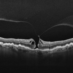 Stage 2 Macular Hole From VMT
Mar 21 2025 by Drew Mitchell
HD 1 line 100x OCT showcasing a full thickness macular hole caused by vitreomacular traction on fovea. Choroidal folds can also be seen on scan.
Photographer: Drew Mitchell OCT-C
Imaging device: Zeiss Cirrus 6000
Condition/keywords: Choroidal Folds, FTMH, macular hole, OCT, PVD
-
 Retinal Macroaneurysm (RAM)
Mar 19 2025 by Drew Mitchell
3x3 OCT-A of a Retinal Macroaneurysm in the left eye along the IT arcade that has surrounding edema and exudates
Photographer: Drew Mitchell, OCT-C
Imaging device: Zeiss Cirrus 5000
Condition/keywords: CIRRUS 5000 ANGIOPLEX, OCT Angiography, RAM, retinal macroaneurysm
-
 Retinal Macroaneurysm (RAM)
Mar 19 2025 by Drew Mitchell
3x3 OCT-A of a Retinal Macroaneurysm in the left eye along the IT arcade that has surrounding edema and exudates.
Photographer: Drew Mitchell OCT-C
Imaging device: Zeiss Cirrus 5000
Condition/keywords: OCT Angiography, RAM, retinal macroaneurysm
-
 Myopic Traction Maculopathy
Mar 17 2025 by Drew Mitchell
HD 1 line 100x 9 mm scan of a right eye with MTM at stage 3c. Macular Schisis Detachment.
Photographer: Drew Mitchell OCT-C
Imaging device: Zeiss Cirrus 5000
Condition/keywords: full thickness macular hole, Macular hole, myopic foveoschisis, myopic macular schisis, myopic traction maculopathy, PVD
-
 Branch Retinal Vein Occlusion with Macular Edema
Mar 14 2025 by Drew Mitchell
Zeiss Montage Angio 8x8 mm OCT Angiography Retina Depth Encoded Angioplex of a New BRVO in the right eye.
Photographer: Drew Mitchell, OCT-C
Imaging device: Zeiss Cirrus 6000
Condition/keywords: branch retinal vein occlusion (BRVO), macular edema, OCT Angiography
-
 Branch Retinal Vein Occlusion with Macular Edema
Mar 14 2025 by Drew Mitchell
Zeiss Montage Angio 8x8 mm OCT Angiography Superficial Angioplex of a New BRVO in the right eye.
Photographer: Drew Mitchell OCT-C
Imaging device: Zeiss Cirrus 6000
Condition/keywords: branch retinal vein occlusion (BRVO), macular edema, OCT Angiography
-
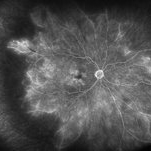 Moderate Nonproliferative Diabetic Retinopathy
Mar 13 2025 by Drew Mitchell
Fluorescein angiography on a patient with Moderate Nonproliferative Diabetic Retinopathy at 5 minutes.
Photographer: Drew Mitchell OCT-C
Imaging device: Optos California
Condition/keywords: Diabetes, ischemia, nonproliferative diabetic retinopathy, OPTOS CALIFORNIA
-
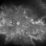 Moderate Nonproliferative Diabetic Retinopathy
Mar 13 2025 by Drew Mitchell
Fluorescein angiography on a patient with Moderate Nonproliferative Diabetic Retinopathy at 5 minutes.
Photographer: Drew Mitchell OCT-C
Imaging device: Optos California
Condition/keywords: Diabetes, nonproliferative diabetic retinopathy, OPTOS CALIFORNIA, retinal ischemia
 Loading…