Search results (247 results)
-
 Astrocytic Hamartoma
Jul 8 2013 by Jason S. Calhoun
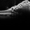
Calcified astrocytic hamartoma of the optic nerve.
Photographer: Jason S. Calhoun, Department of Ophthalmology, Mayo Clinic Jacksonville, Florida
Condition/keywords: hamartoma
-
 Cavernous Hem of Retina
Oct 9 2012 by Alan D. Letson, MD
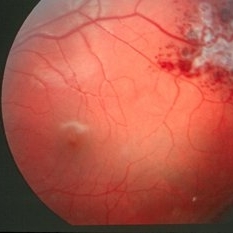
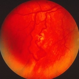
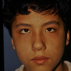
12-year-old boy with cavernous hemangioma of the retina.
Photographer: Beverly Radcliffe
Condition/keywords: cavernous hemangioma of the retina, hamartoma, phakoma
-
 Combined Hamartoma of the Retina and Retinal Pigment Epithelium
Apr 26 2020 by Dipak Nag, MBBS, FCPS, MSc, FRF
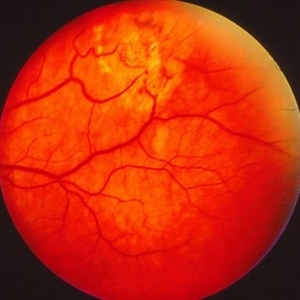
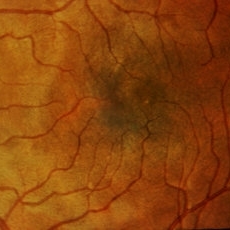
A 28-year-old female presented with a deeply pigmented gray- brown, elevated lesion extending from the temporal side of the disc to the macula (OU). Remarkable retinal vasculature with straightening of the distal vessels and dilatation as well as tortuousity of the peri-lesional vessels. The vitreoretinal interface shows gliosis and epi-retinal membrane (ERM) formation.
Photographer: Dipak Nag
Condition/keywords: hamartoma, retinal pigment epithelium
-
 Combined Hamartoma of the Retina and RPE
Apr 16 2015 by Rita Couceiro, MD, MS
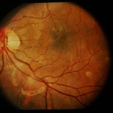
Fundus photograph, red-free picture (top images) and fluorescein angiography pictures (bottom images) of a 6-year-old girl with a combined hamartoma of the retina and RPE in the right eye.
Condition/keywords: hamartoma
-
 Combined Hamartoma of the Retinal Pigment Epithelium Case 1
Oct 5 2012 by Ronald C. Gentile, MD
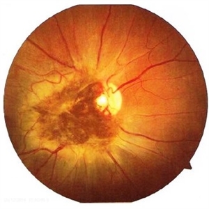
A peripapilary combined hamartoma of the retinal pigment epithelium involving the nasal disc margin. This tumor is slightly elevated, charcoal grey in color with grey-white tissue on it surface. The underlying retinal vessels are obscured.
Photographer: The New York Eye & Ear Infirmary Department of Medical Imaging
Condition/keywords: hamartoma, retinal pigment epithelium
-
 Combined Hamartoma of the Retinal Pigment Epithelium Case 2
Oct 5 2012 by Ronald C. Gentile, MD
A peripapilary combined hamartoma of the retinal pigment epithelium involving the inferior disc margin. This tumor and slightly elevated, charcoal grey to light grey in color with grey-white tissue on it surface. The underlying retinal vessels are obscured with some epiretinal membrane and some striae extending to the inferior nasal macula.
Photographer: The New York Eye & Ear Infirmary Department of Medical Imaging
Condition/keywords: hamartoma, retinal pigment epithelium
-
 Combined Hamartoma of the Retinal Pigment Epithelium Case 2
Oct 5 2012 by Ronald C. Gentile, MD
Magnified view of the peripapilary combined hamartoma of the retinal pigment epithelium involving the inferior disc margin. This tumor and slightly elevated, charcoal grey to light grey in color with grey-white tissue on it surface. The underlying retinal vessels are obscured with some epiretinal vitreous membranes.
Photographer: The New York Eye & Ear Infirmary Department of Medical Imaging
Condition/keywords: hamartoma, retinal pigment epithelium
-
 Combined Harmartoma of the Retina and RPE
Apr 4 2023 by Jeffrey Barker
32 Year old Male with a Combined Hamartoma of the Retina and RPE.
Photographer: Jeffrey P. Barker, B.S.
Condition/keywords: hamartoma
-
 Combined Harmartoma of the Retinal Pigment Epithelium Case 1
Oct 5 2012 by Ronald C. Gentile, MD
Magnified view of the nasal peripapilary combined hamartoma of the retinal pigment epithelium. The tumor is slightly elevated, charcoal grey in color with grey-white tissue on its surface.
Photographer: The New York Eye & Ear Infirmary Department of Medical Imaging
Condition/keywords: hamartoma, retinal pigment epithelium
-
 Congenital Simple Hamartoma of RPE
Aug 3 2015 by Bindu Rajesh
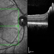
OCT line scan through the hamartoma in a 26-year-old male, showing increased hyperreflectivity in the area of lesion with backshadowing and minimal protrusion into vitreous.
Imaging device: Heidelberg Spectralis
Condition/keywords: congenital, hamartoma, retinal pigment epithelium
-
 Congenital Simple Hamartoma of the RPE Autofluorescence
Aug 3 2015 by Bindu Rajesh
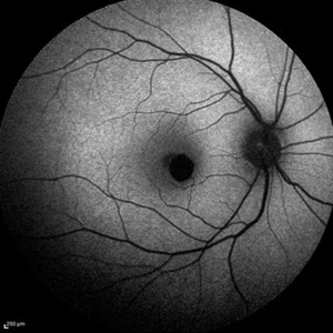
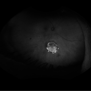
Fundus autofluorescence image of a 26-year-old male depicting hypoautofluorescence nasal to the fovea corresponding to the hamartoma visible clinically.
Imaging device: Heidelberg Spectralis
Condition/keywords: congenital, hamartoma, retinal pigment epithelium
-
 Congenital Simple Hamartoma of the RPE Fundus Photo
Aug 3 2015 by Bindu Rajesh
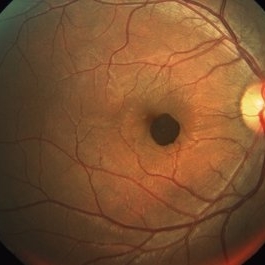
Fundus photograph of a 26-year-old male ,showing a well defined pigmented lesion inferonasal to the fovea suggestive of simple hamartoma.
Imaging device: Visupac
Condition/keywords: congenital, hamartoma, retinal pigment epithelium
-
 Congenital Simple Hamartoma RPE
Jun 5 2020 by stephen oconnell
Congenital simple hamartoma RPE.
Condition/keywords: hamartoma, retinal pigment epithelium
-
 Congenital Simple Hemartoma of the RPE
Jun 8 2016 by John S. King, MD
Congenital simple hamartoma of the RPE.
Condition/keywords: hamartoma, retinal pigment epithelium
-
 Endophytic Vascular Hamartoma
Nov 20 2019 by McGill University Health Centre
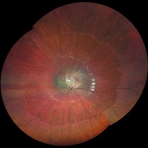
39 year-old Caucasian man with a diagnosis of Von Hippel-Lindau disease. Eyes were obtained post-mortem. Endophytic vascular hamartomas were identified in both eyes.
Condition/keywords: hamartoma, Von Hippel-Lindau
-
 Endophytic Vascular Hamartoma
Nov 20 2019 by McGill University Health Centre
39-year-old Caucasian man with a diagnosis of Von Hippel-Lindau disease. Eyes were obtained post-mortem . Endophytic vascular hamartomas were identified in both eyes.
Condition/keywords: hamartoma, Von Hippel-Lindau
-
 Hamartoma
Nov 8 2019 by Stephanie Burke
Fundus Auto-fluorescence image of an 76-year-old man with hamartoma.
Photographer: Stephanie Burke, CRA, OCT-C
Imaging device: Optos
Condition/keywords: hamartoma
-
 Hamartoma
Nov 8 2019 by Stephanie Burke
Early frame FA image of a 76-year-old man with hamartoma.
Photographer: Stephanie Burke, CRA, OCT-C
Imaging device: Optos
Condition/keywords: hamartoma
-
 Hamartoma of the Retina and Retina Pigment Epithelium
Jun 22 2021 by Giselle DeOliveira
Fundus montage of 9-year-old boy with hamartoma of the retina and retina pigment epithelium.
Photographer: Giselle DeOliveira, University of Miami, Bascom Palmer Eye Institute
Imaging device: Zeiss Clarus
Condition/keywords: hamartoma
-
 Hamartoma of the Retina and Retinal Pigment Epithelium
Jan 5 2025 by César Adrián Gómez Valdivia, MD
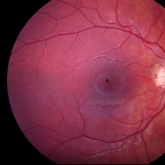
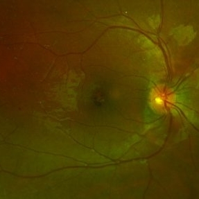
Hamartoma of the retina and retinal pigment epithelium found in a 10 year-old male patient with type 2 neurofibromatosis history. Overlaying fibrous proliferation can be appreciated. Findings were unilateral.
Photographer: @eyemissu2
Imaging device: TOPCON TRC-50DX
Condition/keywords: hamartoma, retinal pigment epithelium (RPE) hamartoma
-
 Hamartoma of the Retinal Pigment Epithelium
Apr 2 2024 by José Laércio Araújo Filho
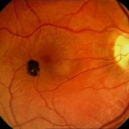
Fundus photograph of a 36-year-old man with a Hamartoma of the retinal pigment epithelium.
Photographer: José Laércio de Araújo Filho, Universidade de São Paulo, São Paulo
Imaging device: Optos Daytona P200T / A10600
Condition/keywords: hamartoma
-
 Hamartoma Tuberous Sclerosis
Jun 7 2016 by Nelson Chamma Capelanes, MD
Fundus photograph of an 52-year-old man with tuberous sclerosis and retinal hamartoma.
Photographer: Nelson Chamma Capelanes, Fundação Hilton Rocha, Promédica Indaiatuba, Brazil
Imaging device: Heidelberg Spectralis
Condition/keywords: hamartoma, tuberous sclerosis
-
 Neurofibromatosis II with RPE Hamartoma
Sep 8 2015 by David Callanan, MD
Neurofibromatosis II with RPE hamartoma.
Condition/keywords: hamartoma, neurofibromatosis, retinal pigment epithelium
-
 Neurofibromatosis II with RPE Hamartoma
Sep 8 2015 by David Callanan, MD
Neurofibromatosis II with RPE hamartoma.
Condition/keywords: hamartoma, neurofibromatosis, retinal pigment epithelium
-
 Neurofibromatosis II with RPE Hamartoma
Sep 8 2015 by David Callanan, MD
Neurofibromatosis II with RPE hamartoma.
Condition/keywords: hamartoma, neurofibromatosis, retinal pigment epithelium
 Loading…