Search results (8 results)
-
 Retinal Astrocytic Hamartoma
Feb 5 2025 by Rinat Sutiushev
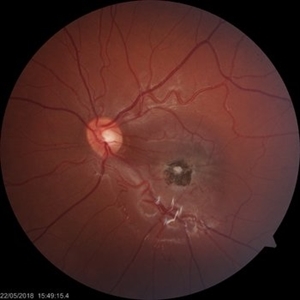
Fundus photograph of a 42-year-old man with retinal astrocytic hamartoma type 3.
Photographer: Rinat Sutiushev, Ophthalmological center “Vision”, Saint Petersburg
Imaging device: Heidelberg Spectralis
Condition/keywords: retina
-
 Combined Hamartoma of the Retina and Retinal Pigment Epithelium (CHRRPE)
Jan 21 2020 by Pierre-Henry Gabrielle, MD
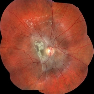
Fundus photograph of a 17-year-old man with combined hamartomas of the retina and retinal pigment epithelium (CHRRPE) at the posterior pole of the left eye.
Photographer: Pierre-Henry Gabrielle, Ophthalmology department, Dijon University Hospital, France
Imaging device: Zeiss Visucam
Condition/keywords: combined hamartoma, fundus photograph
-
 Retina Hamartomas in Tuberous Sclerosis
Jan 8 2019 by Sofia Mano
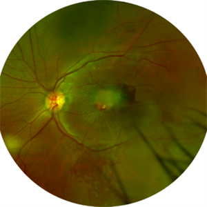
Female 19-years-old with tuberous sclerosis. BCVA LE 10/10. Fundus LE shows three multinodular hamartomas.
Photographer: Sofia Sousa Mano
Imaging device: Canon CR 2 plus
Condition/keywords: hamartoma, tuberculous chorioretinitis
-
 Astrocytic Hamartoma
Jan 4 2019 by Netan Choudhry, MD, FRCS(C) FASRS
Fundus photograph montage of a 7-year-old boy with an astrocytic hamartoma involving the macula.
Photographer: John Golding BA, Vitreous Retina Macula Specialists of Toronto
Imaging device: Topcon TRC-50 Dx
Condition/keywords: astrocytic hamartoma
-
 Combined Hamartoma
Oct 5 2016 by Guruprasad S. Ayachit, MBBS,MS
Fundus photograph of a 9-year-old boy with an ill-defined lesion extending from nasal to the disc going on to include the papillomacular bundle; 14X10 mm in greatest dimensions. There is a thick epiretinal membrane causing distortion and straightening of temporal vascular arcade.
Photographer: Shravan Masurkar, M M Joshi Eye Institute, Hubli
Imaging device: Topcon TRC50DX
Condition/keywords: combined hamartoma
-
 Combined Hamartoma
Feb 29 2016 by Andrea Arriola-Lopez, MD MSc
40 year-old man with diminished VA since 6 month ago. Fundus examination revealed macular folds, yellow-whitish elevated lesion at the fovea and a subretinal hemorrhage.
Photographer: Andrea Elizabeth Arriola-Lopez MD, MSc
Imaging device: OPTOS Dakota
Condition/keywords: combined hamartoma, macula, subretinal hemorrhage
-
 Congenital Simple Hamartoma of RPE
Aug 3 2015 by Bindu Rajesh
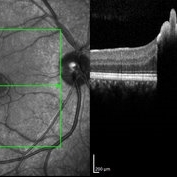
OCT line scan through the hamartoma in a 26-year-old male, showing increased hyperreflectivity in the area of lesion with backshadowing and minimal protrusion into vitreous.
Imaging device: Heidelberg Spectralis
Condition/keywords: congenital, hamartoma, retinal pigment epithelium
-
 Epiretinal Membrane/Macular Pucker With Combined Hamartoma of Retina and RPE
Jul 8 2015 by Emmanuel Chang, MD PhD FACS FASRS
10-year-old with history of progressive severe distortion in the left eye over the past year.
Photographer: Retina and Vitreous of Texas
Imaging device: Heidelberg Autofluorescence
Condition/keywords: combined hamartoma, epiretinal membrane (ERM), retinal pigment epithelium (RPE) hamartoma
 Loading…