 Initializing download.
-
 By Ronald C. Gentile, MD
The New York Eye and Ear Infirmary of Mount Sinai - Uploaded on Oct 5, 2012.
- Last modified by Ronald C. Gentile, MD on Oct 21, 2012.
- Reviewed by Jennifer Hicks
- Rating
- Appears in
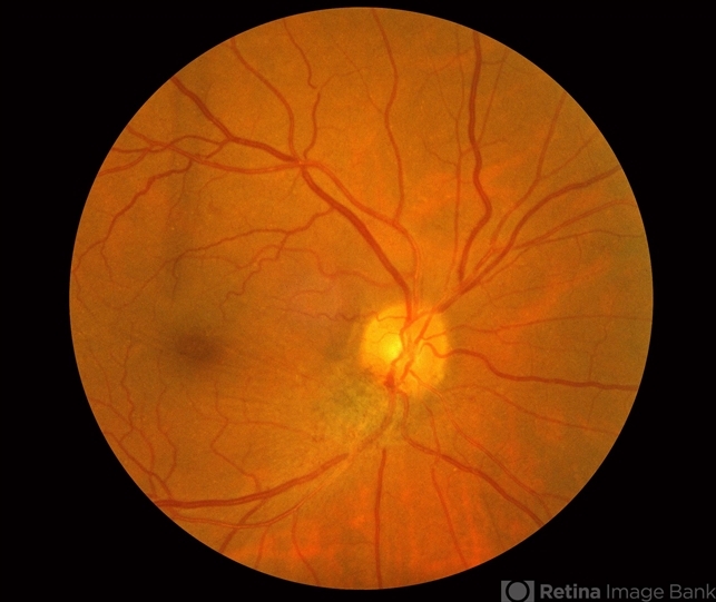
- Combined Hamartoma of the Retinal Pigment Epithelium Case 2
- Condition/keywords
- hamartoma, retinal pigment epithelium
- Photographer
- The New York Eye & Ear Infirmary Department of Medical Imaging
- Imaging device
- Fundus camera
- Description
- A peripapilary combined hamartoma of the retinal pigment epithelium involving the inferior disc margin. This tumor and slightly elevated, charcoal grey to light grey in color with grey-white tissue on it surface. The underlying retinal vessels are obscured with some epiretinal membrane and some striae extending to the inferior nasal macula.


