Search results (247 results)
-
---thumb.jpg/image-square;max$300,300.ImageHandler) Astrocytic Hamartoma
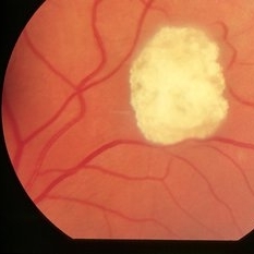
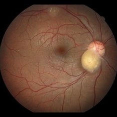
Astrocytic Hamartoma
Feb 20 2013 by From the Collections of Thomas M. Aaberg, MD and Thomas M. Aaberg Jr., MD
Mulberry lesion.
Condition/keywords: tuberous sclerosis
-
 RPE Hamartoma
Oct 18 2012 by Raj K. Maturi, MD
Photographer: Tom Steele, CRA
Imaging device: Topcon 50dx
Condition/keywords: hamartoma, retinal pigment epithelium
-
 Epiretinal Membrane/Macular Pucker With Combined Hamartoma of Retina and RPE
Jul 8 2015 by Emmanuel Chang, MD PhD FACS FASRS
10-year-old with history of progressive severe distortion in the left eye over the past year.
Photographer: Retina and Vitreous of Texas
Imaging device: Heidelberg Autofluorescence
Condition/keywords: combined hamartoma, epiretinal membrane (ERM), retinal pigment epithelium (RPE) hamartoma
-
 Tuberous Sclerosis
Oct 9 2012 by Alan D. Letson, MD
Small astrocytic hamartoma in asymptomatic 65-year-old woman with Tuberous sclerosis.
Photographer: Beverly Radcliffe
Condition/keywords: astrocytoma, hamartoma, tuberous sclerosis
-
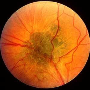 Combined Hamartoma of Retina and RPE
Mar 29 2013 by Henry J. Kaplan, MD
Greenish lesion on the arcade with epiretinal membrane formation, vessels inside the lesion are contracted and those outside are distracted.
Condition/keywords: combined hamartoma
-
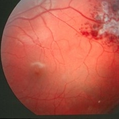 Cavernous Hem of Retina
Oct 9 2012 by Alan D. Letson, MD
12-year-old boy with cavernous hemangioma of the retina.
Photographer: Beverly Radcliffe
Condition/keywords: cavernous hemangioma of the retina, hamartoma, phakoma
-
 RPE Hamartoma
Oct 18 2012 by Raj K. Maturi, MD
Photographer: Tom Steele, CRA
Imaging device: Topcon 50dx
Condition/keywords: hamartoma, red-free, retinal pigment epithelium
-
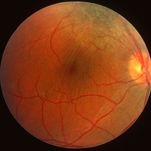
 Retinal Astrocytoma
Apr 16 2015 by Rita Couceiro, MD, MS
Fundus photograph of a 72-year-old man with a retinal astrocytoma of the left eye.
Condition/keywords: astrocytic hamartoma
-
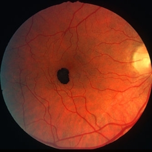 Congenital RPE hamartoma
Jan 11 2013 by Alex P. Hunyor, MD
Congenital RPE hamartoma, right eye.
Condition/keywords: retinal pigment epithelium (RPE) hamartoma, retinal pigment epithelium (RPE) tumor
-
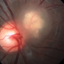
 Astrocytic Hamartoma
Oct 10 2012 by K. Bailey Freund, MD
Fundus photograph of an 85-year-old woman with an astrocytic hamartoma and type 2 choroidal neovascular membrane.
Condition/keywords: choroidal neovascularization (CNV)
-
 Combined Hamartoma of the Retinal Pigment Epithelium Case 1
Oct 5 2012 by Ronald C. Gentile, MD
A peripapilary combined hamartoma of the retinal pigment epithelium involving the nasal disc margin. This tumor is slightly elevated, charcoal grey in color with grey-white tissue on it surface. The underlying retinal vessels are obscured.
Photographer: The New York Eye & Ear Infirmary Department of Medical Imaging
Condition/keywords: hamartoma, retinal pigment epithelium
-
 Astrocytic hamartoma of the retina
Jan 11 2013 by Alex P. Hunyor, MD
Astrocytic hamartoma in a patient with tuberous sclerosis. Note also vascular sheathing inferior to optic disc
Condition/keywords: tuberous sclerosis
-
RPE Hamartoma
Oct 18 2012 by Raj K. Maturi, MD
Photographer: Stephanie Morrow
Imaging device: HRA
Condition/keywords: hamartoma, red-free, retinal pigment epithelium
-
 Retina Hamartomas in Tuberous Sclerosis
Jan 8 2019 by Sofia Mano
Female 19-years-old with tuberous sclerosis. BCVA LE 10/10. Fundus LE shows three multinodular hamartomas.
Photographer: Sofia Sousa Mano
Imaging device: Canon CR 2 plus
Condition/keywords: hamartoma, tuberculous chorioretinitis
-
 Combined Hamartoma of the Retina and Retinal Pigment Epithelium
Dec 22 2015 by P. Mahesh Shanmugam, MBBS, DO, FRCSEd, PhD, FAICO
A fundus photo of 11-year-old boy with CHRRPE, gliotic epiretinal membrane overlying deep greyish lesion. Retinal wrinkling and dragging of macula is seen.
Condition/keywords: combined hamartoma, retinal pigment epithelium (RPE) hamartoma
-
 Optic Nerve Malformation/Possible Combined Hamartoma of Retinal Pigment Epithelium
Feb 19 2013 by From the Collections of Thomas M. Aaberg, MD and Thomas M. Aaberg Jr., MD
No history; Possible Bergmeister's papilla.
Condition/keywords: optic nerve malformation, retinal pigment epithelium
-
 Astrocytoma
Feb 9 2015 by Patricia Araújo
Fundus photography of an 15-year-old boy with tuberous sclerosis.
Photographer: Dr Patricia Correa
Condition/keywords: astrocytic hamartoma, tuberous sclerosis
-
 RPE Hamartoma
Oct 18 2012 by Raj K. Maturi, MD
Photographer: Stephanie Morrow
Imaging device: HRA
Condition/keywords: hamartoma, retinal pigment epithelium
-
 Combined Hamartoma of Retina and RPE
Mar 29 2013 by Henry J. Kaplan, MD
Hamartoma visible as a grreen lesion on superior arcade with ERM formation and dragging of the macula.
Condition/keywords: combined hamartoma
-
 RPE Hamartoma
Oct 18 2012 by Raj K. Maturi, MD
Photographer: Stephanie Morrow
Imaging device: HRA
Condition/keywords: hamartoma, retinal pigment epithelium
-
 Combined Hamartoma of the Retinal Pigment Epithelium Case 2
Oct 5 2012 by Ronald C. Gentile, MD
Magnified view of the peripapilary combined hamartoma of the retinal pigment epithelium involving the inferior disc margin. This tumor and slightly elevated, charcoal grey to light grey in color with grey-white tissue on it surface. The underlying retinal vessels are obscured with some epiretinal vitreous membranes.
Photographer: The New York Eye & Ear Infirmary Department of Medical Imaging
Condition/keywords: hamartoma, retinal pigment epithelium
-
 Gardner Syndrome
Dec 12 2018 by John S. King, MD
66-year-old white male with Gardner Syndrome (colon resection in 1991), who has two children with Gardner Syndrome, presented to Dr. Zocchi with an RD in the fellow eye that was successfully repaired with a pneumatic retinopexy. Currently 20/20 OU with IOP of 7 OD and 14 OS; no RAPD; PCIOL OU. Dr. Zocchi got oral permission by the patient to have these put into the Retina Image Bank. Although the CHRPE like lesions (2 OD) are not bilateral, we both think these lesions represent "retinal pigment epithelial hamartomas associated with familial adenomatous polyposis (RPEH-FAP)" as Shields described in their Intraocular Tumors book. One lesion is located superiorly and is pigmented with depigmented margins; the temporal lesion is atrophic with minimal remaining pigment hypertrophy.
Photographer: Karin Aletter
Imaging device: Optos CA
Condition/keywords: Gardner Syndrome, RPEH-FAP
-
 Astrocytic Hamartoma
Jul 8 2013 by Jason S. Calhoun
Calcified astrocytic hamartoma of the optic nerve.
Photographer: Jason S. Calhoun, Department of Ophthalmology, Mayo Clinic Jacksonville, Florida
Condition/keywords: hamartoma
-
 Astrocytic Hamartoma
Oct 10 2012 by Anat Loewenstein, MD
Five year-old girl came for regular eye examination with no complaints. Medical and ophthalmic history was unremarkable. On examination, visual acuity was 20/25 in both eyes. A Retinal Astrocytic Hamartoma was seen adjacent to the right optic nerve (picture). The rest of the eye examination was normal and no Lisch nodules were seen. The patient was referred for a pediatric neurologist examination and MRI scan of the brain.
Photographer: Galit Yair-Pur
Imaging device: ZEISS FF450 PLUS IR
-
 Combined Harmartoma of the Retinal Pigment Epithelium Case 1
Oct 5 2012 by Ronald C. Gentile, MD
Magnified view of the nasal peripapilary combined hamartoma of the retinal pigment epithelium. The tumor is slightly elevated, charcoal grey in color with grey-white tissue on its surface.
Photographer: The New York Eye & Ear Infirmary Department of Medical Imaging
Condition/keywords: hamartoma, retinal pigment epithelium
 Loading…