Search results (247 results)
-
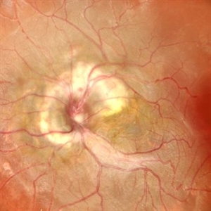
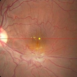
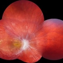
 Retinal Astrocytic Hamartoma
Dec 4 2025 by Abraham Vargas
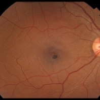
Fundus photograph of an 45 year old man with a Retinal Astrocytic Hamartoma.
Photographer: Abraham Eliazib Vargas, APEC México
Imaging device: Zeiss Clarus 500
Condition/keywords: RAH Retinal Astrocytic Hamartoma
-
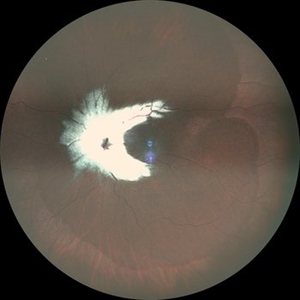
 Combined Hamartoma of Retina and Retinal Pigment Epithelium
Dec 4 2025 by Abraham Vargas
Fundus photograph of an 48 year old woman with a Combined Hamartoma of Retina and Retinal Pigment Epithelium.
Photographer: Abraham Eliazib Vargas, APEC México.
Imaging device: Visucam Zeiss 224
Condition/keywords: CHRRPE
-
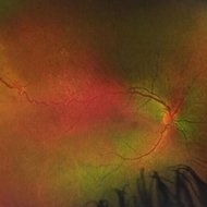
 Goldmann-Favre Syndrome
Aug 19 2025 by Debarun Sharma
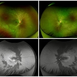
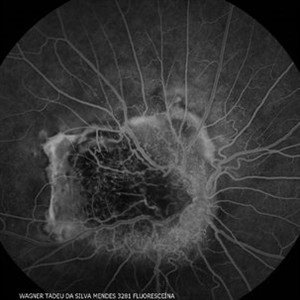
Fundus photograph of a 17 year-old female showing circumferential nummular opacities surrounding the vascular arcades. Fundus autoflourescence shows hypo-autoflourescent circumferential opacities with hyper-autoflourescent ring surrounding macula. Left eye also shows hyper-autoflourescent lesion on the optic nerve head suggestive of astrocytic hamartoma. ERG showed reduced cone response with extinguished rod response. OCT showed schisis of macular area. These features are suggestive of Goldmann-Favre Syndrome.
Photographer: Dr. Debarun Sharma, Sri Sankardeva Nethralaya, Guwahati
Imaging device: Optos
Condition/keywords: Goldmann-Favre Syndrome
-
 Combined Hamartoma of Retina and Retinal Pigment Epithelium
Aug 13 2025 by Drew Mitchell
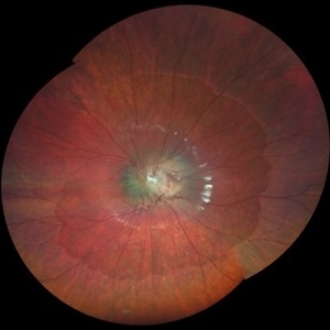
Optos color photograph of a 45 year old male with a combined hamartoma of retina and RPE. Epiretinal membrane formation present.
Photographer: Drew Mitchell OCT-C
Imaging device: Optos Silverstone
Condition/keywords: combined hamartoma of retina and RPE, epiretinal membrane formation, ERM, OPTOS, uwf
-
 Astrocytic Hamartoma
Feb 27 2025 by Daniel Davis, OCT-C
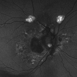
Fundus autofluorescence photo of 55-year-old female with astrocytic hamartoma in association with tuberous sclerosis. No treatment options available, benign. Other findings include; Posterior Vitreous Detachment, Vitreous Hemorrhage, Hereditary Retinal Dystrophy, Vitreous Opacities, Hypertensive Retinopathy.
Photographer: Daniel Davis, OCT-C
Imaging device: Optos California
Condition/keywords: astrocytic hamartoma, fundus autofluorescence (FAF)
-
 Astrocytic Hamartoma
Feb 27 2025 by Daniel Davis, OCT-C
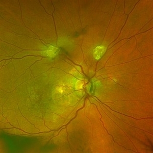
Color fundus photo of 55-year-old female with Astrocytic Hamartoma in association with tuberous sclerosis. No treatment options available, benign. Other findings include; Posterior Vitreous Detachment, Vitreous Hemorrhage, Hereditary Retinal Dystrophy, Vitreous Opacities, Hypertensive Retinopathy.
Photographer: Daniel Davis, OCT-C
Imaging device: Optos California
Condition/keywords: color fundus photograph
-
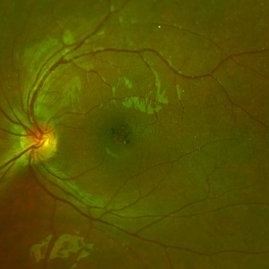
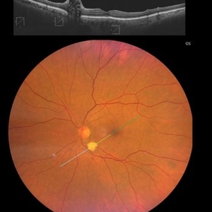
 Retinal Astrocytic Hamartoma
Feb 5 2025 by Rinat Sutiushev
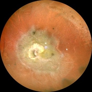
Fundus photograph of a 42-year-old man with retinal astrocytic hamartoma type 3.
Photographer: Rinat Sutiushev, Ophthalmological center “Vision”, Saint Petersburg
Imaging device: Heidelberg Spectralis
Condition/keywords: retinal astrocytic hamartoma
-
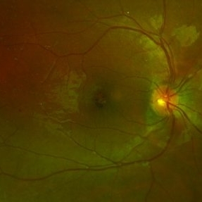
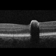
 Retinal Astrocytic Hamartoma
Feb 5 2025 by Rinat Sutiushev
Fundus photograph of a 42-year-old man with retinal astrocytic hamartoma type 3.
Photographer: Rinat Sutiushev, Ophthalmological center “Vision”, Saint Petersburg
Imaging device: Heidelberg Spectralis
Condition/keywords: retina
-
 Combined Hamartoma of the Retina and RPE
Jan 23 2025 by Tejaswita Verma
A 10 year old boy presented with 6/60 vision and LE exotropia with the fundus lesion suggesting a chronic etiology and ILM folds.
Photographer: DR. TEJASWITA VERMA
Imaging device: MIRANTE
Condition/keywords: combined hamartoma of retina and RPE
-
 Hamartoma of the Retina and Retinal Pigment Epithelium
Jan 5 2025 by César Adrián Gómez Valdivia, MD
Hamartoma of the retina and retinal pigment epithelium found in a 10 year-old male patient with type 2 neurofibromatosis history. Overlaying fibrous proliferation can be appreciated. Findings were unilateral.
Photographer: @eyemissu2
Imaging device: TOPCON TRC-50DX
Condition/keywords: hamartoma, retinal pigment epithelium (RPE) hamartoma
-
 Myelinated Nerve Fibres With Combined Hamartoma of Retina and RPE
Jul 31 2024 by Tejaswita Verma
Fundus image of a 20 year old female who presented with metamorphopsia ,slightly blurred vision. BCVA was 6/9, epiretinal membrane present on central fundus examination with myelinated nerve fibres.
Photographer: DR. TEJASWITA VERMA
Imaging device: MIRANTE
Condition/keywords: combined hamartoma of retina and RPE, myelinated nerve fibers
-
 Congenital Hamartoma of Retina and RPE
May 23 2024 by ARVIND JAIN M
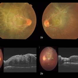
Bilateral involving CHRRPE lesion in a 26 year old gentleman who came with complains of defective vision in both eyes since childhood. His BCVA was Right eye 5/60 and left eye 6/60. His anterior segment examination showed no abnormality with posterior segment examination showed both eyes (Fig.1a and 1b) greyish white elevated lesion involving the macula with thick fibrotic membrane causing the macular drag temporally in right eye and supero-temporally in left eye. (Fig.2a and 2b) OCT showing the thick ERM with the disorganized inner retinal layers suggestive of hamartoma of the Retina and Retinal Pigment Epithelium.
Photographer: Dr. Arvind Jain M, MBBS,MS Ophthal, FVRS
Condition/keywords: CHRRPE
-
 Hamartoma of the Retinal Pigment Epithelium
Apr 2 2024 by José Laércio Araújo Filho
Fundus photograph of a 36-year-old man with a Hamartoma of the retinal pigment epithelium.
Photographer: José Laércio de Araújo Filho, Universidade de São Paulo, São Paulo
Imaging device: Optos Daytona P200T / A10600
Condition/keywords: retinal pigment epithelium (RPE) hamartoma
-
 Hamartoma of the Retinal Pigment Epithelium
Apr 2 2024 by José Laércio Araújo Filho
Fundus photograph of a 36-year-old man with a Hamartoma of the retinal pigment epithelium.
Photographer: José Laércio de Araújo Filho, Universidade de São Paulo, São Paulo
Imaging device: Optos Daytona P200T / A10600
Condition/keywords: hamartoma
-
 Combined hamartoma of retina and retinal pigment epithelium
Aug 8 2023 by Navneet Mehrotra, DNB
A 20 year old female presented with decreased vision and metamorphopsia noticed in her left eye for one year. Other eye was normal. BCVA was 6/12 in her left eye.
Photographer: Dharti, Retina Care , Ahmedabad
Condition/keywords: Combined pigment epithelial and retinal hamartoma
-
 RCH-OD
Jul 28 2023 by Mohammadkarim Johari
15 year old girl with bilateral vision loss, in right eye a nodular, orange-colored lesions that grow in the outer layers of the retina is seen in supra-temporal quadrant of retina. Retinal capillary hemangioma is a benign retinal hamartoma that may be associated with von Hippel-Lindau (VHL) disease
Photographer: Mohammadkarim Johari, Shiraz university of medical science
Condition/keywords: retinal capillary hemangioblastoma
-
 Retinal Astrocytoma
May 9 2023 by JEFFERSON R SOUSA, Tecg.º (Biomedical Systems Technology)
Retinal astrocytoma, also known as astrocytic hamartoma, is a rare benign tumor that occurs in the retina of the eye. It is a type of hamartoma, which means that it is made up of normal tissue that is growing in an abnormal way. Retinal astrocytomas are typically found in children and young adults and may be associated with a genetic condition called tuberous sclerosis complex. They can cause vision problems such as decreased visual acuity, visual field defects, and retinal detachment.
Photographer: JEFFERSON ROCHA DE SOUSA - Retinal Department at Institute Dr. Suel Abujamra Sao Paulo-Brazil
Imaging device: Clarus 700 - Zeiss, 135 degree images end CIRRUS 5000, Protocol, HD 5 Line
Condition/keywords: astrocytic hamartoma, astrocytoma
-
 Combined Harmartoma of the Retina and RPE
Apr 4 2023 by Jeffrey Barker
32 Year old Male with a Combined Hamartoma of the Retina and RPE.
Photographer: Jeffrey P. Barker, B.S.
Condition/keywords: hamartoma
-
 Congenital Simple Hamartoma of the Retinal Pigment Epithelium
May 16 2022 by David C Sousa, MD PhD FRANZCO
A 49-year-old man was referred after an incidental finding in the right eye macula. Best-corrected visual acuity was 20/25. Anterior segment examination was unremarkable. Fundoscopy revealed a juxta-foveal heavily pigmented well-demarcated slightly elevated lesion measuring 0.4 x 0.4 mm. No other changes were observed adjacent to the lesion or elsewhere in either eye. Optical coherence tomography revealed an area of retinal elevation with high optical reflectivity and posterior shadowing. The findings are consistent with congenital simple hamartoma of the retinal pigment epithelium. Given the benign, non-progressive and usually asymptomatic nature of this condition, most patients are diagnosed in adulthood.
Imaging device: Topcon Maestro2
Condition/keywords: retinal pigment epithelium (RPE) hamartoma
-
 Combined Pigment Epithelial and Retinal Hamartoma
Oct 3 2021 by Luiz A Zago, PhD
23-year-old male with a combined pigment epithelial and retinal hamartoma and a secondary neovascularization and evolution with a glial tissue.
Photographer: Luiz Alberto Zago Filho
Imaging device: Topcon 50IX
Condition/keywords: Combined pigment epithelial and retinal hamartoma
-
 Hamartoma of the Retina and Retina Pigment Epithelium
Jun 22 2021 by Giselle DeOliveira
Fundus montage of 9-year-old boy with hamartoma of the retina and retina pigment epithelium.
Photographer: Giselle DeOliveira, University of Miami, Bascom Palmer Eye Institute
Imaging device: Zeiss Clarus
Condition/keywords: hamartoma
-
 Combined Hamartoma of Retina and Retinal Pigment Epithelium
Apr 30 2021 by ARVIND JAIN M
A 26-year-old gentlemen came with complains of defective vision in both eyes since childhood. His BCVA was right eye 5/60 and left eye 6/60. His anterior segment examination showed no abnormality with posterior segment examination showed both eyes (1a and 1b) greyish white elevated lesion involving the macula with thick fibrotic epiretinal membrane causing the macular drag temporally in right eye and supero-temporally in left eye. (2a and 2b) showing the thick ERM with the hamartoma of the retina and RPE.
Photographer: DR ARVIND JAIN, ARAVIND EYE HOSPITAL, COIMBATORE,INDIA
Condition/keywords: combined hamartoma, congenital hypertrophy of the retinal pigment epithelium (CHRPE), epiretinal membrane (ERM), retinal pigment epithelium (RPE) hamartoma
-
 Presumed Combined Hamartoma of the Retina and the Retinal Pigment Epithelium
Dec 7 2020 by Martin J Siemerink, MD, PhD
7-year-old girl, right eye VA 20/200, left eye unremarkable.
Photographer: Faroch Payman, Bergman Clinics Doetinchem
Condition/keywords: abnormal retinal vessel, combined hamartoma
 Loading…