 Initializing download.
-
 By Dipak Nag, MBBS, FCPS, MSc, FRF
Govt.
Co-author(s): Dr Rinku Paul - Uploaded on Apr 26, 2020.
- Last modified by Caroline Bozell on Apr 28, 2020.
- Rating
- Appears in
- Miscellaneous
- Condition/keywords
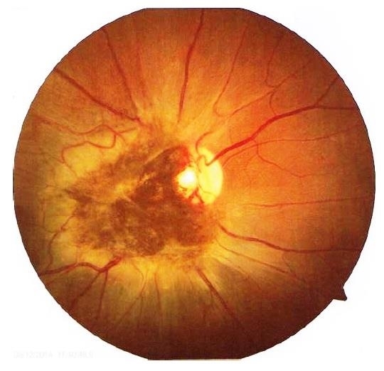
- hamartoma, retinal pigment epithelium
- Photographer
- Dipak Nag
- Imaging device
- Fundus camera
- Description
- A 28-year-old female presented with a deeply pigmented gray- brown, elevated lesion extending from the temporal side of the disc to the macula (OU). Remarkable retinal vasculature with straightening of the distal vessels and dilatation as well as tortuousity of the peri-lesional vessels. The vitreoretinal interface shows gliosis and epi-retinal membrane (ERM) formation.


