Search results (247 results)
-
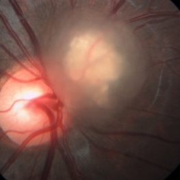
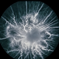
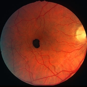
 Epiretinal Membrane/Macular Pucker With Combined Hamartoma of Retina and RPE
Jul 8 2015 by Emmanuel Chang, MD PhD FACS FASRS
10-year-old with history of progressive severe distortion in the left eye over the past year.
Photographer: Retina and Vitreous of Texas
Imaging device: Heidelberg Autofluorescence
Condition/keywords: combined hamartoma, epiretinal membrane (ERM), retinal pigment epithelium (RPE) hamartoma
-
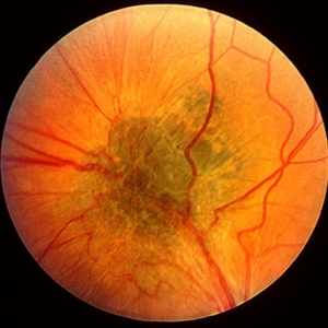
 Combined Harmartoma of the Retina and RPE
Apr 4 2023 by Jeffrey Barker
32 Year old Male with a Combined Hamartoma of the Retina and RPE.
Photographer: Jeffrey P. Barker, B.S.
Condition/keywords: hamartoma
-
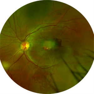
 Astrocytic Hamartoma
Oct 10 2012 by Anat Loewenstein, MD
Five year-old girl came for regular eye examination with no complaints. Medical and ophthalmic history was unremarkable. On examination, visual acuity was 20/25 in both eyes. A Retinal Astrocytic Hamartoma was seen adjacent to the right optic nerve (picture). The rest of the eye examination was normal and no Lisch nodules were seen. The patient was referred for a pediatric neurologist examination and MRI scan of the brain.
Photographer: Galit Yair-Pur
Imaging device: ZEISS FF450 PLUS IR
-
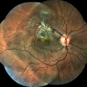
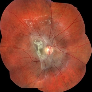
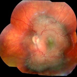
 Combined Hamartoma
Feb 20 2013 by From the Collections of Thomas M. Aaberg, MD and Thomas M. Aaberg Jr., MD
No history.
Condition/keywords: combined hamartoma
-
 Combined Hamartoma of Retina and RPE
Mar 29 2013 by Henry J. Kaplan, MD
Greenish lesion on the arcade with epiretinal membrane formation, vessels inside the lesion are contracted and those outside are distracted.
Condition/keywords: combined hamartoma
-
 Combined Hamartoma
Oct 5 2016 by Guruprasad S. Ayachit, MBBS,MS
Fundus photograph of a 9-year-old boy with an ill-defined lesion extending from nasal to the disc going on to include the papillomacular bundle; 14X10 mm in greatest dimensions. There is a thick epiretinal membrane causing distortion and straightening of temporal vascular arcade.
Photographer: Shravan Masurkar, M M Joshi Eye Institute, Hubli
Imaging device: Topcon TRC50DX
Condition/keywords: combined hamartoma
-
 Combined hamartoma - FA 2
Jan 11 2013 by Alex P. Hunyor, MD
Combined hamartoma of retina and RPE, right eye - late phase fluorescein angiogram. Note: scanned negative film FA
Condition/keywords: combined hamartoma
-
 Combined Hamartoma of Retina and Retinal Pigment Epithelium
Mar 26 2018 by Hashim Ali Khan, OD, FAAO
Fundus photograph of a 12-year-old boy with combined hamartoma of retina and retinal pigment epithelium.
Condition/keywords: combined hamartoma, retinal pigment epithelium
-
 Combined Retinal / RPE Hamartoma
May 6 2014 by David Callanan, MD
7-year-old black male with combined retinal / RPE hamartoma.
Condition/keywords: combined hamartoma, retinal pigment epithelium (RPE) hamartoma
-
 Combined Retinal / RPE Hamartoma
May 6 2014 by David Callanan, MD
7-year-old black male with combined retinal / RPE hamartoma.
Condition/keywords: combined hamartoma, retinal pigment epithelium (RPE) hamartoma
-
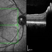 Congenital Simple Hamartoma of RPE
Aug 3 2015 by Bindu Rajesh
OCT line scan through the hamartoma in a 26-year-old male, showing increased hyperreflectivity in the area of lesion with backshadowing and minimal protrusion into vitreous.
Imaging device: Heidelberg Spectralis
Condition/keywords: congenital, hamartoma, retinal pigment epithelium
-
 Retinal Astrocytic Hamartoma
Feb 5 2025 by Rinat Sutiushev
Fundus photograph of a 42-year-old man with retinal astrocytic hamartoma type 3.
Photographer: Rinat Sutiushev, Ophthalmological center “Vision”, Saint Petersburg
Imaging device: Heidelberg Spectralis
Condition/keywords: retina
-
 Retinal Astrocytoma
Apr 16 2015 by Rita Couceiro, MD, MS
Fundus photograph of a 72-year-old man with a retinal astrocytoma of the left eye.
Condition/keywords: astrocytic hamartoma
-
 Astrocytic Hamartoma
Jan 4 2019 by Netan Choudhry, MD, FRCS(C) FASRS
Fundus photograph montage of a 7-year-old boy with an astrocytic hamartoma involving the macula.
Photographer: John Golding BA, Vitreous Retina Macula Specialists of Toronto
Imaging device: Topcon TRC-50 Dx
Condition/keywords: astrocytic hamartoma
-
 Combined Hamartoma of the Retina and Retinal Pigment Epithelium (CHRRPE)
Jan 21 2020 by Pierre-Henry Gabrielle, MD
Fundus photograph of a 17-year-old man with combined hamartomas of the retina and retinal pigment epithelium (CHRRPE) at the posterior pole of the left eye.
Photographer: Pierre-Henry Gabrielle, Ophthalmology department, Dijon University Hospital, France
Imaging device: Zeiss Visucam
Condition/keywords: combined hamartoma, fundus photograph
-
 Congenital RPE hamartoma
Jan 11 2013 by Alex P. Hunyor, MD
Congenital RPE hamartoma, right eye.
Condition/keywords: retinal pigment epithelium (RPE) hamartoma, retinal pigment epithelium (RPE) tumor
-
 Retina Hamartomas in Tuberous Sclerosis
Jan 8 2019 by Sofia Mano
Female 19-years-old with tuberous sclerosis. BCVA LE 10/10. Fundus LE shows three multinodular hamartomas.
Photographer: Sofia Sousa Mano
Imaging device: Canon CR 2 plus
Condition/keywords: hamartoma, tuberculous chorioretinitis
-
 Hamartoma of the Retina
May 29 2018 by JEFFERSON R SOUSA, Tecg.º (Biomedical Systems Technology)
A 4-year-old male patient attended the clinic for evaluation. In the mapping examination and retina and retinography, important alterations were observed in the posterior pole of the left eye. This in turn was sent to perform the ocular ultrasonography examination, which together with the previous examinations, confirmed changes that suggested diagnosis of: COMBINED HAMARTOMA OF RETINA AND PIGMENTARY EPITHELIUM.
Photographer: JEFFERSON R SOUSA - Study Center and Ophthalmological Research Dr. Andre M V Gomes, Institute Dr. Suel Abujamra São Paulo-Brazil
Imaging device: Topcon TRC-50 DX, Imaginet 5.0, angle de 35º . Flash 36 / Mosaic with 9 images.
Condition/keywords: combined hamartoma, retinal pigment epithelium (RPE) hamartoma, tumor
-
 Astrocytic Hamartoma from Tuberous Sclerosis
Sep 18 2016 by John T. Thompson, MD
OCT of astrocytic hamartoma in child with tuberous sclerosis.
Imaging device: Heidelberg Spectralis
Condition/keywords: astrocytic hamartoma, tuberous sclerosis
-
 Astrocytic hamartoma of the retina
Jan 11 2013 by Alex P. Hunyor, MD
Astrocytic hamartoma in a patient with tuberous sclerosis. Note also vascular sheathing inferior to optic disc
Condition/keywords: tuberous sclerosis
-
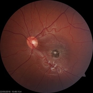
 Combined Hamartoma
Feb 29 2016 by Andrea Arriola-Lopez, MD MSc
40 year-old man with diminished VA since 6 month ago. Fundus examination revealed macular folds, yellow-whitish elevated lesion at the fovea and a subretinal hemorrhage.
Photographer: Andrea Elizabeth Arriola-Lopez MD, MSc
Imaging device: OPTOS Dakota
Condition/keywords: combined hamartoma, macula, subretinal hemorrhage
-
 Combined Hamartoma of Retina and RPE
May 10 2019 by Deepak Bhojwani, MS
A 31-year-old male came with incidental finding of poor vision in left eye on his screening eye examination (done for job purpose). His left eye depicts classic combined hamartoma of retina and RPE in left eye. Right examination was unremarkable.
Photographer: Deepak Bhojwani, Raghudeep Eye Hospital, Ahmedabad
Imaging device: Zeiss Viscam 500
Condition/keywords: combined hamartoma, retina, retinal pigment epithelium, tumor
-
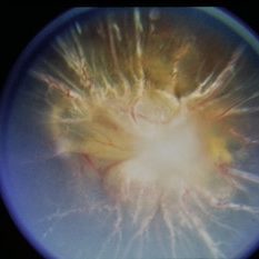
 Combined Hamartoma of the Retinal Pigment Epithelium Case 1
Oct 5 2012 by Ronald C. Gentile, MD
A peripapilary combined hamartoma of the retinal pigment epithelium involving the nasal disc margin. This tumor is slightly elevated, charcoal grey in color with grey-white tissue on it surface. The underlying retinal vessels are obscured.
Photographer: The New York Eye & Ear Infirmary Department of Medical Imaging
Condition/keywords: hamartoma, retinal pigment epithelium
-
 RPE Hamartoma
Oct 18 2012 by Raj K. Maturi, MD
Photographer: Tom Steele, CRA
Imaging device: Topcon 50dx
Condition/keywords: hamartoma, red-free, retinal pigment epithelium
-
 RPE Hamartoma
Oct 18 2012 by Raj K. Maturi, MD
Photographer: Stephanie Morrow
Imaging device: HRA
Condition/keywords: hamartoma, retinal pigment epithelium
 Loading…