 Initializing download.
-
 By Ronald C. Gentile, MD
The New York Eye and Ear Infirmary of Mount Sinai - Uploaded on Oct 5, 2012.
- Last modified by Ronald C. Gentile, MD on Nov 18, 2012.
- Reviewed by Jennifer Hicks
- Rating
- Appears in
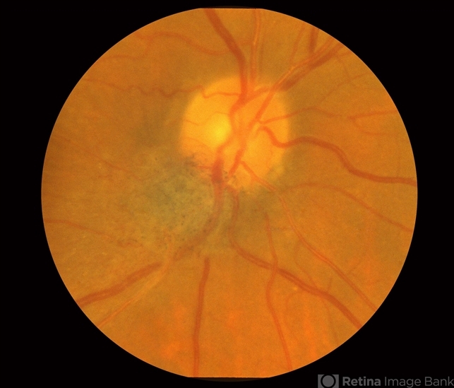
- Combined Hamartoma of the Retinal Pigment Epithelium Case 2
- Condition/keywords
- hamartoma, retinal pigment epithelium
- Photographer
- The New York Eye & Ear Infirmary Department of Medical Imaging
- Description
- Magnified view of the peripapilary combined hamartoma of the retinal pigment epithelium involving the inferior disc margin. This tumor and slightly elevated, charcoal grey to light grey in color with grey-white tissue on it surface. The underlying retinal vessels are obscured with some epiretinal vitreous membranes.


