Search results (216 results)
-
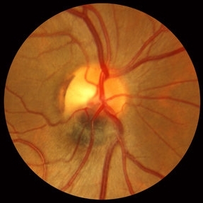
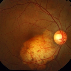
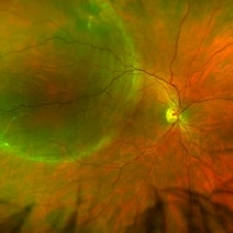 Retinal Detachment Right Eye Optomap
Mar 31 2014 by James B. Soque, CRA, OCT-C, COA, FOPS
36-year-old white male presented with non traumatic retinal detachment OD, with six very distinct demarcation lines and isolated tear, and detachment parameters. Patient underwent PPV OD on 12/3/13 with 20% SF6 gas placement and face down in his first 1 month post op period.
Photographer: James Soque, CRA, COA
Imaging device: Optos Daytona
Condition/keywords: Cryopexy, demarcation line, gas pneumatic displacement, Optomap, Optos, pars plana vitrectomy (PPV), retinal tear, scanning laser ophthalmoscope
-
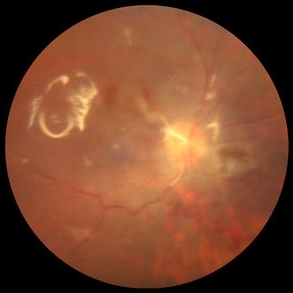
 Giant Retinal Tear Slide 1
Oct 22 2012 by Ronald C. Gentile, MD
Acute loss of vision in a myopic man with flashes and floaters in the right eye. The giant retinal tear is flapped over with the macula detached. The undersurface of the retina can be seen temporally.
Photographer: The New York Eye & Ear Infirmary Department of Medical Imaging
Condition/keywords: retinal tear, vitrectomy
-
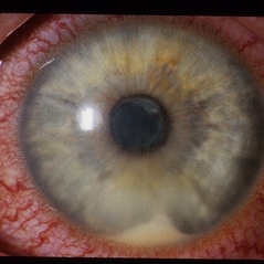 HLA-B27 Associated Uveitis
Jun 4 2014 by Henry J. Kaplan, MD
Severe anterior uveitis with fibrinous reaction and hypopyon formation related to HLA-B27. Notice the membrane on the lens surface.
Condition/keywords: acute anterior uveitis, HLA-B27, hypopyon
-
---thumb.jpg/image-square;max$300,300.ImageHandler) Retinoblastoma To Chemothermotherapy
Retinoblastoma To Chemothermotherapy
Oct 4 2013 by Maurice F. Rabb
A 7 week old girl with a family history of retinoblastoma was found to have a small retinoblastoma in each eye. In the right eye the tumor was adjacent to the optic disc in the papillomacular bundle and measured 2 X 2 X 2 mm. Its temporal margin was 1.0 mm from the foveola and it overhung 20% of the optic disc surface. There was not clinical or ultrasonographic evidence of vitreous seeking or optic nerve invation. In the left eye there was a solitary tumor 1mm superonasal to the optic disc. The tumor measured 1 X 1 X 1 mm. The foveal reflex was normal in both eyes. Both tumors showed a fluorescein angiographic pattern compatible with retinoblastoma with rapid filling and late hyperfluorescence.
Condition/keywords: retina
-
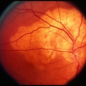 Choroidal Osteoma
Mar 29 2013 by Henry J. Kaplan, MD
Typical choroidal osteoma as yellow subretinal lesion around optic nerve with scalloped border and mild pigmentation on the surface.
Condition/keywords: choroidal osteoma
-
 Russell bodies Observed in a Patient with Fuchs' Heterochromic Iridocyclitis
Nov 1 2016 by PAVEL FLORES-MORENO
Anterior chamber shows in iris surface: small, refractile iris crystals.
Photographer: Pavel Flores-Moreno
Imaging device: Anterior chamber camera
Condition/keywords: Fuchs, Russell bodies
-
 Lattice Lesion
Nov 9 2012 by Norman Byer
This lattice lesion in a 36-year-old woman has remained unchanged over a period of 13 years. It shows a moderate snailtrack feature with discrete yellow dots visible on the surface of the lesion and especially along the posterior border. One of these can be well seen just below the lesion superimposed over the dark shadow of the scleral indentation. The exact nature of these yellow dots is still not entirely clear.
Condition/keywords: lattice degeneration, moderate snail track, scleral indentation, yellow dots
-
---thumb.jpg/image-square;max$300,300.ImageHandler) Retinoblastoma To Chemothermotherapy
Retinoblastoma To Chemothermotherapy
Oct 4 2013 by Maurice F. Rabb
A 7 week old girl with a family history of retinoblastoma was found to have a small retinoblastoma in each eye. In the right eye the tumor was adjacent to the optic disc in the papillomacular bundle and measured 2 X 2 X 2 mm. Its temporal margin was 1.0 mm from the foveola and it overhung 20% of the optic disc surface. There was not clinical or ultrasonographic evidence of vitreous seeking or optic nerve invation. In the left eye there was a solitary tumor 1mm superonasal to the optic disc. The tumor measured 1 X 1 X 1 mm. The foveal reflex was normal in both eyes. Both tumors showed a fluorescein angiographic pattern compatible with retinoblastoma with rapid filling and late hyperfluorescence.
Condition/keywords: retina
-
 Herpes Zoster of Face
-
 Melanocytoma Case 1 Slide 2
Oct 5 2012 by Ronald C. Gentile, MD
Magnified view of the melanocytoma involving the optic nerve. The tumor is black and has a smooth surface involving the inferior portion of the optic disc.
Photographer: The New York Eye & Ear Infirmary Department of Medical Imaging
Condition/keywords: melanocytoma
-
 Combined Hamartoma of the Retinal Pigment Epithelium Case 1
Oct 5 2012 by Ronald C. Gentile, MD
A peripapilary combined hamartoma of the retinal pigment epithelium involving the nasal disc margin. This tumor is slightly elevated, charcoal grey in color with grey-white tissue on it surface. The underlying retinal vessels are obscured.
Photographer: The New York Eye & Ear Infirmary Department of Medical Imaging
Condition/keywords: hamartoma, retinal pigment epithelium
-
 PVR Retinal Detachment with subretinal bands Slide 2
Oct 22 2012 by Ronald C. Gentile, MD
Postoperative fundus photo of the posterior pole with flat retina. As noted by the retinal surface reflex, silicone oil tamponade was used.
Photographer: The New York Eye & Ear Infirmary Department of Medical Imaging
Condition/keywords: proliferative vitreoretinopathy (PVR), subretinal bands
-
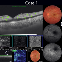 Figure-1 Paracentral Acute Middle Maculopathy (PAMM)
Dec 21 2018 by Fawwaz F Al Mamoori, MD, Medical Retina Consultant
25-year-old male patient medically free, had sudden deterioration in his left eye vision. Visual acuity on presentation was counting fingers at 3 meter distance. Marked Relative Pupillary Afferent Defect (RAPD) was detected and fundoscopic exam showed abnormal foveal reflex. SS OCT B scan: showed a hypereflectivity of the inner plexiform layer (IPL), inner nuclear layer (INL) and OPL layer (fig 1, A).FA images were normal (fig 1, B). Angiography shows remarkable perifoveal capillary drop out within middle retinal layer correlating with perfusion density map which reveals significant decrease in capillary density at the same level (Fig 1, C). Enface ads more proof to PAMM by delineating ischemic distribution in a fern like pattern of hyper reflective areas within DCP (fig1, D).
Photographer: Dr.Fawwaz Al Mamoori (Al Mamoori Eye Clinic)
Imaging device: Triton Swept Source OCT (TOPCON)
Condition/keywords: optical coherence tomography (OCT), paracentral acute middle maculopathy
-
---thumb.jpg/image-square;max$300,300.ImageHandler) Floaters
Floaters
Oct 9 2013 by Maurice F. Rabb
KR is a 25 year old white female who presented with a one month history of floaters OD. Past ocular and systemic history were unremarkable. On clinical examination, the visual acuity was 20/20 OU, and the anterior segments were normal. There was a very mild degree of vitreous cell OD, though no cystoid macular edema nor vasculitis. A lobulated white mass was noted overlying the vitreous base inferotemporally OD (thickness 3.3mm). There was no calcification, though prominent cysts were noted on the surface of the lesion. A fluorescein angiogram, echogram, and CT scan were obtained, along with a thorough systemic evaluation.
Condition/keywords: floaters
-
 Senile Retinoschisis
Nov 9 2012 by Norman Byer
This 48-year-old woman has senile retinoschisis involving the most common location, the lower temporal quadrant. The lesion shown here illustrates one of the two clinical features which are most often responsible for attracting the attention of the examiner to such lesions, namely the multitude of yellow flecks lying on the inner surface of the inner layer. The nature of these flecks is not known, but it seems clear that they do not originate in the schisis cavity for they do not represent remnants of ruptured Miller’s fibers. In this photograph you can easily detect the fluid space which separates the inner and outer retinal layers.
Condition/keywords: lower temporal quadrant, senile retinoschisis, yellow flecks
-
 Scleral Indentation
Nov 9 2012 by Norman Byer
On the crest of this indentation, one can see a small fragment of residual vitreous blood lying close to the retinal surface. The center is becoming typically white as the blood is becoming depigmented.
Condition/keywords: depigmented vitreous blood, scleral indentation, vitreous blood
-
---thumb.jpg/image-square;max$300,300.ImageHandler) Silicone Oil Surface
Silicone Oil Surface
Nov 14 2013 by Hamid Ahmadieh, MD
OCT image of the silicone oil surface bridging an enhanced foveal depression. There is the history of a rhegmatogenous retinal detachment associated with full thickness macular hole repair.
Photographer: Naghmeh Nozhat, Negah Eye Center, Tehran
Condition/keywords: optical coherence tomography (OCT), silicone oil
-
---thumb.jpg/image-square;max$300,300.ImageHandler) Floaters
Floaters
Oct 9 2013 by Maurice F. Rabb
KR is a 25 year old white female who presented with a one month history of floaters OD. Past ocular and systemic history were unremarkable. On clinical examination, the visual acuity was 20/20 OU, and the anterior segments were normal. There was a very mild degree of vitreous cell OD, though no cystoid macular edema nor vasculitis. A lobulated white mass was noted overlying the vitreous base inferotemporally OD (thickness 3.3mm). There was no calcification, though prominent cysts were noted on the surface of the lesion. A fluorescein angiogram, echogram, and CT scan were obtained, along with a thorough systemic evaluation.
Condition/keywords: floaters
-
 Asteroid Hyalosis, Vitreous Face Attached
Dec 10 2012 by Yale L. Fisher, MD
In asteroid hyalosis, accumulations of calcium soaps dispersed throughout the vitreous produce bright echoes in the usually echolucent vitreous. The appearance of asteroid hyalosis should not be confused with that of vitreous hemorrhage or vitritis. Many of the larger aggregates in asteroid hyalosis are easily seen as the gain is reduced to below 60 db, unlike vitreous hemorrhage or vitritis which usually disappears at low gain settings. There is also an area of clear echolucent vitreous between the posterior hyaloid face and the asteroid particles, which is usually not present in vitreous hemorrhage or vitritis.
Condition/keywords: video
-
 Operculated Tear at the Back Surface
Dec 10 2012 by Yale L. Fisher, MD
In this ultrasound movie there is little movement of the vitreous. Observe the operculated, small, relatively strongly reflective tissue on the detached, mildly reflective, vitreous back surface (yellow arrow). There is a sagital view of the superior aspect of the lateral rectus muscle (orange arrow). Operculated tear at 10 o'clock position.
Condition/keywords: operculated tear, ultrasound, video, vitreous
-
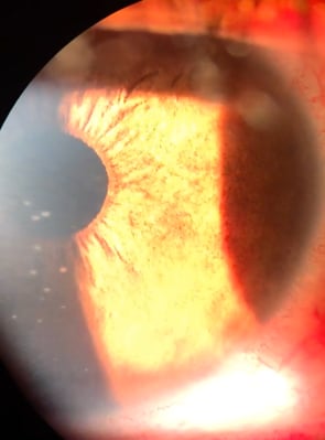
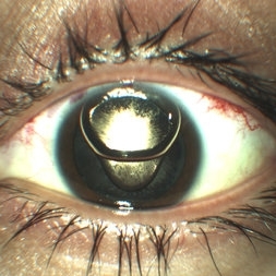 Anterior Chamber Gas and PFC Migration
Jun 21 2018 by Maria Stephanie R. Jardeleza, MD
Anterior segment photographs of 30-year-old male who underwent superior rhegmatogenous retinal detachment repair with intraocular gas tamponade. Perfluorocarbon was used to flatten the macula to prevent a macular fold and was removed during PFC/air exchange. Post operative week two visit shows gas migration into the anterior chamber with retained PFC layered in a tear drop shape posterior to the gas bubble and anterior to the lens. Patient had been maintaining face down positioning.
Photographer: Andy Zepeda, COA, Retina Clinic, San Antonio Eye Center, San Antonio, TX
Condition/keywords: retained perfluorocarbon, retina surgery complications, vitreous substitutes
-
 Lattice Lesion
Nov 9 2012 by Norman Byer
This lattice lesion in a 27-year-old woman shows an interesting change in the middle of the lesion. The predominant feature on the left side of the lesion is the snailtrack appearance while the right side of the lesion shows mainly a reddish crater. Note the many yellow dots above the surface of the retina which are actually located in the vitreous condensation which surrounds the pocket of liquified vitreous over the lesion.
Condition/keywords: lattice lesion, reddish crater, snail track, vitreous condensation, vitreous liquefaction
-
 Combined Hamartoma of the Retinal Pigment Epithelium Case 2
Oct 5 2012 by Ronald C. Gentile, MD
Magnified view of the peripapilary combined hamartoma of the retinal pigment epithelium involving the inferior disc margin. This tumor and slightly elevated, charcoal grey to light grey in color with grey-white tissue on it surface. The underlying retinal vessels are obscured with some epiretinal vitreous membranes.
Photographer: The New York Eye & Ear Infirmary Department of Medical Imaging
Condition/keywords: hamartoma, retinal pigment epithelium
-
 Retinoschisis
Nov 9 2012 by Norman Byer
This 57-year-old man has three large breaks in the outer layer of his retinoschisis. Note the interesting bridges separating the breaks and also note that the intact inner layer is only slightly elevated above the outer layer. This eye has not been treated and has remained essentially the same for eight years. Please note the yellow dots on the surface of the inner layer and the blood vessels running through this layer.
Condition/keywords: intact inner layer, outer layer breaks, retinoschisis, yellow dots
-
 Choroidal Osteoma
Dec 28 2015 by P. Mahesh Shanmugam, MBBS, DO, FRCSEd, PhD, FAICO
Choroidal osteoma - yellow subretinal minimally elevated lesion with scalloped margins and pigmentation on surface. Peripheral part of the lesion is orange in color, the older central part yellow in color.
Condition/keywords: choroidal osteoma
 Loading…