Search results (216 results)
-
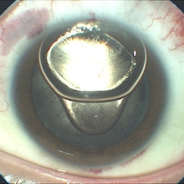
 Wrinkled Anterior Capsule 40X zoom
Feb 18 2023 by Ahmed Abbas Hashmi, OD
Imprint of Iris Pigmentation on Anterior Lens Surface with wrinkled anterior capsule
Photographer: Ahmed Abbas Hashmi
Condition/keywords: lens opacity
-
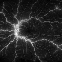 Central Retinal Vein Occlusion
Jan 21 2022 by Olivia Rainey
Ultra-widefield fluorescein angiogram of a 23-year-old female with a Central Retinal Vein Occlusion affecting her left eye. The patient presented on 12/22/2021 cc20/40-2 vision in the left eye. The patient reported recent trauma of being hit with a fist on both sides of face followed by vision loss. The patient has history of Hashimoto's thyroid disease. The following labs have been ordered, PT, PTT, CBC, antithrombin III activity, protein C, protein S, Factor V Leiden mutation, Prothrombin (G20210A), lipid panel, HbA1c, quantiferon gold, RPR, and CXR.
Photographer: Olivia Rainey, OCT-C, COA
Imaging device: Optos California
Condition/keywords: central retinal vein occlusion (CRVO), disc leakage, fluorescein angiogram (FA), fluorescein leakage, left eye, non-ischemic central retinal vein occlusion (CRVO), Optos, trauma, ultra-wide field imaging
-
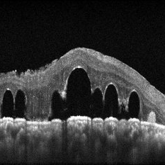 submacular perfluorocarbon liquid
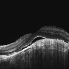
Sep 7 2022 by JEFFERSON R SOUSA, Tecg.º (Biomedical Systems Technology)
A 63-year-old male patient underwent vitreoretinal surgery with the use of perfluorocarbon. From a technological point of view, extended-field retinography presents many points of focus variation due to the difficulty of establishing a diffuse focus, as it is a recent post-operative case. In OCT Fundus Enface, although it has a low resolution, it is extremely important for documenting the presence of perfluor. Best seen in structural OCT.
Photographer: JEFFERSON ROCHA DE SOUSA - Retinal Department at Instituto Dr. Suel Abujamra Sao Paulo-Brazil
Imaging device: Optical Coherence Tomography system OCT CIRRUS 5000, Protocol, HD 5 Line
Condition/keywords: perfluorocarbon fluid, post-vitrectomy, submacular perfluorocarbon liquid (PFO), vitrectomy
-
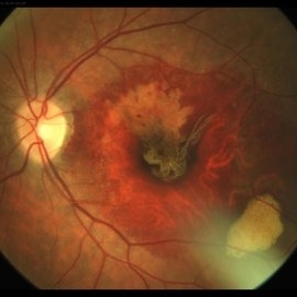
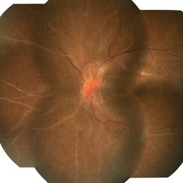 Advanced Proliferative Diabetic Retinopathy With Fibrovascular Proliferation
Jan 4 2019 by Isha Agarwalla
A 29-year-old female with a long-standing history of diabetes mellitus presented with a fibrovascular membrane (FVM) at the viteroretinal interface due to underlying inflammation and angiogenesis induced by ischemia. FVM involved the disc and extended towards the superior and inferior arcades along with extensive capillary drop out areas due to micro aneurysms.
Condition/keywords: fibrovascular proliferation, proliferative diabetic retinopathy (PDR)
-
 Amelanotic Choroidal Melanoma
May 18 2020 by McGill University Health Centre
The enucleation image shows a large amelanotic tumor with large areas of hemorrhage and necrosis. Note the several dilated blood vessels and an adjacent retinal detachment with lipofuscin pigment on its surface (arrow).
Condition/keywords: amelanotic melanoma, enucleation, mushroom-shaped
-
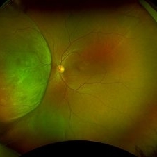 Amelanotic Melanoma
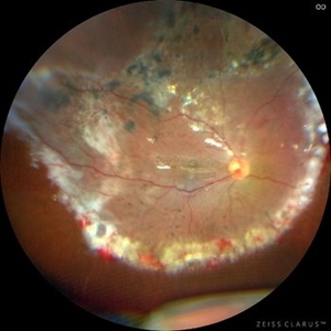
Sep 19 2025 by Aditya S Kelkar, MS, FRCS, FASRS,FRCOphth
Widefield fundus photograph of a 37 year old showing a large, dome-shaped, intraocular mass involving the temporal retina. The lesion appears elevated and lacks surface pigmentation. Overlying retinal vessels are displaced and draped across the tumor surface, with surrounding retinal elevation noted. The appearance is suggestive of amelanotic variant of choroidal melanoma.
Photographer: Dr. Muskan Mangal
Imaging device: Optos Daytona
Condition/keywords: choroidal melanoma, intraocular tumor
-
 Anterior Segment Gas Bubble and PFC Interface
Jun 21 2018 by Maria Stephanie R. Jardeleza, MD
Anterior segment photographs of 30-year-old male who underwent superior rhegmatogenous retinal detachment repair with intraocular gas tamponade. Perfluorocarbon was used to flatten the macula to prevent a macular fold and was removed during PFC/air exchange. Post operative week two visit shows gas migration into the anterior chamber with retained PFC on the posterior aspect of the gas bubble/anterior surface of the lens. Patient had been maintaining face down positioning.
Photographer: Andy Zepeda, COA, Retina Clinic, San Antonio Eye Center, San Antonio, TX
Condition/keywords: retained perfluorocarbon, vitreous substitutes
-
 Ciliary Body Melanoma
Feb 12 2025 by Virginia Gebhart
91 year old female with large collar button tumor emanating from the ciliary body with resolving vitreous hemorrhage. Melanoma cells in the AV as well as studded on the entire retina surface. Pt scheduled for enucleation. CT scans of chest and abdomen showed no evidence of metastatic disease.
Photographer: Virginia Gebhart, Retina Consultants of Carolina
Imaging device: Optos California
Condition/keywords: asteroid hyalosis, ciliary body mass, ciliary body melanoma, vitreous hemorrhage
-
 Expulsion of Retina
Oct 23 2024 by Gustavo Uriel Fonseca Aguirre
Male patient with a history of penetrating keratopathy presents due to blunt ocular trauma. A disruption of the continuity at the interface between the donor and recipient corneas is observed, with expulsion of the lens and retina. Vision is limited to light perception.
Photographer: Lizeth Jiménez Santana, Fundación Hospital Nuestra Señora de la Luz, Ciudad de México
Condition/keywords: ocular trauma, penetrating keratoplasty
-
 Giant Retinal Tear Slide 1
Oct 22 2012 by Ronald C. Gentile, MD
Acute loss of vision in a myopic man with flashes and floaters in the right eye. The giant retinal tear is flapped over with the macula detached. The undersurface of the retina can be seen temporally.
Photographer: The New York Eye & Ear Infirmary Department of Medical Imaging
Condition/keywords: retinal tear, vitrectomy
-
 Hemangioma of Retina
Mar 5 2025 by Virginia Gebhart
64 year old male with choroidal hemangioma in the macula and STA. Persistent IRF and new cuff of SRF compared to previous photos. BCVA CF@face. Pt has had PDT in the past with no significant improvement. Will observe closely
Photographer: Virginia Gebhart, Retina Consultants of Carolina
Imaging device: Optos California
Condition/keywords: hemangioma, inferior subretinal fluid
-
 Hemangioma of Retina (FAF)
Mar 5 2025 by Virginia Gebhart
Fundus autofluorescence of 64 year old male with choroidal hemangioma in the macula and STA. Persistent IRF and new cuff of SRF compared to previous photos. BCVA CF@face. Pt has had PDT in the past with no significant improvement. Will observe closely
Photographer: Virginia Gebhart, Retina Consultants of Carolina
Imaging device: Optos California
Condition/keywords: autofluorescence imaging, hemangioma, inferior subretinal fluid
-
 HRA -OCT of left eye Choroidal Osteoma
Jun 2 2018 by awaneesh m upadhyay, MBBS, DNB
23-year-old male patient's left eye OCT showing undulating surface of osteoma with subretinal fluid and shaggy RPE.
Photographer: Hiteshwar Saikia
Imaging device: Heilderberg spectralis
Condition/keywords: choroidal osteoma
-
 IOFB with BRAO
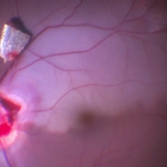
Feb 9 2017 by Manish Nagpal, MD, FRCS (UK), FASRS
Intraoperative photo of a IOFB impacting on the inferotemporal margin of disc leading to a BRAO. This picture is taken moments after dissecting the IOFB from the impact site and bringing it over the retinal surface for eventual removal.
Photographer: Manish Nagpal
Imaging device: Still captured from a 3 chip HD camera on microscope
Condition/keywords: branch retinal artery occlusion (BRAO), intraocular foreign body
-
 Lattice Lesion
Nov 9 2012 by Norman Byer
This lattice lesion in a 36-year-old woman has remained unchanged over a period of 13 years. It shows a moderate snailtrack feature with discrete yellow dots visible on the surface of the lesion and especially along the posterior border. One of these can be well seen just below the lesion superimposed over the dark shadow of the scleral indentation. The exact nature of these yellow dots is still not entirely clear.
Condition/keywords: lattice degeneration, moderate snail track, scleral indentation, yellow dots
-
 Metastatic Breast Carcinoma
Jan 21 2021 by Jamin S. Brown, MD
This anterior segment photograph was taken with a smartphone camera attached to a regular Haag Streit slit lamp ocular demonstrates unusual clustering of white cells on the posterior surface of the intraocular lens. The clinical diagnosis is metastatic breast carcinoma to the vitreous, which is very rare.
Photographer: Stefanie Palmer CRA, Retina Vitreous Surgeons of CNY
Imaging device: Cell phone camera
Condition/keywords: anterior segment, breast cancer, cell phone camera, slit lamp photo
-
 Photic Retinopathy
Jan 30 2025 by Juan Alberto Olivera Cueva
A 23 year-old male with a history of direct exposure to sunlight on several occasions, presenting blurred vision, comes for evaluation due to metamorphopsias of 3 months' evolution. The fundus photograph shows the presence of an epiretinal membrane. The OCT shows a hyperreflective line at the vitreomacular interface that causes traction to the layers of the inner retina, as well as distortion in the architecture of the macular region, with the presence of subfoveal detachment of the RPE.
Photographer: Dr. Juan Alberto Olivera Cueva, Escuela Militar de Medicina, Hospital Militar de Especialidades Oftalmológicas
Condition/keywords: MER, photic retinopathy
-
 RPE Tear After Anti-VEGF Injection
Mar 17 2021 by RAFAEL REIS PEREIRA, MD
Retinal pigment epithelium (RPE) tear is a rare devastating complication of age-related macular degeneration (AMD). The believed mechanism lies in an adherence of the neovascularization to the undersurface of the RPE creating a contractile force that increases after VEGF therapy and causes the tear.
Photographer: Rafael Reis, Retina Clinic, São Paulo
Condition/keywords: retinal pigment epithelium (RPE) contracture
-
 submacular perfluorocarbon liquid
Sep 7 2022 by JEFFERSON R SOUSA, Tecg.º (Biomedical Systems Technology)
A 63-year-old male patient underwent vitreoretinal surgery with the use of perfluorocarbon. From a technological point of view, extended-field retinography presents many points of focus variation due to the difficulty of establishing a diffuse focus, as it is a recent post-operative case. In OCT Fundus Enface, although it has a low resolution, it is extremely important for documenting the presence of perfluor. Best seen in structural OCT.
Photographer: JEFFERSON ROCHA DE SOUSA - Retinal Department at Instituto Dr. Suel Abujamra Sao Paulo-Brazil
Imaging device: Clarus 700 - Zeiss, 135 degree images.
Condition/keywords: perfluorocarbon fluid, post-vitrectomy, submacular perfluorocarbon liquid (PFO), vitrectomy
-
 The Halloween Smile
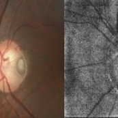
Mar 27 2025 by Shrishti mishra
A 73 year old male with Le optic disc pit . On color fundus photo a single pit can be noted whereas on oct enface is- os interface 2 optic disc pits are noted which resembles a halloween smile .
Photographer: Mr Sudhakar
Imaging device: Zeiss cirrus6000
Condition/keywords: OCT, oct en face, optic disc pit
-
 Traumatic CRAO with Cilioretinal Artery Sparing
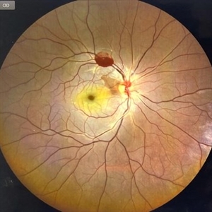
Sep 10 2024 by KRISHNENDU NANDI, MS
A 25 year-old male presented with dimness of vision in the right eye for the last 3 days following blunt trauma. The BCVA of the right eye was CF close to the face and left eye was 6/6, N6 in Snellen’s chart. On examination the retina showed CRAO with cherry red spot and sparing of cilioretinal artery circulation. Traumatic subhyaloid haemorrhage also noted at supero-temporal arcade.
Photographer: Dr Krishnendu Nandi
Condition/keywords: CRAO, subhyaloid hemorrhage, Trauma
-
Vitrectomy for PDR in TRD
Dec 16 2022 by Manish Nagpal, MD, FRCS (UK), FASRS
This is a case of subhyaloid haemorrhage and Tractional retinal detachment in a diabetic patient. The subhyaloid haemorrhage is aspirated using the cutter . 25 gauge bevelled cutter is used to dissect all the epiretinal proliferations and tractional components. The ports of these cutters can reach very close to the retinal surface and cut flush without causing any iatrogenic damage to the retinal surface. Bleeders are stopped raising pressure and applying diathermy. Once the retina is flattened endolaser is done 360 degree to achieve long term regression.
Condition/keywords: cutter, proliferative diabetic retinopathy (PDR), video, vitrectomy
-
 Vitreomacular Traction
Jun 15 2022 by Zach Seim
Optical Coherence Tomography (OCT) of a 69 year old male with Vitreomacular Traction affecting his right eye. Patient was referred to this office for Choroidal Melanoma in his right eye in May 2021. The patient was treated with Brachytherapy in July 2021 and this OCT was taken at a follow-up appointment in May 2022. Patient's vision was 20/30-2 at the time this OCT was taken. Patient states that his vision was better since his last visit, and that he sees floaters occasionally.
Photographer: Zach Seim
Imaging device: Heidelberg Spectralis
Condition/keywords: heidelberg spectralis, OD, optical coherence tomography (OCT), right eye, subretinal fluid, vitreomacular adhesion, vitreomacular interface disorders, vitreomacular traction (VMT)
-
 iOCT of Dislocated IOL
Dec 20 2017 by Sidney A Schechet, MD
Intraoperative optical coherence tomography image of a dislocated IOL being safely grasped and lifted of the surface of the retina with microforceps.
Imaging device: Leica EnFocus intraoperative optical coherence tomography
Condition/keywords: dislocated posterior chamber intraocular lens (PCIOL), optical coherence tomography (OCT), pars plana vitrectomy (PPV)
 Loading…