Search results (216 results)
-
 Herpes Zoster of Face
-
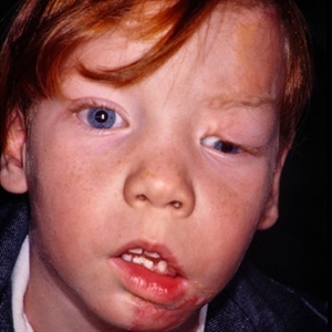 Linear Nevus Sebaceous Syndrome
Feb 20 2015 by H. Michael Lambert, MD
External photo of face in linear nevus sebaceous syndrome.
Condition/keywords: external, face, linear nevus sebaceous syndrome, linear sebaceous nevus of Jadassohn
-
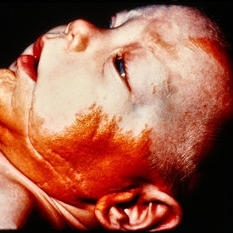 Skin Photo of Linear Nevus Sebaceous Syndrome
Feb 20 2015 by H. Michael Lambert, MD
External photo of face in Linear Nevus Sebaceous syndrome.
Condition/keywords: external, face, linear nevus sebaceous syndrome, linear sebaceous nevus of Jadassohn
-
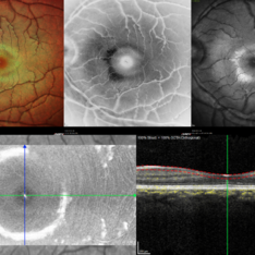
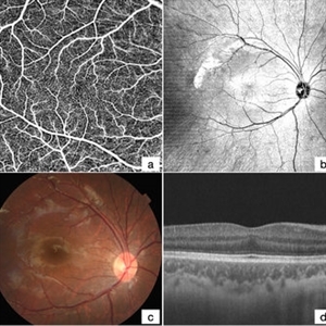 Absence of Macular FAZ in a Child After Laser Therapy for Retinopathy of Prematurity
Dec 24 2024 by Guoming Zhang
OCT angiography of a 5-year-old male child with a history of laser therapy for retinopathy of prematurity, demonstrating the absence of macular FAZ (a), en-face images, fundus visualization, and increased macular retinal thickness.
Photographer: Xinyu Zhao, Shenzhen Eye Hospital, Shenzhen, China
Imaging device: BM-400k BMi zar TowardPi Medical Technology.
Condition/keywords: OCT angiography, OCTA
-
 Acute Macular Neuroretinopathy
Apr 12 2021 by Iuri Golubev, MD
46-year-old female with sudden onset paracentral scotoma below the central point of fixation in her left eye. Enface image shows a wedge shaped lesion pointing towards the fovea (top left). The lesion was spanning outer retinal layers from OPL to RPE (top left insert). One month later, the lesion has diminished in size, and was only involving retinal layers from ellipsoid zone to RPE(top right). At 4 months since presentation, the patient did not have any signs of AMN identifiable on enface or b-scan images (bottom center). Patient's symptoms has slowly improved and eventually resolved over the course of the next 4 years.
Imaging device: Zeiss Cirrus 5000
Condition/keywords: acute macular neuroretinopathy, acute macular outer retinopathy
-
 Acute Partial Vitreous Separation
Dec 10 2012 by Yale L. Fisher, MD
This is a partial vitreous separation that demonstrates a mildly reflective curvilinear shape of the partially separated vitreous face. The vitreous is attached inferonasally and inferotemporally, but detached and freely mobile at 6 o'clock.
Condition/keywords: video
-
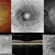
 Acute Posterior Multifocal Placoid Pigment Epitheliopathy
Jan 4 2019 by Cláudia Farinha
Composite image of both eyes of a 27-year-old male with APMPPE. In the fundus photograph, multiple yellowish placoid lesions are observed in the posterior pole in both eyes. The ICGA revealed more lesions than those observed in fundoscopy, and these were hypofluorescent through the angiogram as expected. The en face OCTA segmented at the level of the choriocapillaris revealed areas of ischemia in close correspondence with the hypofluorescent lesions (image superimposed in ICGA ). The OCT b-scan with superimposed flow shows disruption and hyperreflectivity of the external retinal layers in the affected areas and again the absence of flow in the choriocapillaris underneath. A systemic study was carried out to exclude other inflammatory and infectious causes of placoid retinochoroidopathy. The clinical picture resolved after approximately one month from the onset, without recurrence.
Photographer: Pedro Melo, Ophthalmology Department, Centro Hospitalar e Universitário de Coimbra, Coimbra Portugal
Condition/keywords: acute posterior multifocal placoid pigment epitheliopathy (APMPPE), white dot syndrome
-
 Acute Toxoplasmosis in AIDS
Apr 8 2019 by Gary R. Cook, MD, FACS
Left eye of a white male with AIDS and an optic neuritis secondary to ocular toxoplasmosis infection. The patient had no pre-existing chorioretinal scars secondary to Toxo. An edematous optic nerve with a focus of active retinitis inferonasally, small surface hemorrhage above it, and surrounding peripapillary edema is visible.
Imaging device: Topcon VT-50
Condition/keywords: AIDS, ocular toxoplasmosis, optic neuritis, toxoplasmosis
-
 Advanced Proliferative Diabetic Retinopathy
Apr 9 2025 by Gustavo Uriel Fonseca Aguirre
B-mode ultrasound of a patient with long-standing poorly controlled diabetes demonstrates characteristic findings of advanced proliferative diabetic retinopathy. The examination reveals moderate vitreous hemorrhage appearing as diffuse hyperechoic opacities throughout the vitreous cavity, along with a posterior hyaloid membrane densely infiltrated by hemorrhagic material, showing irregular thickening and increased reflectivity. A mild subhyaloid hemorrhage is visible as a subtle hyphema-like space anterior to the retinal surface. The study documents a total tractional retinal detachment, evidenced by rigid retinal folds with clear insertion points of vitreous strands, accompanied by a significant subretinal hemorrhage seen as a prominent hyperechoic collection beneath the elevated retina. These findings collectively illustrate the severe vitreoretinal interface pathology characteristic of end-stage diabetic eye disease, with predominant tractional components and distinct echographic stratification of hemorrhagic layers - from anterior vitreous involvement to deeper subretinal blood accumulation.
Photographer: Gustavo U. Fonseca Aguirre, Hospital Conde de Valenciana, Ciudad de México
Condition/keywords: diabetic retinopathy, tractional retinal detachment, Vitreous hemorrhage
-
 Advanced Proliferative Diabetic Retinopathy With Fibrovascular Proliferation
Jan 4 2019 by Isha Agarwalla
A 29-year-old female with a long-standing history of diabetes mellitus presented with a fibrovascular membrane (FVM) at the viteroretinal interface due to underlying inflammation and angiogenesis induced by ischemia. FVM involved the disc and extended towards the superior and inferior arcades along with extensive capillary drop out areas due to micro aneurysms.
Condition/keywords: fibrovascular proliferation, proliferative diabetic retinopathy (PDR)
-
 Advanced Proliferative Diabetic Retinopathy With Fibrovascular Proliferation
Jan 4 2019 by Isha Agarwalla
A 29-year-old female with a long-standing history of diabetes mellitus presented with a fibrovascular membrane(FVM) at the viteroretinal interface due to underlying inflammation and angiogenesis induced by ischemia. FVM involved the disc and extended towards the superior and inferior arcades along with extensive capillary drop out areas due to micro aneurysms.
Condition/keywords: fibrovascular proliferation, fluorescein angiogram (FA), proliferative diabetic retinopathy (PDR)
-
 AIDS/Toxoplasmosis
Apr 8 2019 by Gary R. Cook, MD, FACS
Laminar venous phase FA of white male with AIDS and optic neuritis secondary to ocular toxoplasmosis OS showing dilated capillaries on the surface of the optic nerve and relative hypofluoescence due to peripapillary edema.
Imaging device: Topcon VT-50
Condition/keywords: AIDS, ocular toxoplasmosis, optic neuritis, toxoplasmosis
-
 Allergic Reaction
Jan 26 2017 by JEFFERSON R SOUSA, Tecg.º (Biomedical Systems Technology)
Male patient, 25-years-old, presented an allergic reaction to the contrast of sodium fluorescein angiographic during the procedure. In the first minute after intravenous injection of contrast, noticed some rashes on the face, hyperfluorescent and evolution of edema of eyelids and lower lip.
Photographer: JEFFERSON R SOUSA
Imaging device: Topcon TRC-50 Dx IA - Angulation of field photo of 35 Degrees, flash 36, Digital system Imaginet
Condition/keywords: allergic reaction
-
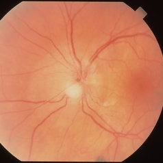
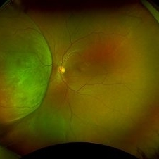
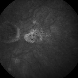 AMD
AMD
Jun 21 2014 by Robert T. Wendel, MD
AMD, smiley face or Casper the Ghost.
Condition/keywords: dry age-related macular degeneration (dry AMD)
-
 Amelanotic Choroidal Melanoma
May 18 2020 by McGill University Health Centre
The enucleation image shows a large amelanotic tumor with large areas of hemorrhage and necrosis. Note the several dilated blood vessels and an adjacent retinal detachment with lipofuscin pigment on its surface (arrow).
Condition/keywords: amelanotic melanoma, enucleation, mushroom-shaped
-
 Amelanotic Melanoma
Sep 19 2025 by Aditya S Kelkar, MS, FRCS, FASRS,FRCOphth
Widefield fundus photograph of a 37 year old showing a large, dome-shaped, intraocular mass involving the temporal retina. The lesion appears elevated and lacks surface pigmentation. Overlying retinal vessels are displaced and draped across the tumor surface, with surrounding retinal elevation noted. The appearance is suggestive of amelanotic variant of choroidal melanoma.
Photographer: Dr. Muskan Mangal
Imaging device: Optos Daytona
Condition/keywords: choroidal melanoma, intraocular tumor
-
 Amyloid Faces
-
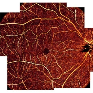 AngioOCT Normal Widefield Scan
May 8 2015 by Timur Shaimov
Optical coherence tomography angiography of a 28-year-old woman without any macular pathology. Seven 6x6mm angioOCT EnFace images used to merge into widefield view. We used the Optovue RTVue XR Avanti (Optovue, USA) optical coherence tomography system with split-spectrum amplitude decorrelation angiography algorithm (SSADA).
Photographer: Timur Shaimov
Imaging device: Optovue RTVue XR Avanti
Condition/keywords: optical coherence tomography (OCT)
-
 Anterior Chamber Gas and PFC Migration
Jun 21 2018 by Maria Stephanie R. Jardeleza, MD
Anterior segment photographs of 30-year-old male who underwent superior rhegmatogenous retinal detachment repair with intraocular gas tamponade. Perfluorocarbon was used to flatten the macula to prevent a macular fold and was removed during PFC/air exchange. Post operative week two visit shows gas migration into the anterior chamber with retained PFC layered in a tear drop shape posterior to the gas bubble and anterior to the lens. Patient had been maintaining face down positioning.
Photographer: Andy Zepeda, COA, Retina Clinic, San Antonio Eye Center, San Antonio, TX
Condition/keywords: retained perfluorocarbon, retina surgery complications, vitreous substitutes
-
 Anterior Segment Gas Bubble and PFC Interface
Jun 21 2018 by Maria Stephanie R. Jardeleza, MD
Anterior segment photographs of 30-year-old male who underwent superior rhegmatogenous retinal detachment repair with intraocular gas tamponade. Perfluorocarbon was used to flatten the macula to prevent a macular fold and was removed during PFC/air exchange. Post operative week two visit shows gas migration into the anterior chamber with retained PFC on the posterior aspect of the gas bubble/anterior surface of the lens. Patient had been maintaining face down positioning.
Photographer: Andy Zepeda, COA, Retina Clinic, San Antonio Eye Center, San Antonio, TX
Condition/keywords: retained perfluorocarbon, vitreous substitutes
-
 Asteroid Hyalosis, Posterior Vitreous Separation
Dec 10 2012 by Yale L. Fisher, MD
VA 20/20. In asteroid hyalosis, there is a space between the posterior hyaloid face and the calcified asteriod lesions. Use gain to differentiate between VH and asteroid hyalosis (asteroid still present when gain is decreased).
Condition/keywords: video
-
 Asteroid Hyalosis, Vitreous Face Attached
Dec 10 2012 by Yale L. Fisher, MD
In asteroid hyalosis, accumulations of calcium soaps dispersed throughout the vitreous produce bright echoes in the usually echolucent vitreous. The appearance of asteroid hyalosis should not be confused with that of vitreous hemorrhage or vitritis. Many of the larger aggregates in asteroid hyalosis are easily seen as the gain is reduced to below 60 db, unlike vitreous hemorrhage or vitritis which usually disappears at low gain settings. There is also an area of clear echolucent vitreous between the posterior hyaloid face and the asteroid particles, which is usually not present in vitreous hemorrhage or vitritis.
Condition/keywords: video
-
 Berlins
Jun 25 2025 by Shivankar Sen, MS, FVRS
A 22 year old female came with history of injury to her left eye with a badminton racquet butt cap an hour before presentation On examination, she was found to have right eye 6/6;N6 vision and within normal limits, left eye 6/9;N6 vision, cells1+ in the anterior chamber, brisk pupillary response, no vitreous reaction and sub-clinical berlin's edema at the posterior pole. Multimodal imaging revealed frank boundaries of Berlin's edema more pronounced in the nasal parafoveal region. Figure details Top (Left to Right) Multicolor Reflectance showing bright yellow ring surrounding the perifovea; Blue Reflectance (Black on white contrast) showing corresponding black ring; Green Reflectance showing a characteristic white ring (all pronounced nasally); Bottom (Left-Right) Transverse structural OCT enface image showing white ring consistent with edema OCTA inner layer segmentation from ILM to GCL
Photographer: Gayathri M S
Imaging device: Heidelberg Spectralis HRA+OCT
Condition/keywords: blue reflectance, En Face OCTA, multicolor
-
 Berlins Edema - Multimodal Imaging
Jun 25 2025 by Shivankar Sen, MS, FVRS
A 22 year old female came with history of injury to her left eye with a badminton racquet butt cap an hour before presentation On examination, she was found to have right eye 6/6;N6 vision and within normal limits, left eye 6/9;N6 vision, cells1+ in the anterior chamber, brisk pupillary response, no vitreous reaction and sub-clinical berlin's edema at the posterior pole. Multimodal imaging revealed frank boundaries of Berlin's edema more pronounced in the nasal parafoveal region. Figure details Top (Left to Right) Multicolor Reflectance showing bright yellow ring surrounding the perifovea; Blue Reflectance (Black on white contrast) showing corresponding black ring; Green Reflectance showing a characteristic white ring (all pronounced nasally); Bottom (Left-Right) Transverse structural OCT enface image showing white ring consistent with edema OCTA inner layer segmentation from ILM to GCL Transverse corresponding OCTA revealing faint hypo ring within perifoveal capillary bed
Photographer: Gayathri M S
Imaging device: Heidelberg Spectralis HRA+OCT
Condition/keywords: blue reflectance, En Face OCTA, enface imaging, multicolor, oct, reflectance
-
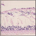 Bilateral Calcific Retina Arteriolar Occlusions in a Patient with Metastatic Ovarian Carcinoma
Dec 10 2020 by McGill University Health Centre
47-year-old female with cough and fever. Imaging showed a right pulmonary infiltrate. Transbronchial needle biopsy revealed lymphangitic spread of papillary adenocarcinoma with psammoma bodies (MRI of thyroid, CT of abdomen and pelvis were negative) gynecologic evaluation negative at that time . The patient had bilateral floaters, VA: 20/40 OD and 20/20 OS. Fundus examination showed retinal arteriolar sheathing and a flat choroidal lesion OS and vitritis OD. Fluorescein angiogram showed staining of left superior temporal retinal arterioles and bilateral midperipheral patchy hyperfluorescence at RPE. The patient vision in the OD deteriorated to 20/400, and in the OS 20/50. Four months later a new choroidal lesion was diagnosed OS. An abdominal mass consistent with a cystadenoma of the ovary was diagnosed. After a year patient developed systemic metastasis. Autopsy: Metastatic adenocarcinoma to the lung, both adrenals, para-aortic lymph nodes, left hip, right breast, occipital skin, serosal surface of liver, pituitary. In almost all metastatic lesions psammoma bodies were found. Presumptive diagnosis is a primary tumor of the ovary. Histopathologic examination of both eyes disclosed : Bilateral metastatic adenocarcinoma to the vitreous with partially calcified proliferation along internal limiting membrane, OS. Metastatic adenocarcinoma to choroid, OS. Bilateral optic atrophy secondary to retinal arteriolar occlusion with calcification.
Condition/keywords: bilateral, calcification, histopathology, metastatic adenocarcinoma, pathology, retinal arteriolar occlusion
 Loading…