Search results (5 results)
-
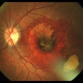
 Amelanotic Melanoma
Sep 19 2025 by Aditya S Kelkar, MS, FRCS, FASRS,FRCOphth
Widefield fundus photograph of a 37 year old showing a large, dome-shaped, intraocular mass involving the temporal retina. The lesion appears elevated and lacks surface pigmentation. Overlying retinal vessels are displaced and draped across the tumor surface, with surrounding retinal elevation noted. The appearance is suggestive of amelanotic variant of choroidal melanoma.
Photographer: Dr. Muskan Mangal
Imaging device: Optos Daytona
Condition/keywords: choroidal melanoma, intraocular tumor
-
 Wrinkled Anterior Capsule 40X zoom
Feb 18 2023 by Ahmed Abbas Hashmi, OD
Imprint of Iris Pigmentation on Anterior Lens Surface with wrinkled anterior capsule
Photographer: Ahmed Abbas Hashmi
Condition/keywords: lens opacity
-
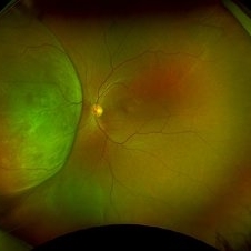
 submacular perfluorocarbon liquid
Sep 7 2022 by JEFFERSON R SOUSA, Tecg.º (Biomedical Systems Technology)
A 63-year-old male patient underwent vitreoretinal surgery with the use of perfluorocarbon. From a technological point of view, extended-field retinography presents many points of focus variation due to the difficulty of establishing a diffuse focus, as it is a recent post-operative case. In OCT Fundus Enface, although it has a low resolution, it is extremely important for documenting the presence of perfluor. Best seen in structural OCT.
Photographer: JEFFERSON ROCHA DE SOUSA - Retinal Department at Instituto Dr. Suel Abujamra Sao Paulo-Brazil
Imaging device: Optical Coherence Tomography system OCT CIRRUS 5000, Protocol, HD 5 Line
Condition/keywords: perfluorocarbon fluid, post-vitrectomy, submacular perfluorocarbon liquid (PFO), vitrectomy
-
 Perforating Ocular Trauma and Choroidal Rupture due to Shotgun Pellet
Mar 31 2022 by Franco Benvenuto, MD
Fundus photograph of a 17-year-old with shotgun injuries with numerous metal pellets in the chest, neck, brain, and face. Fundus exploration showed the left globe with posterior-inferior eye rupture, vitreous hemorrhages and choroidal rupture.
Photographer: Franco Benvenuto, Universidad de Buenos Aires, Argentina. Universidad de Guadalajara, México.
Condition/keywords: choroidal rupture, penetrating trauma, shotgun
-
 RPE Tear After Anti-VEGF Injection
Mar 17 2021 by RAFAEL REIS PEREIRA, MD
Retinal pigment epithelium (RPE) tear is a rare devastating complication of age-related macular degeneration (AMD). The believed mechanism lies in an adherence of the neovascularization to the undersurface of the RPE creating a contractile force that increases after VEGF therapy and causes the tear.
Photographer: Rafael Reis, Retina Clinic, São Paulo
Condition/keywords: retinal pigment epithelium (RPE) contracture
 Loading…