File number: 28295
Comments
-
 CARLA PEREZ MONTAÑO, MD (December 5 2021)
@Maria Stephanie R. Jardeleza
I´m really interested in having a conversation with you about this case. I had a similar case in my practice and I would like to get your valuable insights about your findings. Please contact me at dracarlaperez@gmail.com -
 Suber S. Huang, MD, MBA, FASRS (September 21 2018)
Thanks for contributing this excellent image of this rare complication
-
 Maria Stephanie R. Jardeleza, MD (June 27 2018)
Your comments are much appreciated Dr. Attia. I look forward to seeing how his clinical picture evolves, taking your input in mind!
-
 Hosam Attia, MD (June 27 2018)
You are welcome Dr. Jardeleza. Thank you again for sharing this rarely captured photo and for your response. It's interesting how the PFCL took that tear drop shape under the gas, specially when the pt is in the upright position for photography. It's as if something, almost incomplete doughnut, preventing it from settling down, that's why I was wondering. Thank you
- Maria Stephanie R. Jardeleza, MD (June 26 2018)
Thank you for your comments Dr. Attia. The patient denied any history of trauma and no zonular lysis was noted preoperatively or intraoperatively. The PFO bubbles in the anterior chamber were not visible until the post op week one visit where they were settled inferiorly. However, once the gas bubble in the anterior chamber made an appearance during post op week two, this clinical picture declared itself. The inferior angle was not shallow and ocular tensions were within normal.
- Hosam Attia, MD (June 26 2018)
Nicely Captured!
Thank you for sharing and for the description, since PFCL could be easily missed without that.
was there a history of trauma/ zonulysis , specially w/ PCFL in the A.C in a phakic patient ?
why the PFCL is not settling completely inferiorly in the A.C/ Angle, while patient is upright for photography , specially with the surface tension exerted on it, superiorly form the gas bubble, which is another reason for it to settle down even more !
Sign in to comment.
 Initializing download.
-
 By Maria Stephanie R. Jardeleza, MD
North Carolina Retina Associates
Co-author(s): M. Stephanie R. Jardeleza, M.D., Retina Specialist, San Antonio Eye Center, SA, TX - Uploaded on Jun 21, 2018.
- Last modified by Caroline Bozell on Jun 22, 2018.
- Rating
- Appears in
- Miscellaneous
- Condition/keywords
- vitreous substitutes, retained perfluorocarbon, retina surgery complications
- Photographer
- Andy Zepeda, COA, Retina Clinic, San Antonio Eye Center, San Antonio, TX
- Imaging device
- Photo slit lamp biomicroscope
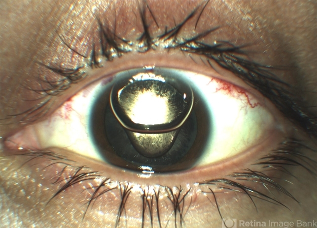
- Description
- Anterior segment photographs of 30-year-old male who underwent superior rhegmatogenous retinal detachment repair with intraocular gas tamponade. Perfluorocarbon was used to flatten the macula to prevent a macular fold and was removed during PFC/air exchange. Post operative week two visit shows gas migration into the anterior chamber with retained PFC layered in a tear drop shape posterior to the gas bubble and anterior to the lens. Patient had been maintaining face down positioning.


