Search results (216 results)
-
 Dissociated Optic Nerve Fiber Layer (DONFL)
Oct 14 2025 by Seif Allah Anwar
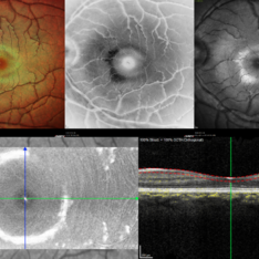
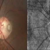
54 year-old male patient, 7 months following PPV with ILM peeling for an idiopathic epiretinal membrane complaining of gradual painless diminution of vision starting 3 month following the operation , en face OCT at the inner retinal layer shows multiple concentric dark spots representing apoptosis of the RNFL and GCL.
Photographer: Dr.Seif Anwar, FRCSEd
Imaging device: Optovue, SOLIX OCT
Condition/keywords: En Face OCTA
-
 Amelanotic Melanoma
Sep 19 2025 by Aditya S Kelkar, MS, FRCS, FASRS,FRCOphth
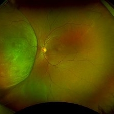
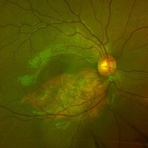
Widefield fundus photograph of a 37 year old showing a large, dome-shaped, intraocular mass involving the temporal retina. The lesion appears elevated and lacks surface pigmentation. Overlying retinal vessels are displaced and draped across the tumor surface, with surrounding retinal elevation noted. The appearance is suggestive of amelanotic variant of choroidal melanoma.
Photographer: Dr. Muskan Mangal
Imaging device: Optos Daytona
Condition/keywords: choroidal melanoma, intraocular tumor
-
Subhyaloid Hemorrhage
Jul 14 2025 by SHRADDHA ASHOK CHANDORKAR, DNB DO FVRS
19 year old female presented with sudden blurring of vision in her right eye since few hours after she attended a DJ party the previous night. On examination Vision was counting fingers close to face and Retina showed Subhyaloid hemorrhage with some RPE damage. YAG hyaloidotomy was performed and the subhyaloid hemorrhage was drained. Need for injections if RPE damage and development of CNV in future was explained. Patient was apprehensive as the vision was not restored immediately after the blood was drained. On subsequent follow ups slowly patient’s vision was restored to 6/6N6 after about a month.
Condition/keywords: subhyaloid hemorrhage
-
 Subhyaloid Hemorrhage
Jul 12 2025 by SHRADDHA ASHOK CHANDORKAR, DNB DO FVRS
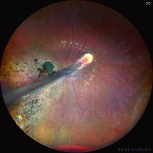
19 year old female presented with sudden blurring of vision in her right eye since few hours after she attended a DJ party the previous night. On examination Vision was counting fingers close to face and Retina showed Subhyaloid hemorrhage with some RPE damage. YAG hyaloidotomy was performed and the subhyaloid hemorrhage was drained. Need for injections if RPE damage and development of CNV in future was explained. Patient was apprehensive as the vision was not restored immediately after the blood was drained. On subsequent follow ups slowly patient’s vision was restored to 6/6N6 after about a month.
Photographer: Dr.Shraddha Chandorkar
Imaging device: Zeiss
Condition/keywords: subhyaloid hemorrhage
-
 CRAO With Cilio-retinal Sparing-MMI
Jun 25 2025 by Shivankar Sen, MS, FVRS
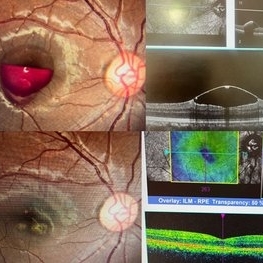
A 41 year old male came with complaints of Right eye blurring of vision since a day associated with watering and redness. He had no systemic illness, though gave a history of fall from bike 1 month back at the time of which he had blunt force trauma to the right side of the face. BCVA was 3/60, less than N36 in the right eye and 6/6, N6 in the left eye. Right eye had Marcus Gunn Pupil with clear lens, Left eye was within normal limits. IOP was normal; 16 in OD and 18 in OS. Retina evaluation revealed CRAO in the right eye with cilio-retinal artery sparing. Left eye was unremarkable Image Details Left to Right (Top 2 rows) Multicolor Reflectance Image (Blue-green enhanced 55 degree) revealing cilioretinal spared retinal stroma and a characteristic Cherry Red Spot; Green Reflectance showing corresopnding dark gray area with spared perfusion and black spot consistent with Cherry Red Spot on multicolor 2nd Row - 35 degree image (Multicolor Standard Reflectance and Green Reflectance) 3rd Row - SD-OCT revealing acute moderate CRAO findings with Middle retinal layer opacification and prominent middle limiting membrane (p-MLM) sign; Inner retinal layer opacification and prominent retinal pigment epithelium at the fovea with Diminished inner retinal layer stratification
Photographer: Gayathri M S
Imaging device: Heidelberg Spectralis HRA+OCT
Condition/keywords: CRAO with cilioretinal sparing, multicolor, multimodal imaging, OCT biomarkers, reflectance
-
 Berlins Edema - Multimodal Imaging
Jun 25 2025 by Shivankar Sen, MS, FVRS
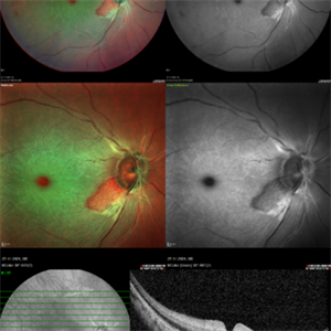
A 22 year old female came with history of injury to her left eye with a badminton racquet butt cap an hour before presentation On examination, she was found to have right eye 6/6;N6 vision and within normal limits, left eye 6/9;N6 vision, cells1+ in the anterior chamber, brisk pupillary response, no vitreous reaction and sub-clinical berlin's edema at the posterior pole. Multimodal imaging revealed frank boundaries of Berlin's edema more pronounced in the nasal parafoveal region. Figure details Top (Left to Right) Multicolor Reflectance showing bright yellow ring surrounding the perifovea; Blue Reflectance (Black on white contrast) showing corresponding black ring; Green Reflectance showing a characteristic white ring (all pronounced nasally); Bottom (Left-Right) Transverse structural OCT enface image showing white ring consistent with edema OCTA inner layer segmentation from ILM to GCL Transverse corresponding OCTA revealing faint hypo ring within perifoveal capillary bed
Photographer: Gayathri M S
Imaging device: Heidelberg Spectralis HRA+OCT
Condition/keywords: blue reflectance, En Face OCTA, enface imaging, multicolor, oct, reflectance
-
 Berlins
Jun 25 2025 by Shivankar Sen, MS, FVRS
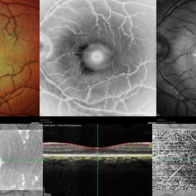
A 22 year old female came with history of injury to her left eye with a badminton racquet butt cap an hour before presentation On examination, she was found to have right eye 6/6;N6 vision and within normal limits, left eye 6/9;N6 vision, cells1+ in the anterior chamber, brisk pupillary response, no vitreous reaction and sub-clinical berlin's edema at the posterior pole. Multimodal imaging revealed frank boundaries of Berlin's edema more pronounced in the nasal parafoveal region. Figure details Top (Left to Right) Multicolor Reflectance showing bright yellow ring surrounding the perifovea; Blue Reflectance (Black on white contrast) showing corresponding black ring; Green Reflectance showing a characteristic white ring (all pronounced nasally); Bottom (Left-Right) Transverse structural OCT enface image showing white ring consistent with edema OCTA inner layer segmentation from ILM to GCL
Photographer: Gayathri M S
Imaging device: Heidelberg Spectralis HRA+OCT
Condition/keywords: blue reflectance, En Face OCTA, multicolor
-
 Commotio Retinae
Jun 10 2025 by CUI YUELING
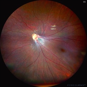
The patient presented 2 hours after sustaining a left eye injury caused by a stick. Visual acuity in the left eye was 0.2 without improvement upon correction, and intraocular pressure measured 15 mmHg. Examination of the anterior segment revealed ciliary conjunctival injection accompanied by patchy subconjunctival hemorrhage. The corneal surface remained smooth, and the anterior chamber was deep with hyphema characterized by blood-tinged aqueous humor predominantly settled inferiorly. The pupil was slightly irregular, approximately 3 mm in diameter, with a superotemporal notch; pupillary light reflex was intact. The lens appeared clear. Fundus examination showed well-defined optic disc margins with normal coloration and a cup-to-disc ratio of 0.2. Retinal arteries and veins were normally distributed with an artery-to-vein ratio of 2:3. At the posterior pole, the foveal reflex exhibited concentric ripple-like changes centered on the fovea, accompanied by localized pigment attenuation and reduced reflex intensity. Irregular reflectivity was noted in the superotemporal and inferotemporal nerve fiber layers.
Photographer: Yueling Cui
Imaging device: Zeiss Clarus 500
Condition/keywords: commotio retinae
-
 Neovascularization of the Disc
Jun 3 2025 by Scott D Walter, MD, MSc, FASRS
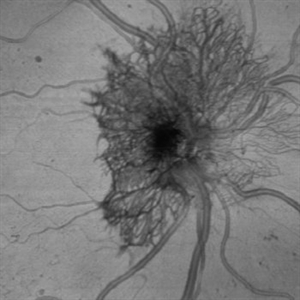
Near-infrared (NIR) en face OCT image showing neovascularization of the disc (NVD) in a patient with type II diabetes mellitus, complicated by proliferative diabetic retinopathy (PDR).
Imaging device: Heidelberg Spectralis
Condition/keywords: Diabetes, Heidelburg Spectralis, microaneurysms, Neovascularisation at the Disc (NVD), NEOVASCULARISATION OF DISC, OCT EN FACE, proliferative diabetic retinopathy (PDR)
-
 Retinal Detachment with Giant Retinal Tear
May 14 2025 by Kimberly Wakester
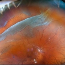
Optomap RGB of an 66-year-old man with a retinal detachment with a giant retinal tear in the right eye. Surgery was recommended. Patient is to continue follow up care post operatively. Also noted in the image is a vitreous opacity that was caught at the right moment and appears to look like a smiley face.
Photographer: Kimberly Wakester, COA, OCT-C
Imaging device: Optos California
Condition/keywords: giant retinal tear, RD
-
 Retromode Image of Dry AMD
May 13 2025 by Anupama Kiran Kumar
A retromode pseudo3D image of a case of dry AMD(NIDEK Mirante ). Retromode deviated left depicts depressed (pseudo concave) medium to large drusen. Here the drusenoid deposits appear to have a spectacular "moon surface" like appearance.
Photographer: Mr Pratap
Imaging device: Mirante SLO/OCT (Nidek Co., Gamagori, Japan)
Condition/keywords: AMD, dry age-related macular degeneration (dry AMD), retro mode
-
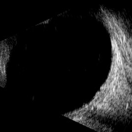 Macular Hole
May 9 2025 by Gustavo Uriel Fonseca Aguirre
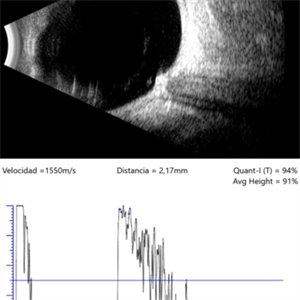
This B-mode longitudinal ultrasound scan demonstrates a full-thickness macular hole, appearing as a well-defined hypoechoic defect in the retinal surface with elevated edges.
Photographer: Gustavo U. Fonseca Aguirre, Hospital Conde de Valenciana, Ciudad de México
Condition/keywords: full thickness macular hole, macular hole
-
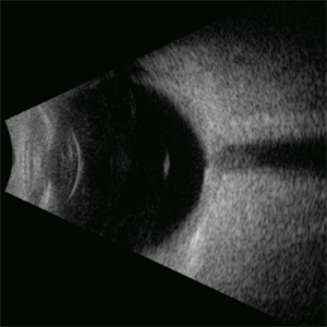 Weiss Ring
Apr 29 2025 by Gustavo Uriel Fonseca Aguirre
This B-mode axial ultrasound scan demonstrates the Weiss ring, visualized as a circular hyperechoic structure in the vitreous cavity, representing the detached posterior vitreous face with the optic disc insertion site. The ring shows mild mobility on dynamic assessment without retinal traction.
Photographer: Gustavo U. Fonseca Aguirre, Hospital Conde de Valenciana, Ciudad de México
Condition/keywords: Weiss ring
-
 Neovascularization of the Disc (NVD)
Apr 28 2025 by Vishal Agrawal, MD, FRCS,FACS,FASRS
Fundus image showing prominent neovascularization of the disc (NVD)- visible as fine, frond-like vascular proliferation extending from the disc surface.
Photographer: Dr Ayushi Gupta
Imaging device: Clarus 700
Condition/keywords: branch retinal vein occlusion (BRVO), NVD
-
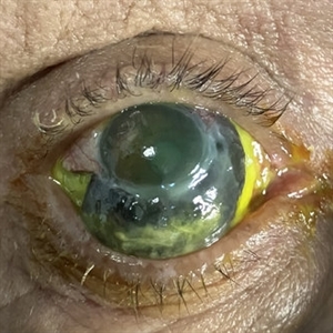 Necrotizing Scleritis
Apr 17 2025 by Gustavo Uriel Fonseca Aguirre
The clinical photograph shows necrotizing scleritis with perilimbal involvement, featuring marked scleral thinning and violaceous episcleral injection in the inferior quadrant. Focal uveal prolapse is visible at the area of maximal scleral necrosis, accompanied by peripheral ulcerative keratitis. Fluorescein staining residue is observed on the ocular surface. Associated findings include mild conjunctival chemosis and dilated episcleral vessels.
Photographer: Gustavo U. Fonseca Aguirre, Hospital Conde de Valenciana, Ciudad de México
Condition/keywords: necrotizing scleritis
-
 Choroidal Osteoma
Apr 17 2025 by Gustavo Uriel Fonseca Aguirre
Scanning laser ophthalmoscopy reveals a well-circumscribed, yellowish-white choroidal osteoma overlying the macular region and extending into the inferior temporal vascular arcade. Retinal vessels course normally over the tumor surface, with no evidence of subretinal fluid or hemorrhage. The surrounding retina shows preserved architecture without secondary degenerative changes.
Photographer: Gustavo U. Fonseca Aguirre, Hospital Conde de Valenciana, Ciudad de México
Condition/keywords: choroidal osteoma, macular choroidal osteoma
-
 Choroidal Osteoma
Apr 17 2025 by Gustavo Uriel Fonseca Aguirre
Top (B-mode): The longitudinal scan reveals a hyperechoic, flat, and well-demarcated macular lesion with posterior acoustic shadowing, pathognomonic for choroidal osteoma. Bottom (A-mode): Standardized tracing shows a tall initial spike (100% reflectivity) at the tumor surface with rapid decay to acoustic silence, confirming sound absorption by calcified tissue. This pattern remains unchanged at variable gain settings.
Photographer: Gustavo U. Fonseca Aguirre, Hospital Conde de Valenciana, Ciudad de México
Condition/keywords: choroidal osteoma, macular choroidal osteoma
-
 Advanced Proliferative Diabetic Retinopathy
Apr 9 2025 by Gustavo Uriel Fonseca Aguirre
B-mode ultrasound of a patient with long-standing poorly controlled diabetes demonstrates characteristic findings of advanced proliferative diabetic retinopathy. The examination reveals moderate vitreous hemorrhage appearing as diffuse hyperechoic opacities throughout the vitreous cavity, along with a posterior hyaloid membrane densely infiltrated by hemorrhagic material, showing irregular thickening and increased reflectivity. A mild subhyaloid hemorrhage is visible as a subtle hyphema-like space anterior to the retinal surface. The study documents a total tractional retinal detachment, evidenced by rigid retinal folds with clear insertion points of vitreous strands, accompanied by a significant subretinal hemorrhage seen as a prominent hyperechoic collection beneath the elevated retina. These findings collectively illustrate the severe vitreoretinal interface pathology characteristic of end-stage diabetic eye disease, with predominant tractional components and distinct echographic stratification of hemorrhagic layers - from anterior vitreous involvement to deeper subretinal blood accumulation.
Photographer: Gustavo U. Fonseca Aguirre, Hospital Conde de Valenciana, Ciudad de México
Condition/keywords: diabetic retinopathy, tractional retinal detachment, Vitreous hemorrhage
-
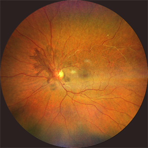
 Comets in the Eye (Retinopathy of Prematurity)
Apr 8 2025 by KANWALJEET HARJOT MADAN, M.S. (Ophthalmology); FAICO (Vitreous - Retina)
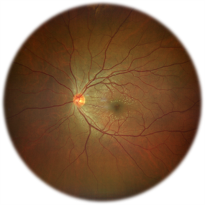
This is the fundus picture of right eye (RE) of a 4 years female child presented with outward deviation of right eye. Her parents also complained of diminution of vision in both eyes. On examination, her best corrected vision in RE was hand movements close to face and was 20/80 in LE. Posterior segment exam revealed presence of macular scar in RE and presence of dry retinal fold with dragging of retinal vessels. LE fundus revealed presence of nasal drag of optic disc. Parents gave history of untreated ROP as an infant. Retinopathy of Prematurity (ROP) is a Vaso proliferative disorder of Retina occurring in premature infants. Advances in neonatal care and ROP treatment has led these babies to live longer with this disease.
Photographer: Dr. Kanwaljeet Harjot Madan, Thind Eye Hospital, Jalandhar City (Punjab) INDIA.
Imaging device: Zeiss Fundus Camera
Condition/keywords: Retinopathy of Prematurity, Vaso proliferative disorder
-
 Comets in the Eye (Retinopathy of Prematurity)
Apr 8 2025 by KANWALJEET HARJOT MADAN, M.S. (Ophthalmology); FAICO (Vitreous - Retina)
This is the fundus picture of right eye (RE) of a 4 years female child presented with outward deviation of right eye. Her parents also complained of diminution of vision in both eyes. On examination, her best corrected vision in RE was hand movements close to face and was 20/80 in LE. Posterior segment exam revealed presence of macular scar in RE and presence of dry retinal fold with dragging of retinal vessels. LE fundus revealed presence of nasal drag of optic disc. Parents gave history of untreated ROP as an infant. Retinopathy of Prematurity (ROP) is a Vaso proliferative disorder of Retina occurring in premature infants. Advances in neonatal care and ROP treatment has led these babies to live longer with this disease.
Photographer: Dr. Kanwaljeet Harjot Madan, Thind Eye Hospital, Jalandhar City (Punjab) INDIA.
Imaging device: Zeiss Fundus Camera
Condition/keywords: Retinopathy of Prematurity
-
 Choroidal Melanoma
Mar 27 2025 by Virginia Gebhart
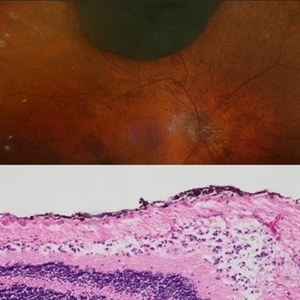
91 year old female with resolved choroidal melanoma. Pathology report confirms dispersed melanoma in the vitreous and on the entire retina surface. Pt is going well 1 month s/p enucleation, once healed she will be referred to an ocularist. No evidence of metastatic disease.
Condition/keywords: biopsy, choroidal melanoma
-
 The Halloween Smile
Mar 27 2025 by Shrishti mishra
A 73 year old male with Le optic disc pit . On color fundus photo a single pit can be noted whereas on oct enface is- os interface 2 optic disc pits are noted which resembles a halloween smile .
Photographer: Mr Sudhakar
Imaging device: Zeiss cirrus6000
Condition/keywords: OCT, oct en face, optic disc pit
-
 Melanocytoma
Mar 25 2025 by Gustavo Uriel Fonseca Aguirre
Longitudinal B-scan echogram shows mildly elevated lesion overlying surface of optic nerve. A-scan shows regular internal structure and high reflectivity of lesion.
Photographer: Gustavo U. Fonseca Aguirre, Hospital Conde de Valenciana, Ciudad de México
Condition/keywords: Melanocytoma
-
 Hemangioma of Retina (FAF)
Mar 5 2025 by Virginia Gebhart
Fundus autofluorescence of 64 year old male with choroidal hemangioma in the macula and STA. Persistent IRF and new cuff of SRF compared to previous photos. BCVA CF@face. Pt has had PDT in the past with no significant improvement. Will observe closely
Photographer: Virginia Gebhart, Retina Consultants of Carolina
Imaging device: Optos California
Condition/keywords: autofluorescence imaging, hemangioma, inferior subretinal fluid
-
 Hemangioma of Retina
Mar 5 2025 by Virginia Gebhart
64 year old male with choroidal hemangioma in the macula and STA. Persistent IRF and new cuff of SRF compared to previous photos. BCVA CF@face. Pt has had PDT in the past with no significant improvement. Will observe closely
Photographer: Virginia Gebhart, Retina Consultants of Carolina
Imaging device: Optos California
Condition/keywords: hemangioma, inferior subretinal fluid
 Loading…