Search results (2010 results)
-
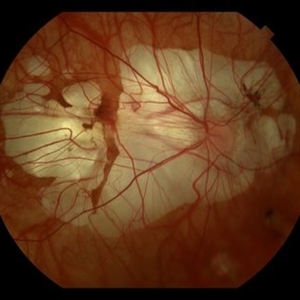
 Intraocular Foreign Body
Jul 9 2012 by George W. Aylward, MD, FRCS, FRCOphth
A metallic IOFB from a chisel being used by a builder with no eye protection. The IOFB was impacted on the retina, but removed via an internal approach. There was no hole in the retina underneath and the patient retained 20/20 vision with no complications.
Condition/keywords: intraocular foreign body
-
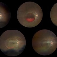
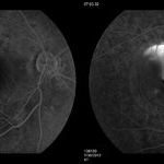
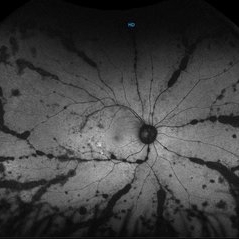 Autofluorescence Stage 3 Vogt-Koyanagi-Harada (VKH) Disease
Oct 20 2021 by Bryon R McKay, MD, PhD, FRCSC, DRCPSC - Retina
27yF presented with sub-acute findings of VKH, she has an interesting pattern of perivascular changes. She was successfully treated with immunosuppressive agents and maintains 20/20 vision.
Photographer: Dr. K. Vaezi, University of British Columbia, Canada
Imaging device: Optos Imaging system
Condition/keywords: Vogt-Koyanagi-Harada
-
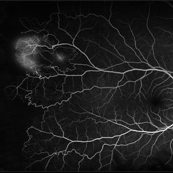
 CRVO
CRVO
Apr 22 2017 by Gabriel Costa Andrade, PhD
Panoramic retinography (Optos® California) of the right eye of a 48-year-old female patient with a history of low-vision in the right eye 2 months ago. At the exam presented visual acuity of 20/200 in the right eye and 20/20 in the left eye. Angiography shows diffuse perivascular leakage associated with areas of hypoperfusion in macula and periphery.
Photographer: Gabriel Andrade
Imaging device: Optos® California
Condition/keywords: central retinal vein occlusion (CRVO)
-
 Central Serous Retinopathy
Mar 19 2024 by Corey Grant
Ultra Wide-Field Fundus Autofluorescence Imaging of a 37 year old female with Central Serous Retinopathy affecting her right eye. Patient Visual Acuity was 20/20 in both eyes. Patient reported black spots in her vision onset three years ago, with associating flashes of light. Patient reports history of cortisone back injections a few years ago and denies Flonase use. The physician stated that there is hyperautofluorescence in the area of gutter of Sub-Retinal Fluid which likely happened from CSR.
Photographer: Corey Grant, OSC
Imaging device: OPTOS CALIFORNIA RGB
Condition/keywords: Central Serous Chorioretinopathy (CSR), central serous retinopathy (CSR), fundus autofluorescence (FAF), Guttering, hyperautofluorescence, inferior retina, OPTOS, Retina, Right Eye, subretinal fluid, ULTRA WIDE FIELD
-
 Coats' Disease
Feb 2 2021 by Niloofar Piri, MD
#1 16-year-old male with abnormal temporal peripheral telangiectatic and aneurysmal vascular lesions associated with hard exudate deposition posteriorly. Vision 20/20. Stage II Coats' disease.
Condition/keywords: Coats' disease, Leber's miliary aneurysm
-
 Diffuse Chorioretinal Atrophy
Feb 21 2024 by Virginia Gebhart
61 year male with myopic degeneration and diffuse chorioretinal atrophy. BCVA 20/200.
Photographer: Virginia Gebhart
Imaging device: Topcon TRC 50DX
Condition/keywords: chorioretinal atrophy, myopic degeneration
-
 Sickle Cell Retinopathy
Feb 15 2021 by Kim Barrett
24-year-old female with Sickle Cell Retinopathy, stage 3. She confirms she has the trait as well as her grandmother, mother and a sibling. She has seafan neovascularization superotemporal OD. Current VA is 20/20. Photo is pre-PRP laser with areas of non-profusion temporally.
Photographer: Kim Barrett C.O.A. Retina Specialist of Michigan, Grand Rapids, MI
Imaging device: Optos California
Condition/keywords: neovascularization (NV), pan-retinal photocoagulation (PRP), sickle cell retinopathy, stage 3, trait
-
 Valsalva retinopathy progression
Oct 4 2023 by Niloofar Piri, MD
Progression fundus photograph images of a 22 yo female with Valsalva retinopathy secondary to violent emesis. Note the sub ILM layered hemorrhage and gradual decrease then disappearance over time. Last image is at 6 month follow up with 20/20 vision. Of note the silhouette of ILM separation is still visible.
Condition/keywords: valsalva retinopathy
-
 Vascular Loop Thrombosis
May 1 2020 by Bianca Susanna
Fundus photograph of a 13-year-old child with central retinal artery occlusion secondary to prepapillary vascular loop complicated by thrombosis. She had visual acuity of 20/20 due to an anomalous artery macular branch.
Photographer: Bianca N. Susanna, Faculdade de Medicina do ABC, Santo André.
Condition/keywords: central retinal artery occlusion (CRAO), prepapillary vascular loop
-
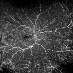
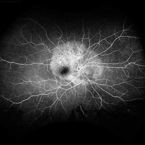
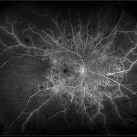
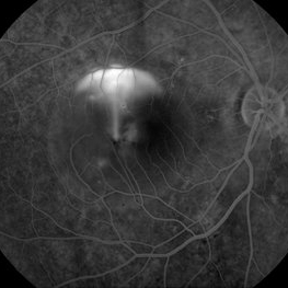
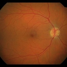
 2min-FA-VKH
Oct 20 2021 by Bryon R McKay, MD, PhD, FRCSC, DRCPSC - Retina
27yF presented with sub-acute findings of VKH, she has an interesting pattern of perivascular changes. She was successfully treated with immunosuppressive agents and maintains 20/20 vision.
Photographer: Dr. K. Vaezi, University of British Columbia, Canada
Imaging device: Optos Imaging system
Condition/keywords: uveitis, Vogt-Koyanagi-Harada
-
 Acute Syphilitic Posterior Placoid Chorioretinitis
May 4 2021 by RAFAEL REIS PEREIRA, MD
A 31-year-old patient with a complaint of photophobia and low visual acuity OD in the previous three weeks. BCVA was 20/60 and 20/20 The fundus examination revealed a placoid white lesion in the posterior pole and vitreous cells in the right eye. The left eye was unremarkable. Fluorescein angiography reveals hyperfluorescent plaque with distinctive “leopard spots” hypofluorescence.
Imaging device: Opto California
Condition/keywords: acute syphilitic posterior placoid chorioretinitis
-
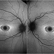 Bilateral “Bull's eye”pattern maculopathy
Mar 14 2023 by Anfisa Ayalon, MD
Both eyes fundus autofluorescence image of a 38-year-old female with “Bull's eye” pattern maculopathy. There is no history of medication use associated with retinal toxicity. BCVA RE 20/25+2, LE 20/20-3
Photographer: Danielle Ferguson and Alec Bertoni, University of Pittsburgh Medical Center
Condition/keywords: bull's eye maculopathy, maculopathy, retina
-
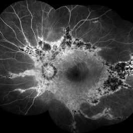
 Central Retinal Vein Occlusion with Severe Retinal Ischemia
Jan 19 2022 by Olivia Rainey
Ultra-widefield fluorescein angiogram of a 56-year-old male with a Central Retinal Vein Occlusion with Severe Retinal Ischemia affecting his right eye. The patient presented on 1/19/2022, sc20/20-2 vision in the right eye. The patient has had a good response to Eylea with complete resolution of edema. The physician is considering PRP to ischemic periphery in the future and given the degree of ischemia in both eyes, she recommends that the patient's PCP check carotid Doppler US.
Photographer: Olivia Rainey, OCT-C, COA
Imaging device: Optos California
Condition/keywords: central retinal vein occlusion (CRVO), FA late phase, fluorescein angiogram (FA), ischemic CRVO, Optos, retinal ischemia, ultra-wide field imaging
-
 Choroidal Metastasis
Apr 11 2024 by Corey Grant
Ultra-Widefield fundus photography and fundus autofluorescence images of a 61 year old female with Choroidal Metastasis affecting both eyes. Patient presented with blurred vision and flashes for a few weeks. Patient visual acuity was cc20/100 PH20/60 in the right eye and cc20/200 in the left eye. Patient admits to history of smoking for many years bit no known history of cancer prior to the visit. Physician recommended going to the ER for full body PET CT and stated that the first line of treatment is usually systemic chemo therapy. Patient will be reassessed in one month.
Photographer: Corey Grant
Imaging device: OPTOS CALIFORNIA RGB
Condition/keywords: cancer, choroidal metastasis, fundus autofluorescence (FAF), fundus photography, hyperautofluorescence, hypoautofluorescence, Optos, OPTOS CALIFORNIA RGB, Retina, ULTRA WIDE FIELD
-
 Congenital Retinal Macrovessel
Oct 13 2023 by Jacob D. Grodsky, MD
41 y/o male who presented with acute onset of blurred vision OD. Visual acuity was 20/200 OD; 20/25 OS. Examination was consistent with congenital retinal macrovessel through the macula with intraretinal hemorrhage as seen in the fundus photo. Intravitreal bevacizumab was injected, and visual acuity improved to 20/40 at 4-week follow-up. MRA head and neck was ordered to rule out other vascular anomalies.
Condition/keywords: congenital retinal macrovessel, RETINAL MACROVESSEL
-
 CSCR Mushroom Cloud
Feb 25 2015 by James J. Bedrick, MD
Late transit FA of a large active subfoveal CSCR leak. Focus is on peri-foveal vessels to give sense of height of large serous RD of macula. This patient presented with a BCVA of 20/200 and fluorescein and historic evidence of prior episodes of leakage. After discussion of known treatment options including observation, he was initially treated with rifampin and had partial resolution to 20/70 BCVA but this was short-lived with reaccumulation of the large serous detachment within 3 months. He then received sub-threshold micro-pulse laser photocoagulation with an 810 nm diode laser which resulted 1 month later in complete drying of the serous detachment and BCVA of 20/25.
Photographer: Diana Bodnar, COT
Imaging device: Topcon 50X with OIS capture station
Condition/keywords: CSCR subfoveal leak
-
 CSCR Mushroom Cloud
Feb 23 2015 by James J. Bedrick, MD
Late transit FA of a large active sub-foveal CSCR leak. You may view this pair in stereo to appreciate the plume of leakage within this large serous RD of the macula. This patient presented with a BCVA of 20/200 and fluorescein and historic evidence of prior episodes of leakage. After discussion of known treatment options including observation, he elected to be treated initially with oral rifampin and BCVA improved to 20/40 with persistent metamorphosis and a shallower persistent macular detachment over several visits. Rifampin was discontinued and he then received sub-threshold micro-pulse laser photocoagulation with an 810 diode which resulted in the patient reporting full restoration of his vision subjectively within a month. He failed to keep his follow-up appointment.
Photographer: Diana Bodnar, COT
Imaging device: Topcon 50X with Merge capture station
Condition/keywords: CSCR subfoveal leak
-
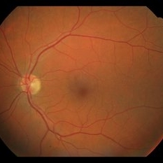
 Degeneration Paravenous
Sep 20 2016 by JEFFERSON R SOUSA, Tecg.º (Biomedical Systems Technology)
Female patient, 32-years-old, Asian, appeared at the clinic with a history of glaucoma. 20/20 visual acuity in both eyes. Examination of color photography, pigmentary changes were observed following the vascular arcades only in the left eye. Suggestive of paravenous degeneration.
Photographer: JEFFERSON R SOUSA - Study Center and Ophthalmological Research Dr. Andre M V Gomes, Institute Dr. Suel Abujamra São Paulo-Brazil
Imaging device: Zeiss / VisuCam-500 - Angulation of field photo of 45 Degrees, flash 24.
Condition/keywords: degeneration paravenous
-
 Drusens
Sep 10 2014 by Mehul A Shah
75-year-old female patient presented with cataract having 20/20 vision post operatively.
Photographer: Drashti Netralaya,Dahod
Imaging device: Zeiss ff450
Condition/keywords: drusen
-
 Epiretinal Membrane
Sep 6 2021 by Ricardo Leitão Guerra
65-year-old woman with an asymptomatic ERM (BCVA=20/20).
Imaging device: Zeiss Clarus 700
Condition/keywords: epiretinal membrane (ERM)
-
 Example of AREDS Category 1 (Small Drusen But Not Considered AMD)
Feb 11 2013 by Neil M. Bressler, MD
Person in AREDS Category 1 were essentially free of age-related macular abnormalities, with a total drusen area less than 5 small drusen (<63 microns) within 3,000 microns of the center of the macula, and visual acuity of 20/32 or better in both eyes1. These are fundus photographs of a 53-year-old man, with visual acuity 20/20 OD and 20/32 OS presenting for evaluation of any diabetic retinopathy. Reference: 1 Age-Related Eye Disease Study Research Group. A randomized, placebo controlled clinical trial of high-dose supplementation with vitamins C and E, beta carotene, and zinc for age-related macular degeneration and vision loss: AREDS report No. 8. Arch Ophthalmol. 2001;119(10):1417-1436.
Condition/keywords: age-related macular degeneration (AMD)
-
 Fibrovascular Retinal Pigment Epithelial Detachment - Color Fundus
Jul 16 2014 by James B. Soque, CRA, OCT-C, COA, FOPS
69-year-old white female with Hx of 10 anti-VEFG treatment injections of right eye, VA 20/200, now stable, off drug for 10 months.
Photographer: James B Soque, CRA COA
Imaging device: Topcon TRC 50 DX with MERGE software, 5 MP dig camera
Condition/keywords: color fundus photograph, fibrovascular pigment epithelial detachment (PED), pigment epithelial atrophy, retina
-
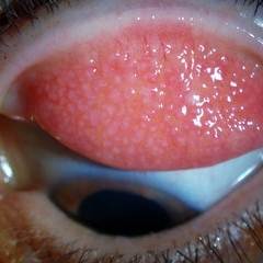 Giant Papillary Conjunctivitis, Left Upper Eyelid
Jul 22 2013 by Jason S. Calhoun
Contact lens wearer, in for exam. Has rough feeling underneath both eyelids. Patient thought it was through SCL wear. Patient VA was 20/20. right eye, 20/30, left eye. Underneath the left upper eyelid, you can see papillary inflammation and redness.
Photographer: Jason S. Calhoun, Department of Ophthalmology, Mayo Clinic Jacksonville, Florida
Imaging device: TOPCON D-90 SL NIKON CAMERA
Condition/keywords: giant papillary conjunctivitis
-
 Hypertensive Retinopathy
Jun 28 2013 by Jason S. Calhoun
Patient came in complaining of spots in vision in both eyes. VA was 20/25 - right eye and 20/20- left eye. Fundus exam reveals little hemorrhages with cotton wool spots due to hypertension and anemia.
Photographer: Jason S. Calhoun, Mayo Clinic Jacksonville, Florida
Imaging device: TOPCON TRC 50-EX
Condition/keywords: hypertensive retinopathy
 Loading…