 Initializing download.
-
 By RAFAEL REIS PEREIRA, MD
Hospital de Olhos do Sul de Minas Gerais
Co-author(s): Hermano Lucio A. Filho; Lucas Mares Guia; Ricardo Nakaghi Okada; André Maia, Retina Clinic- São Paulo. - Uploaded on May 4, 2021.
- Last modified by Caroline Bozell on May 4, 2021.
- Rating
- Appears in
- Miscellaneous
- Condition/keywords
- acute syphilitic posterior placoid chorioretinitis
- Imaging device
- Opto California
- Description
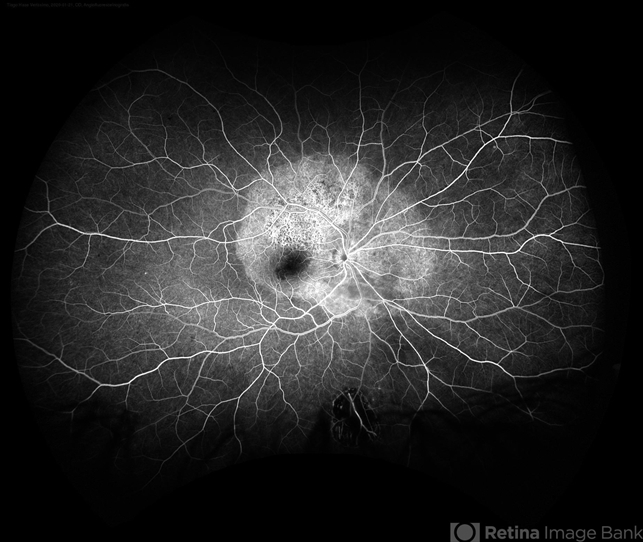
- A 31-year-old patient with a complaint of photophobia and low visual acuity OD in the previous three weeks. BCVA was 20/60 and 20/20 The fundus examination revealed a placoid white lesion in the posterior pole and vitreous cells in the right eye. The left eye was unremarkable. Fluorescein angiography reveals hyperfluorescent plaque with distinctive “leopard spots” hypofluorescence.


---thumb.JPG/image-square;max$79,0.ImageHandler "Syphilitic Maculopathy")


