Search results (2010 results)
-
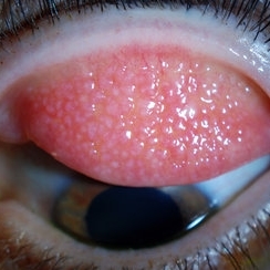 Giant Papillary Conjunctivitis, Left Upper Eyelid
Jul 22 2013 by Jason S. Calhoun
Contact lens wearer in for exam. Has rough feeling underneath both eyelids. Patient thought it was through SCL wear. Patient VA was 20/20. right eye, 20/30, left eye. Underneath the left upper eyelid, you can see papillary inflammation and redness.
Photographer: Jason S. Calhoun, Department of Ophthalmology, Mayo Clinic Jacksonville, Florida
Imaging device: TOPCON D-90 SL NIKON CAMERA
Condition/keywords: giant papillary conjunctivitis
-
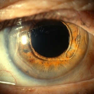
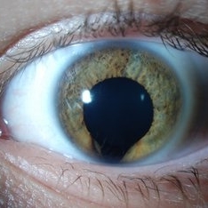 Keyhole Pupil Coloboma
Jul 13 2013 by Jason S. Calhoun
14-year-old male presents with decreased vision in the left eye. Dx with iris and retinal coloboma in the left eye. Patient VA was 20/20, right eye, 20/100 left eye with pinhole improvement 20\50. Patient was fitted for SCL in the left eye.
Photographer: Jason S. Calhoun, Department of Ophthalmology, Mayo Clinic Jacksonville, Florida
Imaging device: TOPCON D-90 SL NIKON CAMERA
Condition/keywords: deformed pupil
-
 Intraocular Foreign Body
Jul 9 2012 by George W. Aylward, MD, FRCS, FRCOphth
A metallic IOFB from a chisel being used by a builder with no eye protection. The IOFB was impacted on the retina, but removed via an internal approach. There was no hole in the retina underneath and the patient retained 20/20 vision with no complications.
Condition/keywords: intraocular foreign body
-
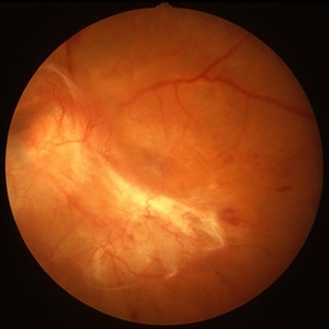 PDR with Traction RD of Macular
Oct 8 2012 by Jeffrey G. Gross, MD, FASRS
PRD, with traction RD of macular, pre-op, 20/200.
Condition/keywords: 20/200, macular, pre-op, tractional retinal detachment
-
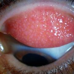 Giant Papillary Conjunctivitis, Left Upper Eyelid
Jul 22 2013 by Jason S. Calhoun
Contact lens wearer, in for exam. Has rough feeling underneath both eyelids. Patient thought it was through SCL wear. Patient VA was 20/20. right eye, 20/30, left eye. Underneath the left upper eyelid, you can see papillary inflammation and redness.
Photographer: Jason S. Calhoun, Department of Ophthalmology, Mayo Clinic Jacksonville, Florida
Imaging device: TOPCON D-90 SL NIKON CAMERA
Condition/keywords: giant papillary conjunctivitis
-
 Anterior Chamber Intraocular Lens
Sep 20 2012 by Jeffrey G. Gross, MD, FASRS
AC-IOL, s/p PPV, lensectomy for dislocated crystalline lens, 20/20
Condition/keywords: anterior chamber, dislocated crystalline lens, intraocular lens (IOL), lensectomy
-
---thumb.JPG/image-square;max$300,300.ImageHandler) Retinal Coloboma
Retinal Coloboma
Jul 8 2013 by Jason S. Calhoun
14-year-old male with decreased vision in the left eye. Dx with iris and retinal coloboma in the left eye. Patient VA was 20/20, right eye, 20/100 left eye with pinhole improvement 20\50. Patient was fitted for SCL in the left eye.
Photographer: Jason S. Calhoun, Department of Ophthalmology, Mayo Clinic Jacksonville, Florida
Condition/keywords: chorioretinal coloboma
-
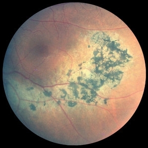
 Pigmented Peripheral Retinal Degeneration
Jun 27 2013 by Jason S. Calhoun
42-year-old male came in for routine eye exam and to follow up on peripheral retinal degeneration in both eyes. VA is 20/20, right eye and 20/25, left eye. Patient is asymptomatic with no visual complaints.
Photographer: Jason S. Calhoun, Mayo Clinic Jacksonville, Florida
Imaging device: TOPCON TRC 50-EX
Condition/keywords: peripheral retinal degeneration
-
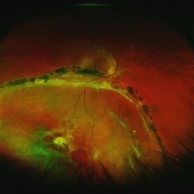
 Asymptomatic Rhegmatogenous Retinal Detachment
Sep 14 2012 by Sharon Fekrat, MD FACS FASRS
Fundus photograph of a 25-year-old emmetropic male graduate student with an inferotemporal phakic chronic asymptomatic rhegmatogenous retinal detachment with a demarcation line in the right eye. His sister who is an ophthalmology resident discovered this incidental finding. Vision 20/20.
Photographer: Brian Lutman CRA, Duke University Eye Center, Durham, NC
Condition/keywords: asymptomatic, demarcation line
-
---thumb.JPG/image-square;max$300,300.ImageHandler) Peripheral Retinal Degeneration
Peripheral Retinal Degeneration
Jul 8 2013 by Jason S. Calhoun
Patient comes in with double vision. VA was 20/20 in both eyes. Fundus exam shows retinal degenerative changes in both eyes. Offer to correct double vision with temporary Fresnel prism.
Photographer: Jason S. Calhoun, Department of Ophthalmology, Mayo Clinic Jacksonville, Florida
Condition/keywords: peripheral retinal degeneration
-
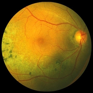
---thumb.jpg/image-square;max$300,300.ImageHandler) Macular CHRPE
Macular CHRPE
Aug 11 2013 by Eric M. Shrier, DO
This a color fundus photograph of a 74-year-old black male with longstanding poor vision os, 20/200. He exhibits mild NPDR additionally.
Photographer: Christopher Bunce
Condition/keywords: congenital hypertrophy of the retinal pigment epithelium (CHRPE)
-
 Presumed Ocular Histoplasmosis Syndrome
May 3 2018 by Nichole Lewis
39-year-old female with presumed ocular histoplasmosis syndrome. Patient presented with vision of 20/200 in 10/2014. Vision CF @ 1 ft on 7/2015. Right eye vision is 20/20.
Photographer: Nichole Lewis
Condition/keywords: ocular histoplasmosis syndrome (OHS), presumed ocular histoplasmosis syndrome (POHS)
-
---thumb.jpg/image-square;max$300,300.ImageHandler) Optic Disc Drusen
Optic Disc Drusen
Jul 10 2013 by Hamid Ahmadieh, MD
SD-OCT image of the left eye of a 24-year-old woman with optic disc drusen and VA 20/20.
Photographer: Solmaz Shahmohammadi, Negah Eye Center, Tehran
Imaging device: Heidelberg Spectralis
Condition/keywords: optic disc drusen, optical coherence tomography (OCT)
-
 Fibrovascular Retinal Pigment Epithelial Detachment - Color Fundus
Jul 16 2014 by James B. Soque, CRA, OCT-C, COA, FOPS
69-year-old white female with Hx of 10 anti-VEFG treatment injections of right eye, VA 20/200, now stable, off drug for 10 months.
Photographer: James B Soque, CRA COA
Imaging device: Topcon TRC 50 DX with MERGE software, 5 MP dig camera
Condition/keywords: color fundus photograph, fibrovascular pigment epithelial detachment (PED), pigment epithelial atrophy, retina
-
 Sector Retinitis Pigmentosa
Oct 8 2012 by Jeffrey G. Gross, MD, FASRS
Sector retinitis pigmentosa, 20/20, left eye.
Condition/keywords: 20/20, left eye, sector retinitis pigmentosa
-
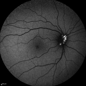 Optic Disc Drusen
Jul 10 2013 by Hamid Ahmadieh, MD
Fundus autofluorescence image of the right eye of a 24-year-old woman with optic disc drusen and VA 20/20.
Photographer: Solmaz Shahmohammadi, Negah Eye Center, Tehran
Imaging device: Heidelberg Spectralis
Condition/keywords: fundus autofluorescence (FAF), optic disc drusen
-
---thumb.jpg/image-square;max$300,300.ImageHandler) Birdshot Choroidopathy
Birdshot Choroidopathy
Oct 9 2013 by Maurice F. Rabb
Forty two year old white female first noted flashing lights in her left eye at the age of 30. Although she had many previous eye examinations for low grade myopia, she had never had a dilated fundus examination. The evaluation twelve years ago disclosed 20/20 acuity in each eye with a myopic correction, an afferent pupillary defect on the left, no evidence of anterior segment inflammation in either eye, a full field on the right and markedly constricted field on the left, fundus pigmentary abnormalities extending beyond the equator in each eye, and narrow vessels with pigment migration into the retina in the left eye only.
Condition/keywords: birdshot choroidopathy
-
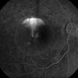
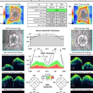 OCT in Patient With IIH Showing Thickened RNFL
Jan 16 2019 by John S. King, MD
18-year-old African American female with increased BMI with a history of headaches, nausea, transient diplopia and vision loss that she notices when getting up from her bed (and goes away after standing upright) for the last two weeks. Went to PCP and was treated for the flu, and after no improvement and visual symptoms known, was sent to ED. MRI did not show any masses and showed empty sella turcia. Vision 20/30 OD and 20/20 OS; no RAPD; IOP 15OU; no anterior segment or vitreous inflammation; discs are elevated with obscuration of the disc margins and some of the smaller vessels; there are no SVPs; there are mild Patton's lines temporally (see Initial Photos). The optic disc cube shows 360 degrees of RNFL thickening (see OCT). Was referred to near-ophthalmologist, Dr. Doyle. She obtained additional work-up, and LP opening pressure was high, and MRV showed bilateral transverse sinus stenosis. Patient showed steady improvement with medical therapy, that included weight loss and oral diamox. On her last visit with Dr. Doyle, vision has remained stable at 20/20-20/25 without an enlarged blindspot; there are SVPs and optic disc edema has resolved (see Post Treatment Photos); she is currently on 1000 mg of diamox and has lost 15 pounds, and no stinting procedure needed.
Imaging device: Cirrus
Condition/keywords: benign idiopatic intracranial hypertension, optic disc edema, papilledema
-
 Chronic Inferior Retinal Detachment
Mar 1 2017 by Philip J. Polkinghorne, MD
Color photograph of chronic retinal detachment with pigment demarcation line and atrophic holes visible. The vision was recorded at 20/20, and follow up is 3 years.
Photographer: Alex Fraser
Condition/keywords: atrophic retinal hole, demarcation line
-
 AZOOR
AZOOR
Aug 24 2012 by Geoffrey G. Emerson, MD, PhD, FASRS
A 17-year-old healthy woman noticed a pacman-shaped scotoma in her temporal right vision. Acuity measured 20/20 and color vision measured 11/11. Angiography showed some late staining of the nasal macula.
Photographer: Geoffrey Emerson, MD, PhD, Retina Center, Minneapolis
Condition/keywords: scotoma
-
 Sector Retinitis Pigmentosa
Oct 8 2012 by Jeffrey G. Gross, MD, FASRS
Sector retinitis pigmentosa, 20/20, right eye.
Condition/keywords: 20/20, sector retinitis pigmentosa
-
---thumb.jpg/image-square;max$300,300.ImageHandler) "Spots" In The Central Visual Zone
"Spots" In The Central Visual Zone
Oct 14 2013 by Maurice F. Rabb
A 26 year old healthy female who had been aware of decreased vision in OS for 5 days before the initial examination. When questioned specifically about OD, she did admit to being aware of some "spots" in the central visual zone. Her past ocular history is negative for eye disease and the family history is negative for retinal and macular disease. The patient is in excellent general health. She had a recent upper respiratory infection and is presently disabled because of a herniated disc. Uncorrected vision OD is 20/20 and OS is 20/400, improving to 20/100- with pinhole. The findings of significance are noted in the posterior poles.
Condition/keywords: spots in the central visual zone
-
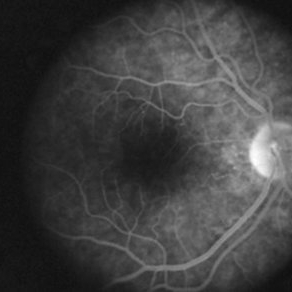
 Acute Syphilitic Posterior Placoid Chorioretinopathy
Feb 9 2015 by Leandro C. Zacharias, MD, PhD
Fundus photograph of a 43-year-old man with syphilitic posterior placoid chorioretinopathy. His visual acuity came back to 20/20 after systemic treatment.
Photographer: Leandro Cabral Zacharias
Imaging device: Zeiss Visucam
Condition/keywords: acute syphilitic posterior placoid chorioretinitis
-
 CSCR Mushroom Cloud
Feb 25 2015 by James J. Bedrick, MD
Late transit FA of a large active subfoveal CSCR leak. Focus is on peri-foveal vessels to give sense of height of large serous RD of macula. This patient presented with a BCVA of 20/200 and fluorescein and historic evidence of prior episodes of leakage. After discussion of known treatment options including observation, he was initially treated with rifampin and had partial resolution to 20/70 BCVA but this was short-lived with reaccumulation of the large serous detachment within 3 months. He then received sub-threshold micro-pulse laser photocoagulation with an 810 nm diode laser which resulted 1 month later in complete drying of the serous detachment and BCVA of 20/25.
Photographer: Diana Bodnar, COT
Imaging device: Topcon 50X with OIS capture station
Condition/keywords: CSCR subfoveal leak
-
---thumb.jpg/image-square;max$300,300.ImageHandler) "Spots" In The Central Visual Zone
"Spots" In The Central Visual Zone
Oct 14 2013 by Maurice F. Rabb
A 26 year old healthy female who had been aware of decreased vision in OS for 5 days before the initial examination. When questioned specifically about OD, she did admit to being aware of some "spots" in the central visual zone. Her past ocular history is negative for eye disease and the family history is negative for retinal and macular disease. The patient is in excellent general health. She had a recent upper respiratory infection and is presently disabled because of a herniated disc. Uncorrected vision OD is 20/20 and OS is 20/400, improving to 20/100- with pinhole. The findings of significance are noted in the posterior poles.
Condition/keywords: spots in the central visual zone
 Loading…