 Initializing download.
-
 By Niloofar Piri, MD
SSM Health Group, St Louis University
Co-author(s): Nicholas K Baugnon, MD, Saint Louis University - Uploaded on Oct 4, 2023.
- Last modified by Joshua Friedman on Oct 5, 2023.
- Rating
- Appears in
- Miscellaneous
- Condition/keywords
- valsalva retinopathy
- Imaging device
- Fundus camera
- Description
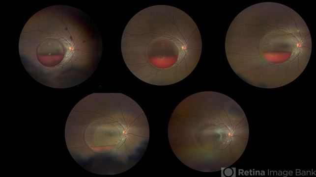
- Progression fundus photograph images of a 22 yo female with Valsalva retinopathy secondary to violent emesis. Note the sub ILM layered hemorrhage and gradual decrease then disappearance over time. Last image is at 6 month follow up with 20/20 vision. Of note the silhouette of ILM separation is still visible.


---thumb.jpg/image-square;max$79,0.ImageHandler "Primary Subhyaloid Hemorrhage Due to Valsalva Retinopathy")
---thumb.jpg/image-square;max$79,0.ImageHandler "Primary Subhyaloid Hemorrhage Due to Valsalva Retinopathy")
---thumb.jpg/image-square;max$79,0.ImageHandler "Primary Subhyaloid Hemorrhage Due to Valsalva Retinopathy")
---thumb.jpg/image-square;max$79,0.ImageHandler "Primary Subhyaloid Hemorrhage Due to Valsalva Retinopathy")
---thumb.jpg/image-square;max$79,0.ImageHandler "Primary Subhyaloid Hemorrhage Due to Valsalva Retinopathy")