Search results (2010 results)
-
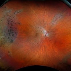
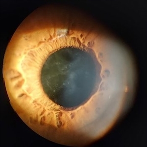
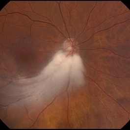 Myelinated Retinal Nerve Fiber Layer
May 20 2025 by Ignacio Leonardo Pueyo Bestue, MD
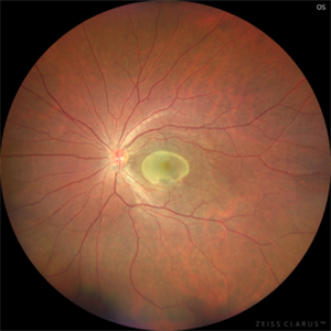
Fundus photo of an 80-year-old woman with myelinated RNFL, 20/20 vision, and mild hyperopia
Photographer: Pueyo-Bestue, I.L., MD, Universite Libre de Bruxelles, Ophthalmology Department
Condition/keywords: myelinated nerve fiber layer
-
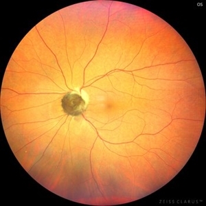 Optic Nerve Melanocytoma
May 4 2025 by KANWALJEET HARJOT MADAN, M.S. (Ophthalmology); FAICO (Vitreous - Retina)
This is a fundus picture of a young 42-year male who visited for a routine eye exam. His BCVA was 20/20 in both eyes. Anterior segment examination was normal. His left eye showed grey-black pigmentation at the infero-nasal margin of the optic disc. Fundus of the right eye was normal. The patient was diagnosed to have optic disc melanocytoma on multimodal imaging and was advised regular follow-up. Optic nerve melanocytoma is typically a benign tumor made up of melanocytes and melanin. It can grow, but rarely transforms into a malignancy. Patients with Optic Nerve Melanocytoma should be periodically examined for evidence of growth, loss of visual field and optic nerve compression.
Photographer: Dr. Kanwaljeet Harjot Madan, Thind Eye Hospital, Jalandhar City (Punjab) INDIA.
Imaging device: Zeiss Fundus Camera
Condition/keywords: melanocytoma, melanoma, optic nerve
-
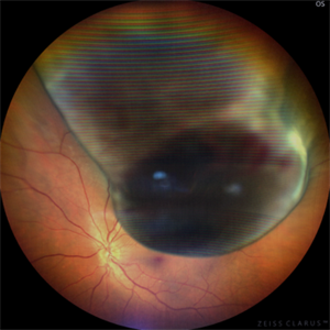 Uveal Melanoma
Apr 26 2025 by Vishal Agrawal, MD, FRCS,FACS,FASRS
A 32 year-old male presented with complaints of perceiving a shadow in OS for 15-20 days. His BCVA was 20/20 OU. On Fundus examination, a large, elevated, well-defined, pigmented choroidal mass with few hemorrhages over the lesion was seen and a provisional diagnosis of uveal melanoma was made. urgent oncological consultation was recommended for further treatment.
Photographer: Dr Ayushi Gupta
Imaging device: Clarus 700
Condition/keywords: melanoma
-
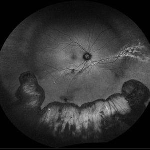
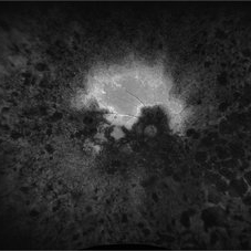 Retinitis Pigmentosa
Apr 17 2025 by Virginia Gebhart
Fundus autofluorescence of 75 year old female with Retinitis Pigmentosa. Pt diagnosed at age 53. Diffuse RPE atrophy with minimal central sparing present in both eyes. Stable and unchanged compared to previous exams. BCVA 20/200 OD, NLP OS
Photographer: Virginia Gebhart, Retina Consultants of Carolina
Imaging device: Optos California
Condition/keywords: autofluorescence imaging, bone spicule, retinitis pigmentosa, RP
-
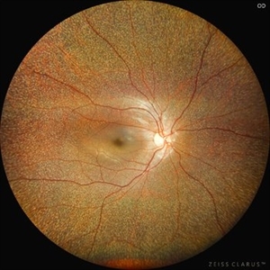
 Retinitis Pigmentosa
Apr 9 2025 by Virginia Gebhart
35 year old female with stable sectoral RP and high myopia OU. RP has not progressed in either eye since initial visit in 2021. Will continue to observe. VA 20/20 OU
Photographer: Virginia Gebhart, Retina Consultants of Carolina
Imaging device: Optos California
Condition/keywords: high myopia, retinitis pigmentosa
-
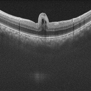 Retinal Fold in Posterior Microphthalmos
Mar 1 2025 by Hemanth Murthy, MBBS, MD, FASRS
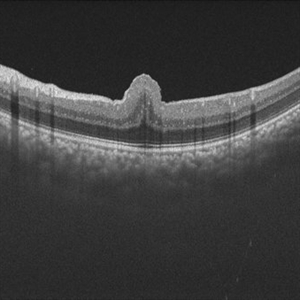
Swept source OCT image of left eye of 34 year male patient with high hypermetropia(+14). BCVA 20/20 in right eye and 20/60 in left eye. Anterior segment was normal. There is loss of foveal pit with omega shaped elevation of inner retinal layers.
Photographer: Mr Veda Vyas
Condition/keywords: posterior microphthalmos
-
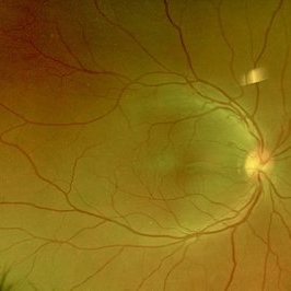
 Retinal Fold in Posterior Microphthalmos
Mar 1 2025 by Hemanth Murthy, MBBS, MD, FASRS
Fundus photo of left eye of 34 year male patient with high hypermetropia(+14). BCVA 20/20 in right eye and 20/60 in left eye. Anterior segment was normal. There is loss of foveal pit with omega shaped elevation of inner retinal layers.
Photographer: Mr Veda Vyas
Condition/keywords: posterior microphthalmos
-
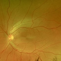
 Retinal Fold in Posterior Microphthalmos
Mar 1 2025 by Hemanth Murthy, MBBS, MD, FASRS
Fundus photo of Right eye of 34 year male patient with high hypermetropia(+14). BCVA 20/20 in right eye and 20/60 in left eye. Anterior segment was normal. There is loss of foveal pit with omega shaped elevation of inner retinal layers.
Photographer: Mr Veda Vyas
Condition/keywords: posterior microphthalmos
-
 Retinal Fold in Posterior Microphthalmos
Mar 1 2025 by Hemanth Murthy, MBBS, MD, FASRS
Swept source OCT image of Right eye of 34 year male patient with high hypermetropia(+14). BCVA 20/20 in right eye and 20/60 in left eye. Anterior segment was normal. There is loss of foveal pit with omega shaped elevation of inner retinal layers.
Photographer: Mr Veda Vyas
Condition/keywords: posterior microphthalmos
-
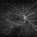
 Bilateral Proliferative Diabetic Retinopathy OU
Feb 21 2025 by Drew Mitchell
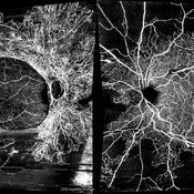
OCT-Angiography 8x8 Montage OU. PDR with active NVE OD. 37 year old male with no visual complaints. Vision is 20/20 in both eyes.
Photographer: Drew Mitchell OCT-C
Imaging device: Zeiss Cirrus 5000
Condition/keywords: CIRRUS 5000 ANGIOPLEX, Diabetes, NVE, OCT Angiography, proliferative diabetic retinopathy (PDR)
-
 Sub Hyaloid Hemorrhage
Jan 29 2025 by Vishal Agrawal, MD, FRCS,FACS,FASRS
A 45-year-old male patient presented with decreased vision in Left Eye. On fundus examination, a boat-shaped sub hyaloid hemorrhage was noted. YAG hyaloidotomy was performed and the patient recovered with a vision of 20/20.
Photographer: Dr Ayushi Gupta
Imaging device: Clarus 700
Condition/keywords: Sub hyaloid haemorrhage, YAG HYALOIDOTOMY
-
 CSR with Fibrin-FFA
Jan 29 2025 by Vishal Agrawal, MD, FRCS,FACS,FASRS
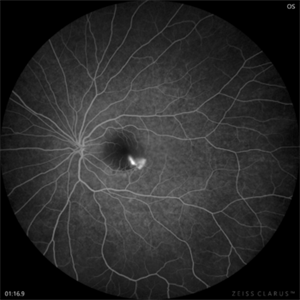
A 31-year-old female was referred with a diagnosis of subretinal cysticercosis. BCVA was 20/200 OS. OCT showed a large subfoveal bacillary layer detachment (BALAD) without any scolex. FFA revealed a smoke-stack appearance. A final diagnosis of CSR with Fibrin was made and was managed conservatively. BCVA at final visit was 20/20.
Photographer: Dr Ayushi Gupta
Imaging device: Clarus 700
Condition/keywords: central serous chorioretinopathy (CSCR)
-
 CSR with Fibrin
Jan 28 2025 by Vishal Agrawal, MD, FRCS,FACS,FASRS
A 31-year-old female was referred with a diagnosis of subretinal cysticercosis. BCVA was 20/200 OS. OCT showed a large subfoveal bacillary layer detachment (BALAD) without any scolex. FFA revealed a smoke-stack appearance. A final diagnosis of CSR with Fibrin was made and was managed conservatively. BCVA at final visit was 20/20.
Photographer: Dr Ayushi Gupta
Imaging device: Clarus 700
Condition/keywords: central serous chorioretinopathy (CSCR)
-
 Vitreous Prolapse
Jan 28 2025 by Korey Starkey
Slit lamp image of a 62-year-old patient presented at first visit with vitreous prolapse due to mechanical complications from IOL placement. IOP was being managed with drops, vision was 20/20, patient opted for surgery due to constant haze in vision.
Photographer: Korey Starkey
Imaging device: Slit lamp camera
Condition/keywords: slit lamp photo, vitreous prolapse
-
 Elmiron Toxicity
Jan 15 2025 by Virginia Gebhart
54 year old female with pigmentary degeneration secondary to Elmiron. Stippled RPE maculopathy has lightly progressed with stable vision compared to previous visits. BCVA 20/200 OU. Pt reports taking Elmiron from 2010 to 2019.
Photographer: Virginia Gebhart
Imaging device: Optos California
Condition/keywords: autofluorescence imaging, Maculopathy, secondary pigmentary degeneration
-
 Non Proliferative Diabetic Retinopathy
Dec 18 2024 by Angela Rico
71 y/o Male with Background DR OD V/A OD 20/200 Scattered Ma's- Non perfusion
Photographer: Angela Rico M.D.
Condition/keywords: Diabetic Retinopathy
-
 Chronic CRVO
Dec 12 2024 by Korey Starkey
Fluorescein Angiography of a 62 year-old man with chronic central retinal vein occlusion. Vision is 20/200.
Photographer: Korey Starkey
Imaging device: Optos
Condition/keywords: capillary nonperfusion, central retinal vein occlusion (CRVO), FLUORESCEIN ANGIOGRAPHY, ischemia, microaneurysms, Optos
-
 Repaired Retinal Detachment with Multiple Breaks
Dec 9 2024 by Virginia Gebhart
FAF in 25 year old female of repaired retinal detachment 1.5 year s/p scleral buckle/cryo. Pt had been having symptoms for over a year, inferior demarcation line from retinal fluid that was present. Retina remains flat and attached under buckle. Treated lattice inferiorly, no new holes or tears. VA 20/20
Photographer: Virginia Gebhart, Retina Consultants of Carolina
Imaging device: Optos California
Condition/keywords: autofluorescence imaging, cryotherapy, demarcation line, lattice degeneration, scleral buckle
-
 Benign Familial Fleck Retina
Dec 2 2024 by KANWALJEET HARJOT MADAN, M.S. (Ophthalmology); FAICO (Vitreous - Retina)
This is fundus picture of a 21 year old female patient who had come for refractive surgery consultation. Her best corrected vision in both eyes was 20/20. She had myopic astigmatism in both eyes. Fundus exam revealed presence of multiple yellowish white flecks spread throughout retina sparing macular area in both eyes. Her color vision was normal. Electroretinogram and electrooculogram were normal. She gave no history of night blindness. A diagnosis of Benign Familial Fleck Retina was made. She was also advised ocular exam of her parents and elder brother which was normal.
Photographer: Dr. Kanwaljeet Harjot Madan, M.S. (Ophthalmologist) Fellow in Vitrous & Retina. Thind Eye Hospital, Jalandhar City. Punjab. India
Imaging device: Zeiss Clarus
Condition/keywords: Benign familial fleck retina, Night Blindness
-
 Valsalva Retinopathy
Oct 28 2024 by Andrew Jin, MD
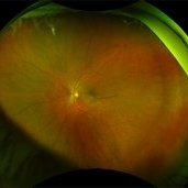
These fundus photos show the progression of subhyaloid hemorrhage due to valsalva retinopathy in a 38-year-old woman with hyperemesis gravidarum. She was managed conservatively with observation alone. The photos depict the initial presentation after 1 day of vision loss, at 1 month, and at 6 months of follow-up. Presenting visual acuity was counting fingers and returned to 20/20.
Condition/keywords: fundus photograph, valsalva retinopathy
-
 Valsalva Retinopathy
Oct 28 2024 by Andrew Jin, MD
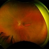
These fundus photos show the progression of subhyaloid hemorrhage due to valsalva retinopathy in a 38-year-old woman with hyperemesis gravidarum. She was managed conservatively with observation alone. The photos depict the initial presentation after 1 day of vision loss, at 1 month, and at 6 months of follow-up. Presenting visual acuity was counting fingers and returned to 20/20.
Condition/keywords: fundus photograph, valsalva retinopathy
-
 Valsalva Retinopathy
Oct 28 2024 by Andrew Jin, MD
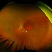
These fundus photos show the progression of subhyaloid hemorrhage due to valsalva retinopathy in a 38-year-old woman with hyperemesis gravidarum. She was managed conservatively with observation alone. The photos depict the initial presentation after 1 day of vision loss, at 1 month, and at 6 months of follow-up. Presenting visual acuity was counting fingers and returned to 20/20.
Condition/keywords: fundus photograph, valsalva retinopathy
-
 Stargardt's Disease
Oct 23 2024 by Virginia Gebhart
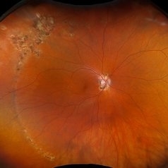
62 year old female with bullseye RPE changes and flecks, mottled FAF, and silent choroid on FA consistent with late onset Stargardt's Disease. Pt is asymptomatic with 20/20 vision OU at this time
Photographer: Virginia Gebhart, Retina Consultants of Carolina
Imaging device: Optos California
Condition/keywords: Stargardt disease, Stargardts Disease
-
 POHS/Schlaegel Lines
Sep 19 2024 by Virginia Gebhart
46 year old female with h/o Histoplasmosis. Multiple punched out chorioretinal scars with Schlaegel lines. No evidence of CNV or active inflammation. VA 20/20
Photographer: Virginia Gebhart, Retina Consultants of Carolina
Imaging device: Optos California
Condition/keywords: chorioretinal scar, histoplasmosis, presumed ocular histoplasmosis syndrome (POHS), Schlaegel
-
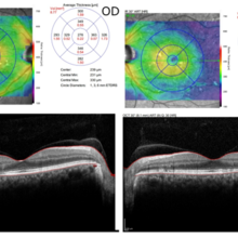 Pattern dystrophies – OCT demonstrates significant RPE irregularities and multiple focal inner segment-outer segment (IS-OS) disruptions with overlying cystic changes in both eyes of a 60-year-old man with PD
Sep 17 2024 by Nicolas A Yannuzzi, MD
Visual acuity was 20/20 OD and 20/25 OS. Genetic testing showed only 1 pathogenic variation in ABCA4, which is atypical for STGD1 Stargardt disease that is inherited in autosomal recessive fashion. (Images courtesy of Byron L. Lam, MD)
Condition/keywords: inherited retinal disease, pattern dystrophy
 Loading…