Search results (35 results)
-
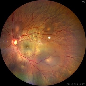
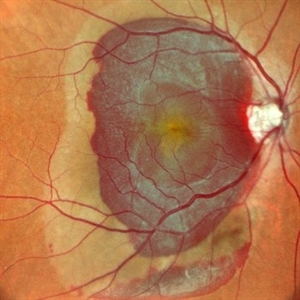
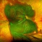
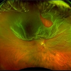
 Idiopathic Uveal Effusion Syndrome
Aug 22 2024 by Jordyn Beckman
61 year old male with Idiopathic Uveal Effusion Syndrome with starry night appearance on fluorescein. 3 weeks s/p single external drainage retinotomy and 9 weeks of oral pred with recurrent choroidal effusions. Has since returned to surgery for secondary drainage retinotomy; subretinal fluid remain persistent.
Photographer: Jordyn Beckman
Imaging device: Optos California
Condition/keywords: chorioretinitis, Choroidal, exudative detachment, window defect
-
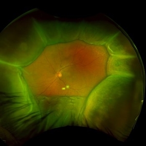
 Central Serous Chorioretinopathy in Pregnancy (OS)
Apr 28 2024 by Vishal Agrawal, MD, FRCS,FACS,FASRS
30-year female with sudden loss of vision came for examination. She was in her first trimester of pregnancy. Examination revealed bilateral bullous NSD with subretinal fibrin s/o CSR.
Photographer: Dr Ayushi
Imaging device: Clarus 700
Condition/keywords: Central Serous Chorioretinopathy (CSR), neurosensory detachment of retina, pregnancy
-
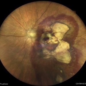
 Optic Nerve Melanocytoma
Apr 3 2023 by Gustavo Aguirre Suarez
Fundus photograph of a 36-year-old female with a lesion dependent on the optic nerve head with subretinal extension, elevated, about 1.5 disc diameters, dark brown to black in color, involving more than three quarters of the neuroretinal ring towards the inferonasal area.
Photographer: Dr. Gustavo Aguirre-Suarez
Imaging device: Zeiss Visucam 500
Condition/keywords: melanocytic lesion, Melanocytoma
-
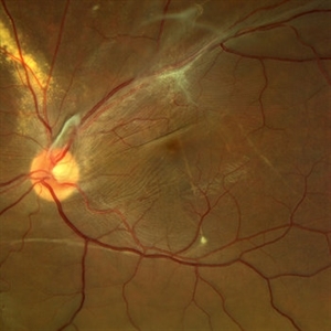
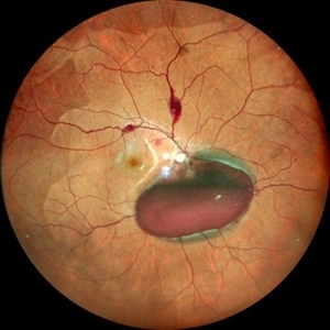
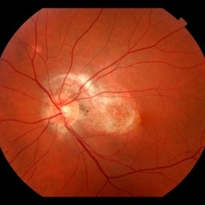
 Idiopathic Uveal Effusion Syndrome
Jun 13 2023 by Ahmad B. Tarabishy, MD
66 year old male presented with a 4 month vision of painless decreased vision in the left eye. Clinical findings consistent with idiopathic uveal effusion syndrome. Fundus photography shows 360 degree choroidal elevation with dependent inferior subretinal fluid.
Photographer: Dr. Angela Rico, Retina Specialists of Tampa
Imaging device: Idiopathic Uveal Effusion Syndrome
Condition/keywords: idiopathic uveal effusion syndrome, uveal effusion
-
 PEHCR (Peripheral Exudative Hemorrhagic Chorioretinopathy)
May 12 2023 by Niloofar Piri, MD
Ultrawide fundus photograph of the left eye demonstrating extensive peripheral hemorrhagic exudative detachment in a 79 yo Caucasian female with prior history of non-exudative AMD. Recent diagnosis of Acute myeloid leukemia with low platelet count which might have contributed to the above presentatuon. Please note the temporal subretinal hemorrhage as well as RPE atrophy and hyperplasia in the macula.
Photographer: Rocio Bentivegna, MD, Saint Louis University; Jessica Maddox, COA, Saint Louis University
Condition/keywords: peripheral exudative hemorrhagic chorioretinopathy (PEHCR)
-
 Subretinal Hemorrhage
Feb 28 2023 by Akansha Sharma
Color fundus photograph of an 84-year old male with subretinal hemorrhage associated with areas of scarring.
Photographer: Dr. Urmil Shah, Dr. Denish Patel, Dr. Akansha Sharma, Bharati Eye Hospital, Ahmedabad, Gujarat
Condition/keywords: choroidal neovascularization (CNV), subretinal hemorrhage
-
 Lady in a dress
Feb 9 2023 by Shelby Helton
Fluorescein Angiography on a 67-year-old male with significant RPE changes secondary to a severe subretinal hemorrhage that required a vitrectomy with subretinal TPA in 2013.
Photographer: Shelby Helton
Imaging device: Heidelberg Spectralis
Condition/keywords: wet age-related macular degeneration (wet AMD)
-
 Choroidal Melanoma
Nov 3 2022 by pedro fernandes souza neto
Transillumination of Enucleation specimen of Choroidal Melanoma: anterior chamber is closed. Total secondary retinal detachment with subretinal serous fluid and some subretinal hemorrhages are present.
Photographer: Eduardo Marback, Federal University of Bahia, Brazil
Condition/keywords: enucleation, melanoma
-
 Displaced & folded macula
Oct 10 2022 by Ricardo Leitão Guerra
Tractional retinal detachment due to sickle cell retinopathy leading to a displaced and folded appearance of the macula in this 36-yo male. Subretinal bands are also noticed crossing the macula towards inferior retinal detachment area.
Photographer: Ricardo Leitão Guerra
Imaging device: Clarus 700 - Zeiss
Condition/keywords: folds, sickle cell retinopathy, subretinal bands, tractional retinal detachment
-
 Subretinal BSS and air
Apr 12 2022 by Nassim Alejandro Abreu Arbaje, MD
67 year old female who presented with complaints of 5 days of decreased vision of her left eye. She underwent PPV + BSS and Air injection in the subretinal space
Photographer: Nassim Abreu, Dr. Elias Santana Hospital
Imaging device: Ngenuity 3D system screenshot
Condition/keywords: subretinal hemorrhage
-
 Subretinal Bleed
Jul 12 2022 by Akansha Sharma
73 year old diabetic and hypertensive female presented with sub-retinal hemorrhage for which she was operated with pars-plana vitrectomy with intra-vitreal anti-VEGF
Photographer: Dr. Akansha Sharma-Retina Foundation, Ahmedabad
Condition/keywords: subretinal hemorrhage, subretinal blood
-
 Valsalva Retinopathy
Dec 20 2021 by Unnati Vishwanath Shukla, M. S. ,DNB, FVRS FNERF, MNAMS,PhD Scholar(Retina)
26-year-old male with Valsalva Retinopathy. History of severe cough for 3 days. All hematological investigations were within normal limits.
Photographer: Dr. Unnati Shukla, Consultant, Retina Foundation, Ahmedabad
Imaging device: Nidek Mirante
Condition/keywords: subhyaloid hemorrhage, subretinal hemorrhage, valsalva retinopathy
-
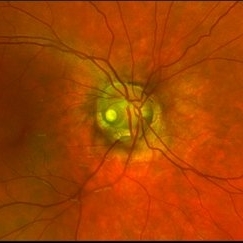 Optic Nerve Pit Right Eye
Feb 15 2021 by Kim Barrett
A 14-year-old male presented with vision loss and VF defect. Patient was treated for presumed amblyopia with patching since age 4. He has had neurologic care for post traumatic skull fracture and brain bleed in 2012. IOP's WNL. OD is without retinoschisis or subretinal fluid. Patient is at risk of serous detachment. Current VA OD 20/200+1 PH 20/80.
Photographer: Kim Barrett C.O.A. Retina Specialist of Michigan, Grand Rapids, MI
Imaging device: Optos California
Condition/keywords: amblyopia, hemifield, Humphrey visual field, nerve, optic nerve pit, visual field defect
-
 Trio of Retinal Hemorrhages
Dec 8 2020 by Priya Rasipuram Chandrasekaran, MBBS, DO, DNB, FRCS
This is the fundus photo of a 29-year-old following blunt trauma showing hemorrhages in all the three layers of the retina (vitreous hemorrhage, subhyaloid hemorrhage and subretinal hemorrhage)
Condition/keywords: blunt trauma, retinal hemorrhage
-
 Blunt Ocular Trauma Due to Firework Injury
Jun 9 2020 by Brittany Rota
Ultra- widefield pseudocolor image of an 18-year-old male with blunt ocular trauma in the right eye due to a firework injury. The patient presented with commotio retinae (sclopteria), an acute vitreous hemorrhage, choroidal rupture, and a subretinal hemorrhage. The referring physician performed surgery on the lateral rectus muscle which was macerated but not severed, and several orbital fibrous foreign bodies were removed from the posterior orbit. The globe was intact. There is no evidence of retinal tear in the region of sclopetaria; however, there is complete necrosis of the temporal peripheral choroid and retina. The vitreous hemorrhage was slowly clearing on his exam 6-9-2020. The patient is developing subretinal fibrosis. The physician is concerned about the choroidal rupture that is visible through the submacular hemorrhage. There is one rupture that appears to course directly under the fovea. The physician states that if this is the case, his vision most likely will be 20/200 or worse. His vision was hand motion in all fields except nasally, which he was unable to see hand motion at his visit on 6-9-2020.
Photographer: Brittany Rota
Imaging device: Optos California
Condition/keywords: blunt trauma, choroidal rupture, commotio retinae, fibrosis, firework injury, fundus photograph, hand motion, necrotizing retina, Optos, pseudocolor, subretinal hemorrhage, vitreous hemorrhage
-
 Subretinal Fibrosis (PPCNVM and POHS) OS
Sep 18 2019 by John S. King, MD
57-year-old white male with history of PPCNVM OS and POHS OU here for a routine visit. History of avastin in 2014, and stable since then. Va OS 20/20. PP scar with macular subretinal fibrosis. No heme or exudates. CR spot supero-nasally.
Photographer: Shelly Blair
Imaging device: Topcon 50
Condition/keywords: choroidal neovascular membrane (CNVM), ocular histoplasmosis syndrome (OHS), peripapillary choroidal neovascularization (PPCNVM), presumed ocular histoplasmosis syndrome (POHS)
-
 Massive SRH in Patient on Coumadin Being Treated for Exudative AMD
Sep 30 2019 by John S. King, MD
78-year-old white female using 1mg of warfarin for atrial fibrillation, who had a large PED, Type 1 lesion from AMD. Noticed acute darkening of vision one week after anti-VEGF injection. Has very large SRH, subRPE heme, and corrugated retinal appearance post RPE tear. Vision HM (from 20/100). 20/25 in the fellow eye that has dry AMD.
Photographer: Shelly Blair
Imaging device: Optos CA
Condition/keywords: subretinal hemorrhage, wet age-related macular degeneration (wet AMD)
-
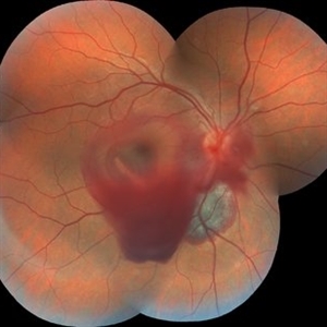
 Ruptured Macroaneurysm
May 22 2019 by Nichole Lewis
FA of a 91-year-old woman with a ruptured macroaneurysm, intraretinal hemorrhage and subretinal hemorrhage. VA 20/400.
Photographer: Nichole Lewis
Condition/keywords: intraretinal hemorrhage, ruptured macroaneurysm, subretinal hemorrhage
-
 Optos Giant Tear within Retinal Detachment
Apr 30 2019 by Lauren Whaley
Noticed an inferior visual field defect on a patient with history of vitreous hemorrhage. Decided to take an Optos image and this is what we found. Doctor performed pneumatic retinopexy in office and patient recovering well.
Photographer: Lauren R. Whaley
Imaging device: Optos
Condition/keywords: Optos, retinal tear, subretinal fluid
-
 Ocular Hypotony Due to Leaking Bleb
Apr 1 2019 by Anfisa Ayalon, MD
81-year-old male who had trabeculectomy in his right eye 4 years ago, presented to the emergency room with complains of decreased vision in that eye for two months. Slit-lamp examination showed cystic bleb with leakage, intraocular pressure was 0 MMHg. Fundus examination showed hypotony maculopathy, peripheral choroidal detachments, multiple chorioretinal folds with subretinal fluid.
Photographer: Anfisa Ayalon, MD., Meir Medical Center, Kfar Saba, Israel.
Imaging device: California, Optos 200 DTX
Condition/keywords: choroidal detachment, hypotonous retinopathy, hypotony maculopathy
-
 Multiple Retinal Lesions Secondary to Blunt Trauma
Jun 19 2018 by Somnath Chakraborty, MD
A montage of the right eye of a 15-year-old boy, who was struck by a football. The image shows multiple choroidal ruptures in the macular area, with sub-retinal blood and multiple, large retinal tears temporally. There is also an area of juxtapapillary, pigmentary changes.
Photographer: Saptarshi Mehta, Retina Institute of Bengal
Condition/keywords: blunt trauma, choroidal rupture, giant retinal tear, subretinal hemorrhage
-
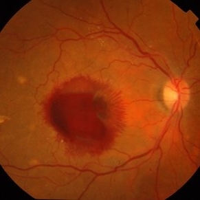
 Submacular Hemorrhage
Apr 24 2018 by Pauline T Merrill, MD, FASRS
Fundus photo of left eye of a 65-year-old AMD patient who presented with sudden drop of vision from 20/30 to CF due to a large submacular hemorrhage, 7 months following her last Eylea injection. She underwent immediate injection of C3F8 in the office, with little effect. 10 days later vitrectomy with subretinal tPA and air-fluid exchange was performed, with successful displacement of the hemorrhage.
Photographer: Ermelinda Diaz, Illinois Retina Associates, Chicago, Illinois
Imaging device: Topcon 50DX
Condition/keywords: neovascular age-related macular degeneration (AMD), submacular hemorrhage
-
 Autosomal Recessive Bestrophinopathy - Color Photo OD
Dec 22 2017 by Tony Tsai, MD, FASRS
11-year-old Asian male with 20/40 vision OU, negative family history for ocular conditions, and bilateral atypical vitelliform deposits and subretinal fluid. EOG confirmed abnormally low Arden ratios OU. Genetic testing revealed homozygous recessive mutation in BEST1 gene (p.L140V:c.418C>G). Also known as p.L80V; Ref: Davidson (2009) Am J Hum Genet 85, 581.
Photographer: San Juanita Zazueta
Imaging device: Topcon
Condition/keywords: Best disease
-
 Central Serous Chorioretinopathy
Apr 19 2017 by Gustavo Barreto de Melo, MD, PhD, FASRS
Fundus photograph of a 32-year-old pregnant woman with a serous detachment and subretinal fibrin deposit surrounding the fovea.
Photographer: Denyson Silva, Sergipe Eye Hospital
Condition/keywords: central serous chorioretinopathy (CSCR), pregnancy
-
 Macroaneurysm
Apr 1 2017 by Manish Nagpal, MD, FRCS (UK), FASRS
Case of a ruptured macroaneurysm with subhyaloid and subretinal blood.
Photographer: Avijit Vishnoi
Condition/keywords: macroaneurysm, ruptured macroaneurysm
 Loading…