Search results (1804 results)
-
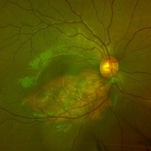
 CSCR
CSCR
Jul 8 2025 by T. P . VIGNESH, MBBS,MS
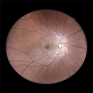
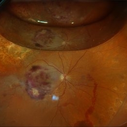
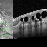
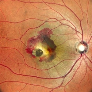
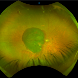
Fundus photograph of an 45 year old man revealing CSCR with subretinal precipitates.
Photographer: Sivanath
Imaging device: EIDON
Condition/keywords: central serous chorioretinopathy (CSCR)
-
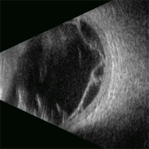
 Choroidal Melanoma (USG)
Jul 5 2025 by Gustavo Uriel Fonseca Aguirre
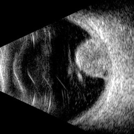
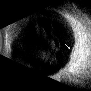
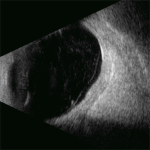
This B-mode transverse ultrasound scan reveals a mushroom-shaped choroidal tumor in the inferior nasal quadrant adjacent to the optic nerve head. The lesion appears solid with homogeneous internal reflectivity and is associated with minimal surrounding subretinal fluid and scleral excavation. It measures 6.54 mm in height × 7.52 mm in base diameter (transverse view) and extends 9.52 mm longitudinally. The vitreous contains abundant punctate opacities consistent with pigment dispersion. The retina and choroid remain attached elsewhere.
Photographer: Gustavo U. Fonseca Aguirre, Hospital Conde de Valenciana, Ciudad de México
Condition/keywords: choroidal melanoma
-
 Giant Retinal Tear
Jul 5 2025 by Gustavo Uriel Fonseca Aguirre
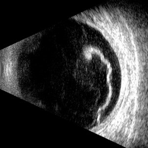
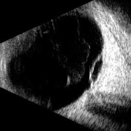
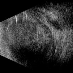
This B-mode longitudinal ultrasound scan reveals a giant retinal tear, demonstrating a circumferential retinal flap with rolled edges extending over M-X to M-I. The vitreous shows diffuse hemorrhage and anterior-posterior traction strands inserting at the tear margins. The remaining retina appears attached without subretinal fluid.
Photographer: Gustavo U. Fonseca Aguirre, Hospital Conde de Valenciana, Ciudad de México
Condition/keywords: giant retinal tear
-
 Retinal Dialysis
Jul 5 2025 by Gustavo Uriel Fonseca Aguirre
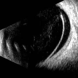
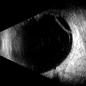
This B-mode longitudinal ultrasound scan demonstrates a retinal dialysis, appearing as a linear discontinuity at the ora serrata with associated vitreous base avulsion. The scan reveals mild subretinal fluid extending from the dialysis site with macular involvement.
Photographer: Gustavo U. Fonseca Aguirre, Hospital Conde de Valenciana, Ciudad de México
Condition/keywords: retinal dialysis
-
 T-Cell Lymphoma
Jul 3 2025 by Virginia Gebhart
78 year old male s/p vitreous biopsy for T-Cell lymphoma. Pt presented with peripheral blot hemorrhages and numerous white subretinal infiltrates. Retinal pallor and thickening temporally. History of cutaneous T-cell lymphoma. PPV/vitreous biopsy performed to find differential diagnosis. Silicone oil was placed for 6 weeks, then removed and exchanged with a gas bubble. Hematology pathologist and Emory reviewed path report and agrees it is consistent with T-cell lymphoma. Pt received intravitreal Methotrexate and will be scheduled for weekly treatments. BCVA CF
Photographer: Virginia Gebhart, Retina Consultants of Carolina
Imaging device: Optos California
Condition/keywords: biopsy, gas bubble, lymphoma
-
 Diabetic Macular Edema
Jul 3 2025 by Gustavo Uriel Fonseca Aguirre
This B-mode longitudinal ultrasound scan demonstrates diabetic macular edema with mild subretinal fluid accumulation, appearing as a subtle hypoechoic space beneath the neurosensory retina. The macular region shows retinal thickening and heterogeneous medium reflectivity, consistent with active exudative changes (arrow). No vitreomacular traction is observed.
Photographer: Gustavo U. Fonseca Aguirre, Hospital Conde de Valenciana, Ciudad de México
Condition/keywords: diabetic macular edema
-
 Macular Retinoschisis
Jul 3 2025 by Gustavo Uriel Fonseca Aguirre
This B-mode longitudinal ultrasound scan reveals macular retinoschisis, demonstrating a characteristic splitting of retinal layers with a smooth, dome-shaped elevation. The lesion shows maintained structural integrity of both inner and outer retinal walls without associated subretinal fluid or vitreous traction.
Photographer: Gustavo U. Fonseca Aguirre, Hospital Conde de Valenciana, Ciudad de México
Condition/keywords: macular retinoschisis
-
 OCT Choroidal Rupture
Jun 26 2025 by Hector Gabriel Moreno Solano, MD, MHA
High-resolution OCT of the right eye shows a localized disruption of the retinal pigment epithelium (RPE)–Bruch’s membrane complex, consistent with a choroidal rupture. There is loss of the normal outer retinal architecture over the lesion, with focal elevation and irregularity of the underlying RPE. Hyperreflective material is noted at the level of the break, without associated subretinal fluid or signs of active choroidal neovascularization.
Photographer: Hector Gabriel Moreno Solano, Instituto Mexicano de Oftalmología “IMO I.A.P”
Imaging device: REVO
Condition/keywords: Choroidal Rupture, OCT
-
 Serpiginous Choroidopathy
Jun 23 2025 by César Adrián Gómez Valdivia, MD
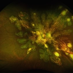
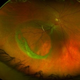
Fundus photograph of a 29 year-old female patient diagnosed with Serpiginous Choroidopathy. Finings were bilateral. The most common complication of SC is choroidal neovascularization affecting up to 35% of patients. Other reported complications are subretinal fibrosis, cystoid macular edema, branch vein occlusion, serous retinal detachment, optic disc neovascularization ,and anterior uveitis.
Photographer: @eyemissu2
Imaging device: TOPCON TRC-50DX
Condition/keywords: serpiginous choroiditis
-
 Serpiginous Choroidopathy
Jun 23 2025 by César Adrián Gómez Valdivia, MD
Fundus photograph of a 29 year-old female patient diagnosed with Serpiginous Choroidopathy. Finings were bilateral. The most common complication of SC is choroidal neovascularization affecting up to 35% of patients. Other reported complications are subretinal fibrosis, cystoid macular edema, branch vein occlusion, serous retinal detachment, optic disc neovascularization, and anterior uveitis.
Photographer: @eyemissu2
Imaging device: California ICG OPTOS
Condition/keywords: serpiginous choroiditis
-
 Subretinal PFO
Jun 18 2025 by Korey Starkey
86-year-old patient had history for retinal detachment surgery x2 and intraocular injections for AMD performed elsewhere. Left eye has PVR developing and subretinal PFO. Due to guarded vision, opting to defer any further treatment at this time.
Photographer: Korey Starkey
Imaging device: Heidelberg
Condition/keywords: AMD, Heidelburg Spectralis, OCT, PFO, PVR, retinal detachment, silicone oil
-
 Complex Retinal Detachment with PVR and Starfold
Jun 6 2025 by Jenn Geelan
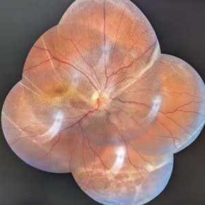
57 year old male with a Complex Retinoschisis related retinal detachment with PVR and a Posterior Star Fold
Photographer: Jenn Geelan, Retina-Vitreous Surgeons of CNY
Imaging device: Optos California
Condition/keywords: proliferative vitreoretinopathy (PVR), rare, Retinal Detachment, retinoschisis, Starfolds, subretinal fluid
-
 Retinal Detachment
Jun 5 2025 by César Adrián Gómez Valdivia, MD
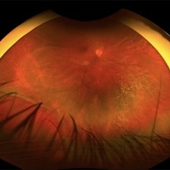
Fundus Photograph of a 19 year-old male patient with a RRD due to a Retinal Dialysis. Subretinal fluid and retinal folding can be appreciated.
Photographer: @eyemissu2
Imaging device: OPTOS
Condition/keywords: retinal detachment
-
 Retinal Detachment
Jun 5 2025 by César Adrián Gómez Valdivia, MD
Fundus Photograph of a 19 year-old male patient with a RRD due to a Retinal Dialysis. Subretinal fluid and retinal folding can be appreciated.
Photographer: @eyemissu2
Imaging device: TOPCON TRC-50DX
Condition/keywords: retinal detachment
-
 When the Macula Decides to Bleed... Artistically (Case of Macular Scar with Subretinal Bleed)
Jun 2 2025 by rohan jain
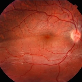
A case of 42 years old male. Color photograph showing macular scar with subretinal bleed.
Photographer: Dr. ROHAN JAIN
Imaging device: mirante
Condition/keywords: CNVM, macular scar, scar, subretinal hemorrhage, subretinal blood
-
 Rhegmatogenous Retinal Detachment
May 19 2025 by Saarang Hansraj
Rhegmatogenous retinal detachment with grade C subretinal PVR
Condition/keywords: proliferative vitreoretinopathy (PVR), Rhegmatogenous retinal detachment
-
 VKH Pseudotumor – Chronic Subretinal Fibrosis
May 11 2025 by Felipe Murati
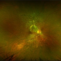
Ultra-widefield fundus image from a 36-year-old woman with chronic VKH syndrome showing a pseudotumor-like subretinal fibrotic lesion in the right eye. The lesion developed after multiple relapses and remained stable over a 1-year follow-up with immunosuppressive treatment including prednisone, mycophenolate mofetil, and adalimumab. No active choroiditis or exudative detachment was observed. Multimodal imaging was essential for disease monitoring.
Photographer: Felipe A. Murati, MD, University of Arizona
Imaging device: Optos California ultra-widefield retinal imaging system, single-capture, color fundus modality.
Condition/keywords: adalimumab, chronic inflammation, granulomatous uveitis, OCT, Optos ultra-widefield imaging, pseudotumor, subretinal fibrosis, VKH, Vogt-Koyanagi-Harada
-
 VKH Pseudotumor – Fluorescein Angiography
May 11 2025 by Felipe Murati
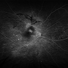
Fluorescein angiography image from a 36-year-old woman with chronic Vogt-Koyanagi-Harada (VKH) syndrome showing a pseudotumor-like lesion with late-phase staining and no active leakage. The image highlights subretinal fibrosis in the right eye, stable under long-term immunosuppressive therapy with mycophenolate mofetil and adalimumab. No signs of active choroiditis are present, confirming a quiescent phase.
Photographer: Felipe A. Murati, MD, University of Arizona
Imaging device: Optos California, fluorescein angiography modality
Condition/keywords: choroiditis, Fluorescein angiography, granulomatous uveitis, Optos FA, pseudotumor, subretinal fibrosis, VKH, Vogt-Koyanagi-Harada
-
 Scleral Rupture
May 9 2025 by Gustavo Uriel Fonseca Aguirre
This B-mode longitudinal ultrasound scan reveals dense vitreous hemorrhage, subretinal fluid, annular choroidal detachment, and scleral wall discontinuity with adjacent scleral folds. These findings indicate severe ocular trauma with probable scleral rupture and multi-compartment involvement.
Photographer: Gustavo U. Fonseca Aguirre, Hospital Conde de Valenciana, Ciudad de México
Condition/keywords: ocular trauma, scleral rupture
-
 Diabetic Macular Edema
Apr 28 2025 by Gustavo Uriel Fonseca Aguirre
This B-mode longitudinal ultrasound scan demonstrates irregular macular thickening with homogeneous medium-to-high internal reflectivity, consistent with diabetic macular edema. The lesion shows poorly defined borders and absence of cystic spaces or subretinal fluid on dynamic evaluation.
Photographer: Gustavo U. Fonseca Aguirre, Hospital Conde de Valenciana, Ciudad de México
Condition/keywords: diabetic macular edema
-
 Sub retinal Bleed
Apr 23 2025 by Anjana Mirajkar, MS Ophthalmology
A widefield image of right eye of a 65 year old male showing large subretinal bleed at the posterior pole most likely in a case of PCV.
Photographer: Dr. Anjana Mirajkar- HV desai eye hospital ,Pune
Imaging device: optos
Condition/keywords: idiopathic polypoidal choroidal vasculopathy, subretinal blood
-
 Retinoschisis
Apr 21 2025 by Gustavo Uriel Fonseca Aguirre
This B-mode longitudinal ultrasound scan reveals a peripheral temporal retinoschisis, demonstrating a characteristic thin, dome-shaped separation of the retinal layers without associated subretinal fluid or vitreous traction. The lesion shows smooth, convex contours with maintained structural integrity of both retinal layers.
Photographer: Gustavo U. Fonseca Aguirre, Hospital Conde de Valenciana, Ciudad de México
Condition/keywords: retinoschisis
-
Vitreous Waltz vs Retinal Rigidity
Apr 18 2025 by Gustavo Uriel Fonseca Aguirre
B-mode dynamic ultrasound of an eye with vitreous hemorrhage shows hyaloid traction inducing retinal detachment in diabetic retinopathy. The video clearly delineates all anatomical compartments: vitreous, subhyaloid, and subretinal spaces. Characteristic movement patterns are observed - the vitreous demonstrates smooth, wide excursions while the detached retina shows shorter, stiffer motions -confirming tractional pathology.
Condition/keywords: diabetic retinopathy, retinal detachment
-
 Proliferative Vitreoretinopathy
Apr 17 2025 by Gustavo Uriel Fonseca Aguirre
This B-mode transverse ultrasound scan depicts a post-vitrectomy eye with recurrent retinal detachment in a patient with diabetic retinopathy history. The image reveals fresh vitreous cavity hemorrhage and subretinal bleeding, along with subretinal proliferative bands (PVR strands). These findings indicate complicated tractional re-detachment with active hemorrhagic components.
Photographer: Gustavo U. Fonseca Aguirre, Hospital Conde de Valenciana, Ciudad de México
Condition/keywords: proliferative vitreoretinopathy (PVR)
-
 Choroidal Osteoma
Apr 17 2025 by Gustavo Uriel Fonseca Aguirre
Scanning laser ophthalmoscopy reveals a well-circumscribed, yellowish-white choroidal osteoma overlying the macular region and extending into the inferior temporal vascular arcade. Retinal vessels course normally over the tumor surface, with no evidence of subretinal fluid or hemorrhage. The surrounding retina shows preserved architecture without secondary degenerative changes.
Photographer: Gustavo U. Fonseca Aguirre, Hospital Conde de Valenciana, Ciudad de México
Condition/keywords: choroidal osteoma, macular choroidal osteoma
 Loading…